Download presentation
Presentation is loading. Please wait.

1
Cardiovascular system By: Dr Hossam El-deen Salem

3
Heart

5
I. Endocardium Endothelium of simple squamous epithelium Subendothelial layer of loose connective tissue Structure of the heart II. Myocardium (the thickest layer of the heart). The contractile cardiac muscle fibers are arranged as sheets in a complex and spiral manner Impulse-conducting system [S-A node, A-V node, A-V bundle, and Purkinje fibers] is formed of specialized (conducting) noncontractile cardiac muscle fibers III. Epicardium layer of connective tissue (rich in fat) and contains blood vessels, lymphatic and nerve fibers. The visceral layer of pericardium
. The contractile cardiac muscle fibers are arranged as sheets in a complex and spiral manner Impulse-conducting system [S-A node, A-V node, A-V bundle, and Purkinje fibers] is formed of specialized (conducting) noncontractile cardiac muscle fibers III. Epicardium layer of connective tissue (rich in fat) and contains blood vessels, lymphatic and nerve fibers. The visceral layer of pericardium.")
6
Blood vessels

7
I. Tunica intima Endothelium of simple squamous epithelium Subendothelial layer of loose connective tissue Internal elastic lamina: elastic fibers (may be absent) General Structure Blood vessels II. Tunica media Circular smooth muscle fibers. External elastic lamina: elastic fibers (may be present) III. Tunica adventitia Loose connective tissue Small blood vessels of the blood vessels (called vasa vasorum) are present in this coat. [because in larger vessels the wall is too thick to be nourished only from diffusion from the blood in the lumen]
 General Structure Blood vessels II. Tunica media Circular smooth muscle fibers. External elastic lamina: elastic fibers (may be present) III. Tunica adventitia Loose connective tissue Small blood vessels of the blood vessels (called vasa vasorum) are present in this coat. [because in larger vessels the wall is too thick to be nourished only from diffusion from the blood in the lumen].")
8
Walls of both arteries and veins have a tunica intima, tunica media, and tunica adventitia, which correspond roughly to the heart's endocardium, myocardium and epicardium. An artery has a thick tunica media and relatively narrow lumen. A vein has a thick tunica adventitia and a larger lumen The tunica intima of veins is often folded to form valves. Capillaries have only an endothelium, with no subendothelial layer or other tunics. Differences between types of blood vessels

9
Large (elastic) arteries Need to be elastic to accommodate systole and diastole Tunica media is rich in elastic fibers Internal and external elastic laminas are present but not easily distinct (because the media is rich in elastic fibers) Medium (muscular) arteries Thick tunica media (well developed muscular wall) Control blood flow to organs by contracting or relaxing the smooth muscle cells of the tunica media Prominent internal elastic lamina Arteriole Narrow lumen (about 0.5 mm) No internal or external elastic laminas Arteries
 arteries Need to be elastic to accommodate systole and diastole Tunica media is rich in elastic fibers Internal and external elastic laminas are present but not easily distinct (because the media is rich in elastic fibers) Medium (muscular) arteries Thick tunica media (well developed muscular wall) Control blood flow to organs by contracting or relaxing the smooth muscle cells of the tunica media Prominent internal elastic lamina Arteriole Narrow lumen (about 0.5 mm) No internal or external elastic laminas Arteries")
10
Capillaries

13
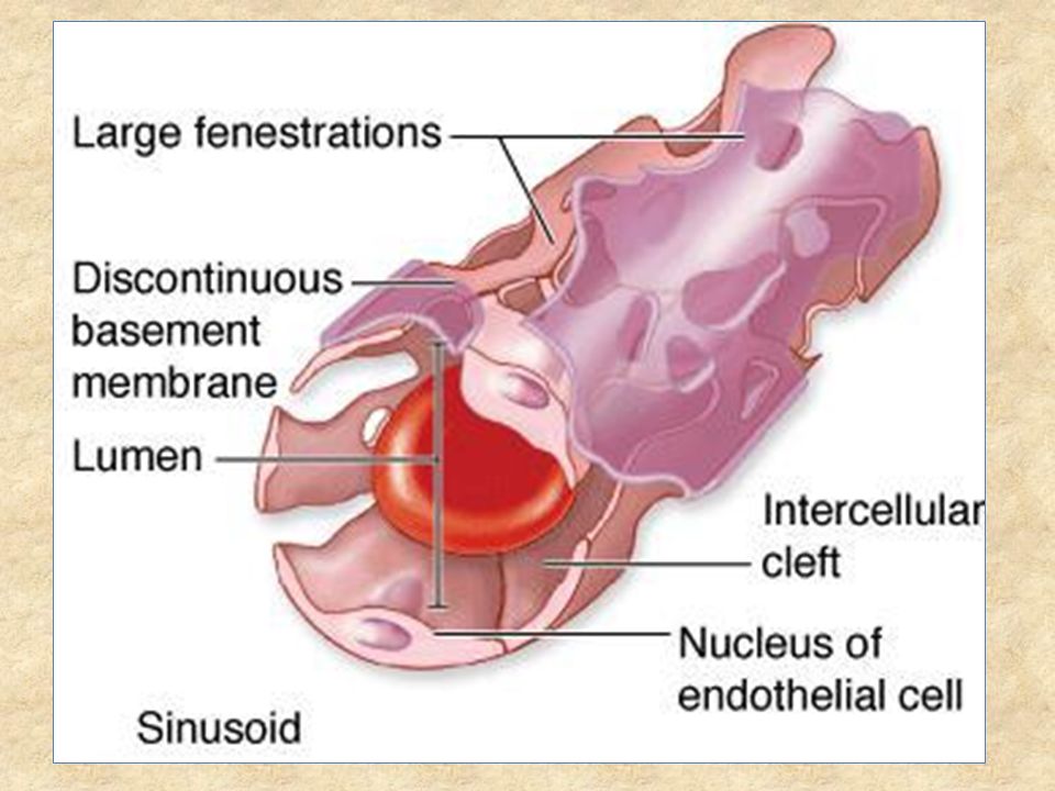
Continuous capillary The most common type of capillary Numerous pinocytotic vesicles are responsible for transfer of macromolecules in both directions across the endothelial cytoplasm. Fenestrated capillary Characterized by the presence of small fenestrae (=perforations), covered by a diaphragm, and the basal lamina is continuous, covering the fenestrae. They are found in tissues where rapid interchange of substances occurs between the tissues and the blood, as in the kidney, the intestine, and the endocrine glands. sinusoid Endothelial cells have large fenestrae without diaphragms, and the basal lamina is discontinuous. So they are discontinuous capillaries that permit maximal exchange of macromolecules and even cells Sinusoids are irregularly shaped and wide (much greater than those of other capillaries). Both causes slow blood flow They found in the liver, spleen, and bone marrow
, covered by a diaphragm, and the basal lamina is continuous, covering the fenestrae. They are found in tissues where rapid interchange of substances occurs between the tissues and the blood, as in the kidney, the intestine, and the endocrine glands. sinusoid Endothelial cells have large fenestrae without diaphragms, and the basal lamina is discontinuous. So they are discontinuous capillaries that permit maximal exchange of macromolecules and even cells Sinusoids are irregularly shaped and wide (much greater than those of other capillaries). Both causes slow blood flow They found in the liver, spleen, and bone marrow.")
Similar presentations


 Dept.of Histology and Embryology.>")


, (10x obj.) TI TM TA elastic fibers.>")


 Arteries; b) Veins; c) Microcirculatory bed 4. Lymphatics 5. Heart.>")