Download presentation
Presentation is loading. Please wait.

1
SKELETAL SYSTEM

2
I’m all about the osseous tissues. But I heard ladies love musicians. So I picked up this Trom-bone.

3
Osseous Tissue and the Skeletal Structure The skeletal system includes: –Bones of the skeleton and associated cartilages –Ligaments and other connective tissues that stabilize and/or connect them

4
Primary Functions Framework: support the body’s muscle fat, and skin (soft tissues). Protection: Surround vital organs to protect them Examples Skull that surrounds brain Ribs that protect heart and lungs. Levers: attach to muscles to help provide movement Produce blood cells: produce red and white blood cells and platelets. Storage: store most of body’s calcium, supply blood

5
Structure of bone Matrix of Bone: -Combination of the two provides strong bones which are somewhat flexible and resistant to shattering. 2/3 of bone weight is calcium phosphate 1/3 of bone weight is collagen fibers

6
Cells in Bones

7
Terms chondro refers to cartilage – chondrocyte – endochondral – perichondrium osteo refers to bone – osteogenesis – osteocyte – periostium blast refers to precursor cell or one that produces something – osteoblast cyte refers to cell – osteocyte

8
Osteocytes= mature bone cells – recycle the calcium salts in the matrix around them – participate in the repair of damaged bone

9
Osteoblasts= responsible for the production of new bone. (osteogenesis) – Elevated local concentrations of calcium phosphate favorable for calcification
 – Elevated local concentrations of calcium phosphate favorable for calcification.")
10
Osteoclasts= giant cells with 50 or more nuclei. – Derived from circulating monocytes (phoagocytic white blood cells) – Important in the regulation of calcium and phosphate concentrations in body fluids Osteoclast dissolving bone
 – Important in the regulation of calcium and phosphate concentrations in body fluids Osteoclast dissolving bone.")
11
Osteoprogenitor= mesenchymal cells that maintain populations of osteoblasts and play an important role in fracture repair

12
Classification of Bones By Shape

13
Long Bones- A long bone is one that is cylindrical in shape, being longer than it is wide. Long bones function as levers; they move when muscles contract. – Examples: humerus, ulna, radius, femur, tibia, fibula). – Keep in mind, however, that the term describes the shape of a bone, not its size.
. – Keep in mind, however, that the term describes the shape of a bone, not its size..")
14
Flat Bones- typically thin and often curved. Serve as points of attachment for muscles and often protect internal organs. – Examples include the cranial (skull) bones, the scapulae (shoulder blades), the sternum (breastbone), and the ribs. Flat bones
 bones, the scapulae (shoulder blades), the sternum (breastbone), and the ribs. Flat bones.")
15
Short Bones- Short bones are shaped similarly to a cube Examples: – The bones in the wrist and ankle

16
Irregular Bones- Bones that do not fall into the category of long, short, or flat are considered irregular bones. Examples: – The vertebrae and some of the skull bones are irregular.

17
Bone Markings Bulges, depressions, and holes that serve as sites of attachment for muscles, ligaments, and tendons, joint surfaces, conduits for blood vessels and nerves.

18
Gross anatomy of bones

19
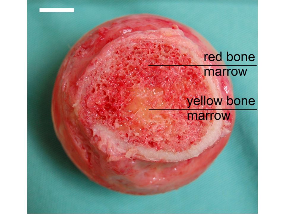
Diaphysis is the tubular shaft that runs between the proximal and distal ends of the bone Medullary cavity- The hollow region in the diaphysis, which is filled with yellow marrow. The walls of the diaphysis are composed of dense and hard compact bone.

20
Long Bone Flat Bone

21
The outer surface of the bone is covered with a fibrous membrane called the periosteum (peri- = “around” or “surrounding”). The periosteum contains blood vessels, nerves, and lymphatic vessels that nourish compact bone. WARNING NEXT SLIDE IS GRAPHIC

23
The medullary cavity has a delicate membranous lining called the endosteum (end- = “inside”; oste- = “bone”), where bone growth, repair, and remodeling occur.
, where bone growth, repair, and remodeling occur.")
24
Tendons and ligaments also attach to bones at the periosteum. The periosteum covers the entire outer surface except where the epiphyses meet other bones to form joints.

25
In this region, the epiphyses are covered with articular cartilage, a thin layer of cartilage that reduces friction and acts as a shock absorber.

26
Epiphysis (plural = epiphyses)- The wider section at each end of the bone, filled with spongy bone. – Red marrow fills the spaces in the spongy bone.

28
Epiphyseal plate (growth plate)- narrow area where the epiphysis meets the diaphysis.Made of a layer of hyaline (transparent) cartilage in a growing bone. – When the bone stops growing in early adulthood (approximately 18–21 years), the cartilage is replaced by osseous tissue and the epiphysealplate becomes an epiphyseal line.
, the cartilage is replaced by osseous tissue and the epiphysealplate becomes an epiphyseal line..")
29
Bone Development and Growth

30
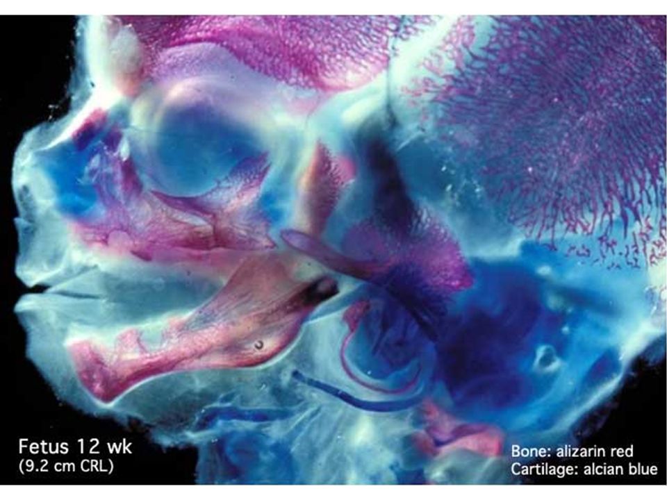
Determines the size and proportions of our body Starts @ 6weeks (cartilaginous) Ends @ ~25 years Growth of the skeleton
 ~25 years Growth of the skeleton")
31
Sultan Kösen 2.51 m (8 ft 3 in) He Pingping 74 cm (2 ft 5 in)
 He Pingping 74 cm (2 ft 5 in)")
32
Growth of the skeleton Ossification= Formation of bone Calcification= deposition of calcium salts within a tissue

33
2 forms of Bone Growth Intramenbranous Ossification deposition of bony matrix in connective tissue membranes Endochondral Ossification replacement of cartilage by bone.

35
Endochondral Ossification 1.Cartilage cells become enlarged in ossification centers – Long bones have three centers: 1 at each end (epiphyses) 1 at the midpoint of diaphysis (shaft) 2.Enlarged cells soon break down leaving spaces filled by osteoblasts (bone forming cells) 3.While organic components of bone are being formed, minerals are being deposited and blood vessels branch throughout the newly forming bone 4.Eventually osteoblasts become osteocytes
 1 at the midpoint of diaphysis (shaft) 2.Enlarged cells soon break down leaving spaces filled by osteoblasts (bone forming cells) 3.While organic components of bone are being formed, minerals are being deposited and blood vessels branch throughout the newly forming bone 4.Eventually osteoblasts become osteocytes")
36
Endochondral ossification Stages 1-3 during fetal week 9 through 9 th month Stage 4 is just before birth Stage 5 is process of long bone growth during childhood & adolescence

37
Intramembranous Ossification 1.Osteoblasts deposit matrix materials in the form of calcium salts 2.Osteoblasts become osteocytes

38
Dynamic Nature of Bone Remodeling- components of bone matrix are continually being recycled and renewed throughout life as part of normal bone maintenance. Young adults remodel 1/5 (20%) of the adult skeleton per year
 of the adult skeleton per year.")
39
Dynamic Nature of Bone Remodeling- components of bone matrix are continually being recycled and renewed throughout life as part of normal bone maintenance. Young adults remodel 1/5 (20%) of the adult skeleton per year
 of the adult skeleton per year.")
40
Diseases and Disorders.

41
Bone Structures

42
Compact Bone Osteon = the functional unit of mature compact bone. (aka Haversian systems/canals) Within osteon, osteocytes are arranged in concentric layers around the central canal. Central Canals run parallel to the surface of the bone, contain one or more blood vessels.
 Within osteon, osteocytes are arranged in concentric layers around the central canal. Central Canals run parallel to the surface of the bone, contain one or more blood vessels..")
43
Nutrients diffuse from vessels in central canal Alternating direction of collagen fibers increases resistance to twisting forces Isolated osteon:

44
Spongy Bone Matrix of spongy bone: – Forms supportive mesh called trabeculae – Seem to align along stress lines – Has no osteons or blood vessels. Nutrients reach osteocytes by diffusion

45
Bones are adaptable, their shapes reflect the forces applied to them Exercise Effects on Bone Heavily stressed bones become thicker and stronger, bones not subject to ordinary stresses will become thin and brittle

46
Moderate amounts of physical activity and weight bearing activities are essential to stimulate normal bone maintenance and maintain adequate bone strength. Exercise Effects on Bone

47
Bone markings reflect the stresses

49
Fracture repair Bones may crack or break if subjected to extreme loads, sudden impacts, or stressed from unusual directions. Fracture categories: Displaced/Non-Displaced Open/Closed.

50
Displaced/ Non-Displaced Displaced fracture- bone ends are not lined up straight. Non-Displaced fracture- bone maintains its proper alignment.

51
Open/Closed Closed fracture- bone breaks but no open wound or puncture in the skin. Open fracture- bone breaks through the skin. (risk of a deep bone infection) X
 X.")
52
WARNING! GRAPHIC CONTENT!

53
Open/Closed Closed Fracture Open Fracture

54
X-Ray of Fracture

55
A. Fracture Hematoma (i.e. blood clot) forms Steps in Bone Healing
 forms Steps in Bone Healing")
56
B. Soft Tissue Callus (1st 3 to 4 weeks) - Fibroblasts and osteoblasts migrate in from the periosteum and endosteum - Fibroblasts lay down a collagen matrix - some of the fibroblasts differentiate into chondroblasts (i.e. cartilage-forming cells) and lay down a fibrocartilage splint (i.e. soft tissue callus)
 - Fibroblasts and osteoblasts migrate in from the periosteum and endosteum - Fibroblasts lay down a collagen matrix - some of the fibroblasts differentiate into chondroblasts (i.e. cartilage-forming cells) and lay down a fibrocartilage splint (i.e. soft tissue callus).")
57
C. Bony Callus (starting 3 to 4 weeks after the injury) - Osteoblasts begin to replace the fibrocartilage splint with spongy and compact bone, forming a bulge that is initially wider than the original bony shaft Steps in Bone Healing
 - Osteoblasts begin to replace the fibrocartilage splint with spongy and compact bone, forming a bulge that is initially wider than the original bony shaft Steps in Bone Healing.")
58
D. Bone Remodeling - As the patient starts to use (or bear weight on) the bone, the bone starts to remodel along lines of maximal stress (this remodeling process requires the activity of both osteoblasts and osteoclasts Steps in Bone Healing
 the bone, the bone starts to remodel along lines of maximal stress (this remodeling process requires the activity of both osteoblasts and osteoclasts Steps in Bone Healing.")
59
END

Similar presentations


 Identifying functions of the skeletal system.>")


 or an organ –Bone referred to as a connective tissue consists of: cells extracellular.>")


 Joints ► Cartilages Ligaments ► Divided.>")


 Joints Cartilages Ligaments Divided into two divisions Axial skeleton –>")

