Download presentation
Presentation is loading. Please wait.

1
The Cellular Level of Organization

2
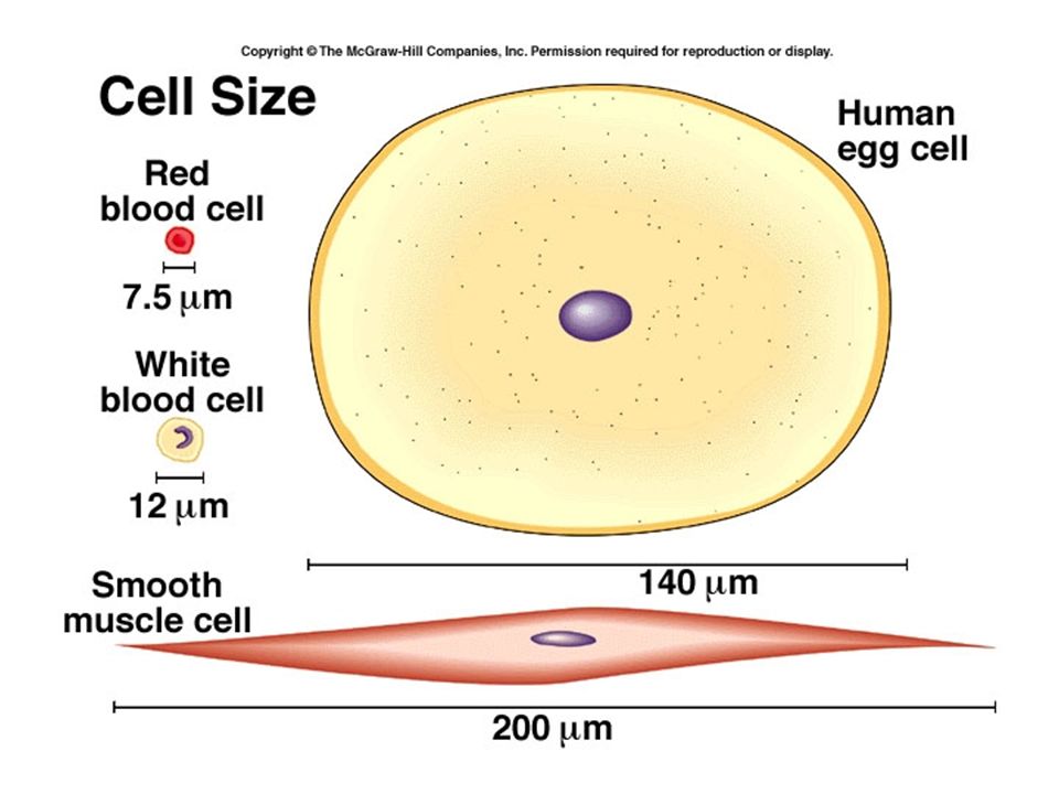
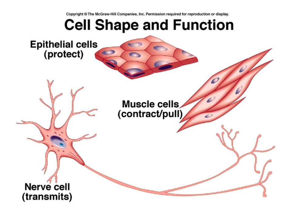
A cell is the basic, living, structural and functional unit of the body. Cells are measured in micrometers. Cells vary in size and shape.

5
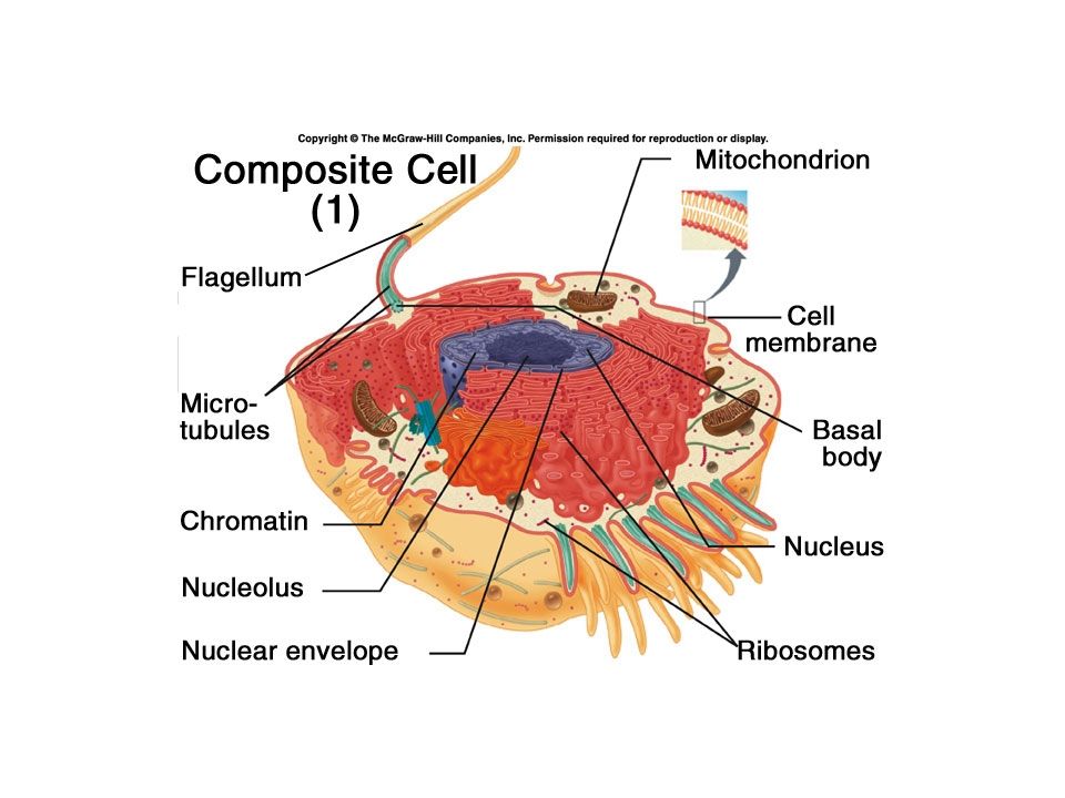
Every Eukaryotic cell has three main parts: Plasma (cell) membrane - separates inside of cell from external environment. Nucleus – organelle that contains the cell’s DNA and is surrounded by a double membrane. Cytoplasm – everything from the nuclear membrane to the plasma membrane

7
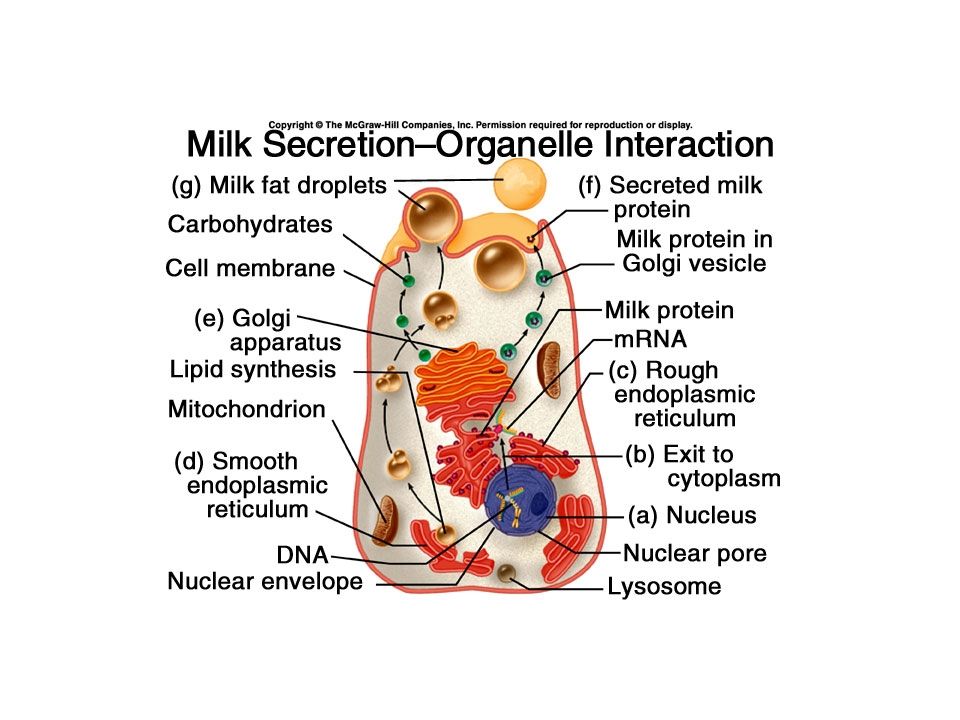
Cytoplasm refers to cytosol plus organelles and inclusions. cytosol - contains proteins, enzymes, nutrients, ions, and other small molecules organelles - highly organized structures with characteristic shapes that are specialized for specific cellular activities. inclusions - are temporary structures in the cytoplasm that contain secretions and storage products of the cell.

8
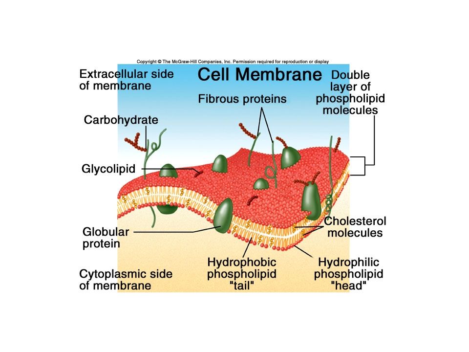
Cell Membrane The plasma membrane does more than just separate the outside of a cell from the inside; it controls what enters and leaves the cell, and much of the activity within the cell. Most of the cell membrane is made of phospholipid. Also find glycolipids and cholesterol. Amphipathic molecule – phosphate heads on the outside and inside, and fatty acid tails in the middle.

10
The membrane is selectively permeable – it allows fat soluble substances to pass through (such as steroid hormones) and some other small, uncharged molecules.. Glycolipids are the targets of certain bacterial toxins, and are important for cell adhesion and communication. Cholesterol is a large molecule, and helps to stabilize the membrane.

11
Fluid mosaic model - proteins float like ice burgs in a sea of phospholipids. Proteins can be integral proteins – go all the way through the membrane, or may be peripheral proteins -bound to the inside or outside membrane.

13
Integral Proteins can be channels or transporters. Peripheral proteins can be receptors, or can be cell identity markers that identify a cell as “self” (like UPC codes). These are often glycoproteins. They may also mark worn out red blood cells or cells that have been infected with a virus.
. These are often glycoproteins. They may also mark worn out red blood cells or cells that have been infected with a virus..")
14
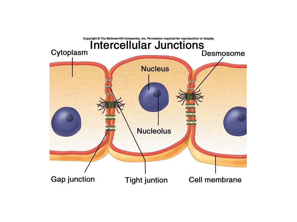
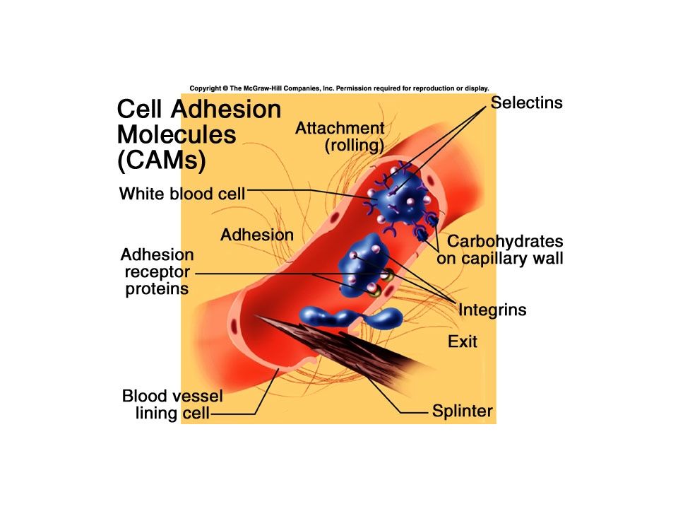
Intercellular junctions: Tight junctions – membranes of adjacent cells come together and fuse, forming a barrier to substances that want to pass between cells. Desmosomes are protein “spot welds” in skin and cardiac muscle. Gap junctions are tubular channels that connect the cytoplasm of one cell with that of another. Cellular Adhesion Molecules help cells form temporary attachments to other cells. CAMs

17
Membrane Physiology Cell membrane function: –Cellular communication –Establish an electrochemical gradient –Are selectively permeable Lipids Size Electrical charge Presence of channels and transporters

18
Movement of materials Passive processes: –Depend on concentration and kinetic energy –Do not require energy –Move substances from an area of high concentration to an area of low concentration Down a concentration gradient

19
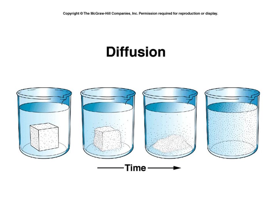
Diffusion Passive process Reaches equilibrium or Physiological steady state

24
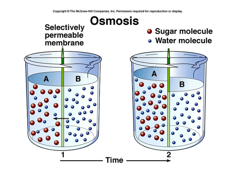
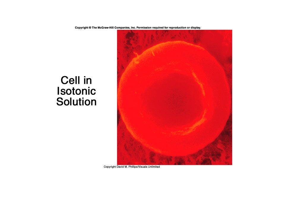
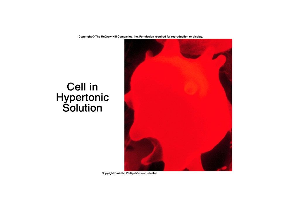
Tonicity Concentration of one solution relative to another Isotonic – equal concentrations Hypertonic – more concentrated Hypotonic – less concentrated

29
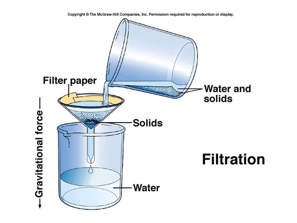
Filtration -a type of bulk flow where the movement of water and dissolved substances across a membrane is due to gravity or hydrostatic pressure (water pressure).
.")
32
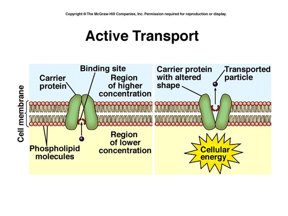
Active Transport Depends on the use of energy (ATP) Moves substances up a concentration gradient (up hill) These systems are often called “pumps” –Na+ / K+ pump
 Moves substances up a concentration gradient (up hill) These systems are often called pumps –Na+ / K+ pump")
34
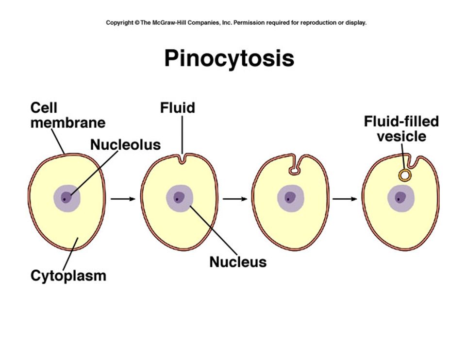
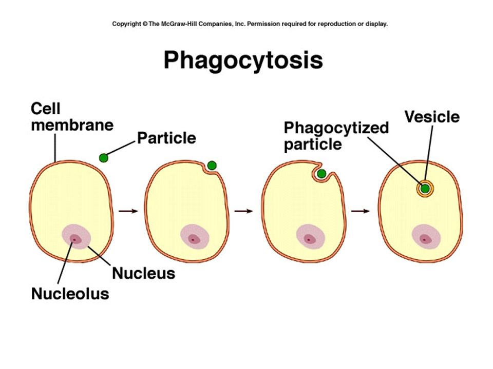
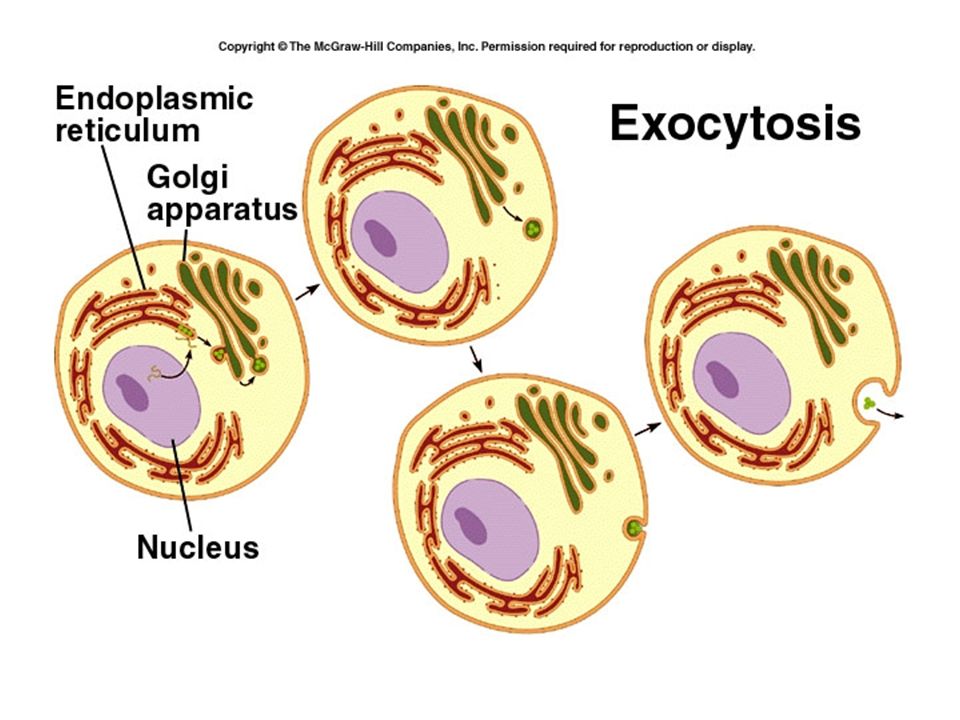
Vesicular Transport Exocytosis – moving substances outside the cell Endocytosis – taking substances into the cell Pinocytosis – “cell drinking” Phagocytosis – “cell eating” Receptor mediated endocytosis

40
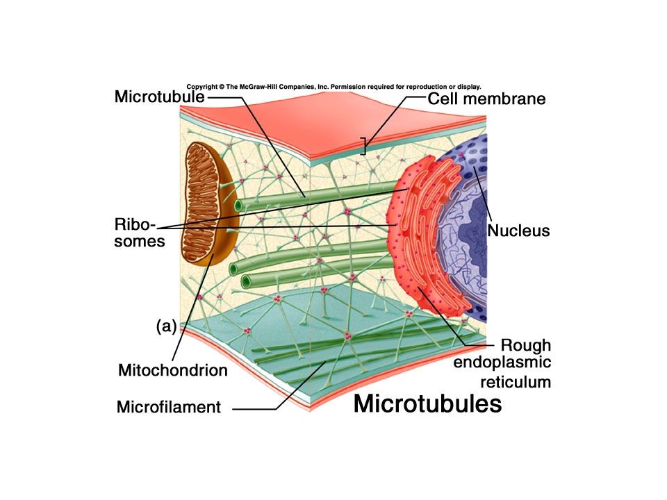
Cytoplasm Cytosol –Semifluid, mostly water –Protein, carbohydrates, lipids, and inorganic substances (ions) –Many important metabolic reactions take place here –Cytoplasm is the cytosol plus the organelles
 –Many important metabolic reactions take place here –Cytoplasm is the cytosol plus the organelles")
41
Organelles “little organs” – have characteristic appearance and have specialized functions. Ribosomes – made of ribosomal RNA and protein, these are the “work benches” where proteins are put together.

42
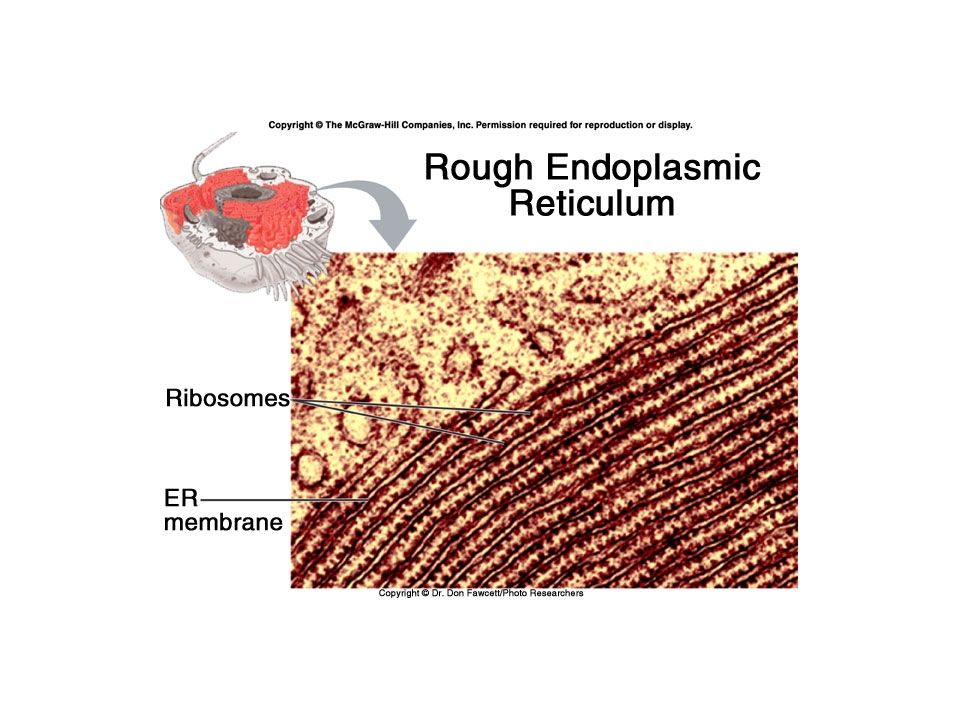
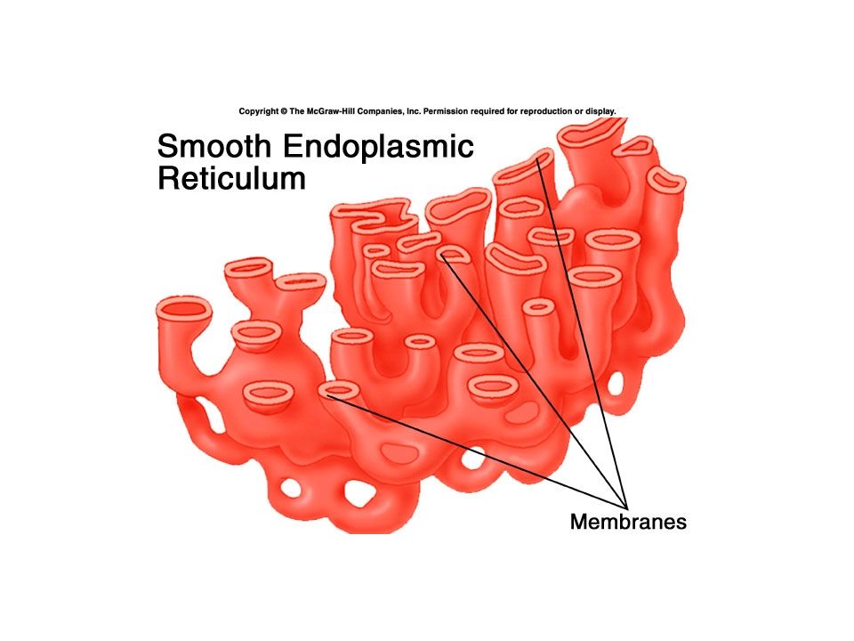
Endoplasmic reticulum (ER) Can be rough or smooth Rough ER has ribosomes, makes proteins for export outside the cell. Smooth ER is the site of fatty acid, phospholipid and steroid synthesis. In certain cells also detoxifies chemicals, such as alcohol and pesticides.

46
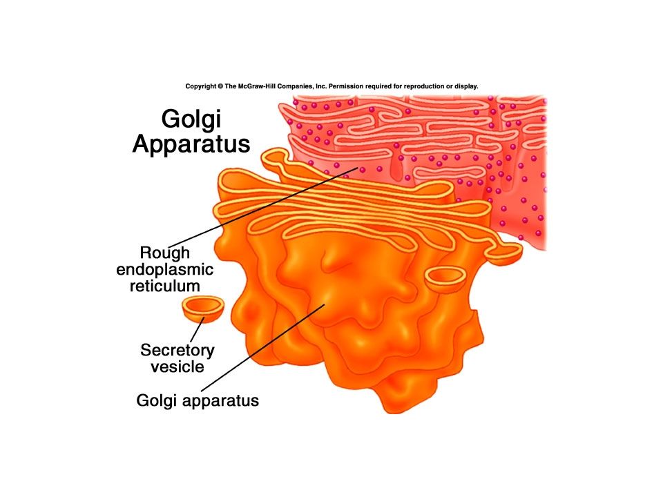
Gogli apparatus (body, complex) Made of flattened sacs called cisternae Process, sort and deliver proteins and lipids to the plasma membrane and forms vesicles and lysosomes. The “UPS” of the cell

50
Mitochondria Mitochondrion – singular Two membranes – inner folds called cristae. Main function is the use of oxygen to produce ATP – cellular (aerobic) respiration These are the “power plants” of the cell.
 respiration These are the power plants of the cell..")
53
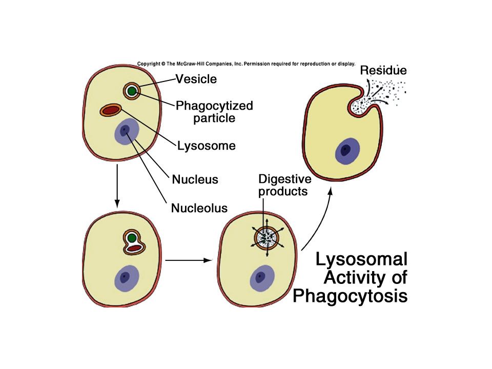
Lysosomes The cell’s “stomach” – vesicles that contain digestive enzymes. Tay-Sachs disease Peroxisomes are vesicles that have enzymes that use oxygen to digest organic substances and produce hydrogen peroxide. Also make catalase to break down H 2 O 2

55
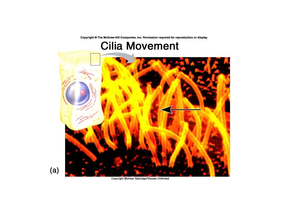
Centrosomes and Centrioles Centrioles are 2 cylinders of tubules arranged at right angles. Form the microtubules of the mitotic spindle during cell division, and also make up a part of cilia and flagella

61
Flagella

62
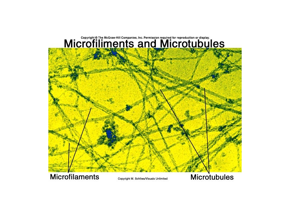
Cytoskeleton Microfilaments and microtubules Give the cell shape, and allow it to move – the Musculoskeletal system of the cell. Myofilaments are made of the protein actin Myotubules are made of the protein tubulin

65
Vesicles Membrane sacs that transport substances within the cell. Vesicle trafficking

66
Inclusions Usually contain chemical substances produced by the cell, these are temporary structures that are not surrounded by a membrane. Melanin, glycogen, triglycerides ribosomes

67
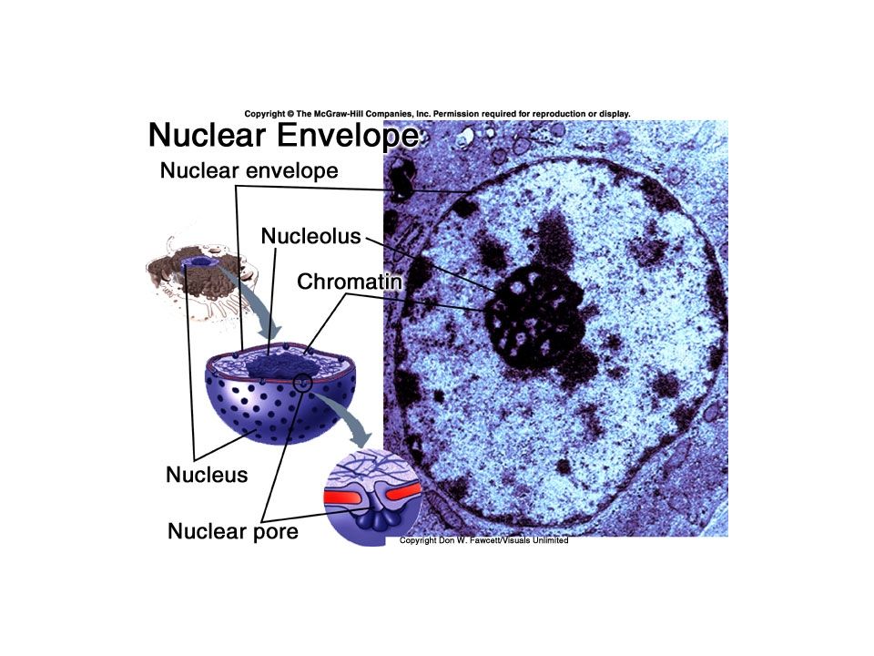
Nucleus Double membrane called the nuclear envelope Nucleoplasm Chromatin granules – unwound DNA Nucleoli – puts RNA and protein together to make ribosomes Nucleus is essential for cell survival

69
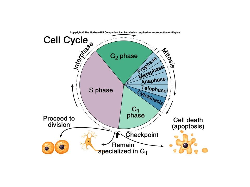
The Cell Cycle Nuclear division (mitosis or meiosis) Cytoplasmic division – cytokinesis Mitosis is somatic (body) cell division Meiosis is reproductive cell division
 Cytoplasmic division – cytokinesis Mitosis is somatic (body) cell division Meiosis is reproductive cell division")
70
Mitosis Homologous chromosomes Cell cycle – from one cell division to the next Interphase – “resting phase” G 1 – Gap1 – growth phase S – Synthesis – replication of DNA G 2 – Gap 2

72
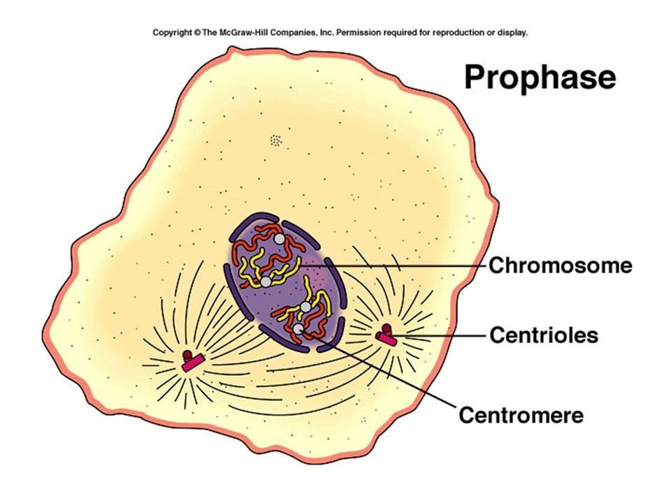
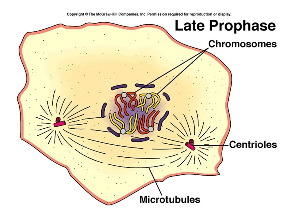
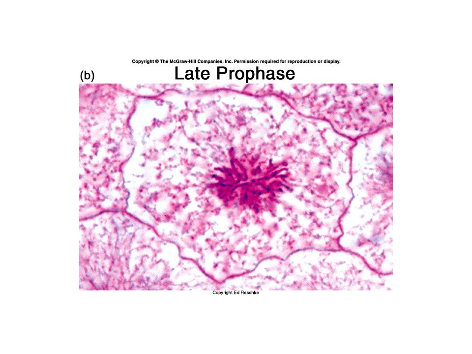
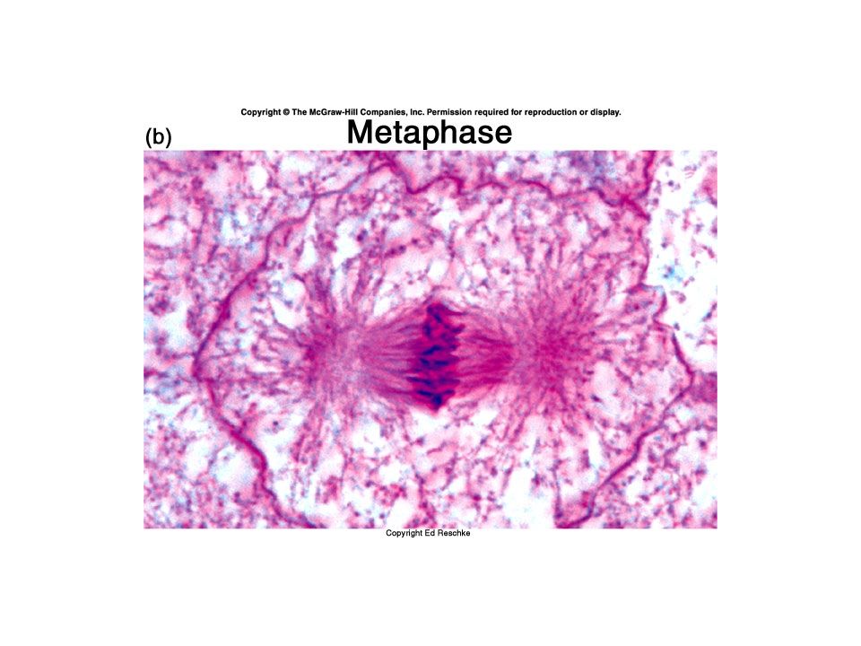
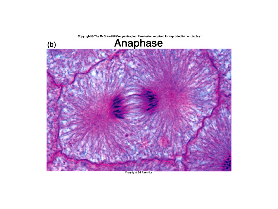
Nuclear division – mitosis or karyokinesis Prophase Metaphase Anaphase Telophase PMAT

85
Cytokinesis Contractile ring of actin microfilament Cleavage furrow Two new daughter cells

90
Control of cell division How many times a cell divides depends on the type of cell Stem cells retain the ability to divide and differentiate Cell senescence Telomeres Levels of proteins called kinases and cyclins

91
Control – contd. Cell size External factors –hormones and growth factors Contact inhibition

92
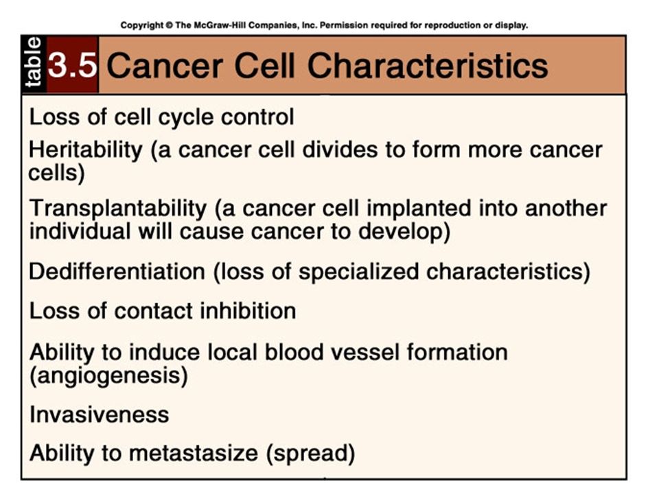
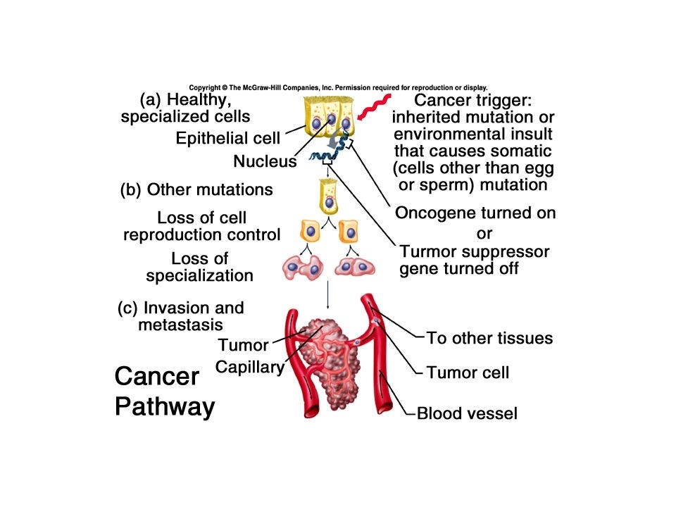
Loss of control over division Neoplasm or tumor Benign tumors remain in a single location Malignant tumors (cancer) can spread or metastasize Oncogenes – want these turned “off” Tumor suppressor genes – want these turned “on” Apoptosis
 can spread or metastasize Oncogenes – want these turned off Tumor suppressor genes – want these turned on Apoptosis")
Similar presentations


>")


 Relationship.>")


