Download presentation
Presentation is loading. Please wait.

1
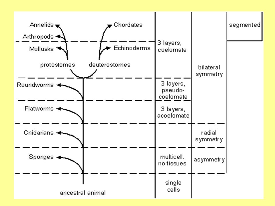
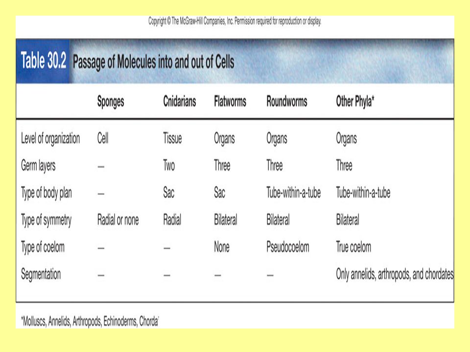
Evolution of Animals Fossil record of invertebrates is incomplete because soft-bodied animals are poorly preserved as fossils. All animals probably evolved from Protists. The classification of animals is based on the level of organization or number of germ layers, symmetry, type of coelom, body plan, and presence or absence of segmentation. The following evolutionary tree is based on these features and shows a possible evolutionary relationship between the animals.

2
Evolutionary tree

5
Levels of Organization Three levels or organization: cell, tissue, or organ One of the main events during animal development is the establishment of germ layers. If two germ layers (ectoderm and endoderm) are present, then the animal has the tissue level of organization. If all three germ layers (ectoderm, endoderm, and mesoderm) are present, then the animals has the organ level of organization.
 are present, then the animal has the tissue level of organization. If all three germ layers (ectoderm, endoderm, and mesoderm) are present, then the animals has the organ level of organization..")
6
Type of Body Plan Two body plans are present in the animal kingdom: Sac plan: Incomplete digestive system with only one opening. Ex: Jellyfish & planaria

7
Tube-within-a-tube plan: Complete digestive system. Two openings allows for specialization along the length of the tube. Ex: Roundworms, earthworms, insects

8
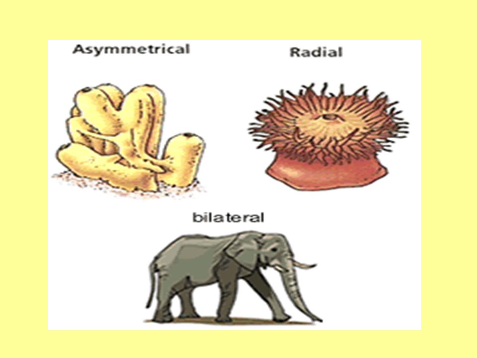
Type of Symmetry Animals can be asymmetrical, radially symmetrical, or bilaterally symmetrical. Asymmetrical animals have no particular symmetry. Radial symmetry means the animal is organized similar to a wheel. Bilateral symmetry means the animal has definite right and left halves. Bilateral symmetry leads to cephalization (brain and sense organs located an anterior end of animal).
..")
11
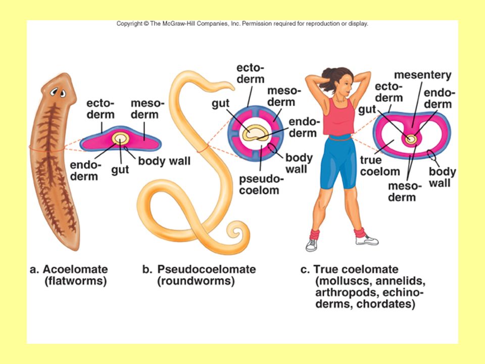
Type of Coelom A true coelom (in coelomates) is an internal body cavity completely lined with mesoderm, where internal organs are found. Benefits: Allows organs to freely move, grow, and develop independently of the body wall Fluid cushions and protects organs from shocks – in some acts as a skeleton Allows for separation of digestion & circulation Allows for increase in size & metabolic rate. Coelomates are either protostomes or deuterostomes (explained a few slides later). Ex: Annelids, mollusks, arthropods
. Ex: Annelids, mollusks, arthropods.")
12
Animals that have a pseudocoelom have a body cavity incompletely lined with mesoderm. No circulatory system – fluid in pseudocoelom transports oxygen & nutrients Pressure of fluid inside also provides support as would a skeleton More mobile, more complex reproductive & digestive systems. They can store wastes for discharge out of the body. Ex: Roundworms, rotifers

13
Acoelomates have mesoderm but no body cavity. Movement squeezes & distorts the body, restricting the flow of nutrients and other materials Have no circulatory system. Must rely either on diffusion or on muscle contractions for the transport of nutrients, respiratory gases, and waste products around the body. (Less efficient than heart & blood vessels which develop in those with a coelom.) Ex: Flatworms, sponges, jellyfish
 Ex: Flatworms, sponges, jellyfish.")
15
Protostomes vs. Deuterostomes During development, when the embryo resembles a tiny globe of cells, a small pucker develops on one side of the embryo. This grows into a pocket, and allows some cells to migrate inside to form an additional layer of cells within the outer layer. In the Protostomes, the mouth develops from the edge of this pocket; the anal opening develops later. In the Deuterostomes, the reverse is true; the pocket edge develops into the anus, and the mouth is formed later.

17
Segmentation Segmentation is the repetition of body parts along the length of the body. Animals can be segmented or nonsegmented. Segmentation leads to specialization of parts because the various segments can become differentiated for specific purposes. Ex: annelids, arthropods, and chordates (includes vertebrates).
..")
18
Phylum Porifera - Sponges Meaning: Pore-bearing Symmetry – none (asymmetrical) Organization – cellular level Acoelomates – no body cavity (so no organs) Non-segmented Habitat – fresh & salt water
 Organization – cellular level Acoelomates – no body cavity (so no organs) Non-segmented Habitat – fresh & salt water")
19
Anatomy (see below and on next slide)
")
20
Flagella Filaments Collar cell Internal cavity Epidermis Flagella Osculum Amebocyte Collar cell Pore channel Pores Spicules

21
Anatomy Epidermis – found along outer body wall Amoeboid cells (amebocytes) – middle layer Transport nutrients Produce spicules Form sex cells Collar cells - inner layer – digest nutrients Collar cells contain the following: Flagella - pull water in through pores and circulate water though the sponge Filaments - trap food particles.
 – middle layer Transport nutrients Produce spicules Form sex cells Collar cells - inner layer – digest nutrients Collar cells contain the following: Flagella - pull water in through pores and circulate water though the sponge Filaments - trap food particles.")
22
Life processes Sponges are classified according to type of spicules Support - spicules (act like bones for the sponge) Diet - filter feeders – filter bacteria, protists, and sometimes small crustaceans Feeding – –Filaments trap food –Collar cells digest food (engulf food particles (endocytosis), digest them, and pass them to amoeboid cells.
 Diet - filter feeders – filter bacteria, protists, and sometimes small crustaceans Feeding – –Filaments trap food –Collar cells digest food (engulf food particles (endocytosis), digest them, and pass them to amoeboid cells.")
23
Movement: - Swim as larva - Sessile (permanently attached to a surface) as adults. Response - no nervous system. Excretion - through the osculum. Respiration - take in oxygen as water passes through body – diffusion. Internal transport - ameboid cells transport nutrients around the body from cell to cell

24
Reproduction – Hermaphrodites (make eggs & sperm). Asexually by: Budding: Produce internal buds called gemmules (cells containing ameboid cells, organic molecules, & spicules) that can grow into new sponges when the conditions are more favorable Regeneration: Growth of a whole organism from a fragment
 that can grow into new sponges when the conditions are more favorable Regeneration: Growth of a whole organism from a fragment.")
25
Sexually: Release sperm into the water Sperm enters pores of another sponge of same species Fertilizes egg within Larva released through osculum (see life cycle diagram – separate sheet). Some will release eggs & sperm into internal cavity, larva develops, and is released into water. Poor reproductive odds!!! http://www.eeob.iastate.edu/faculty/DrewesC/htdocs/Q-sponge.htm

26
5. 6. 7. 1. 2. 3. 4. 1.Sperm 2.Egg 3.Dividing cells (spongocoel) 4.Larva 5.Larva released 6.Flagella (moves) 7.New sponge develops
 4.Larva 5.Larva released 6.Flagella (moves) 7.New sponge develops.")
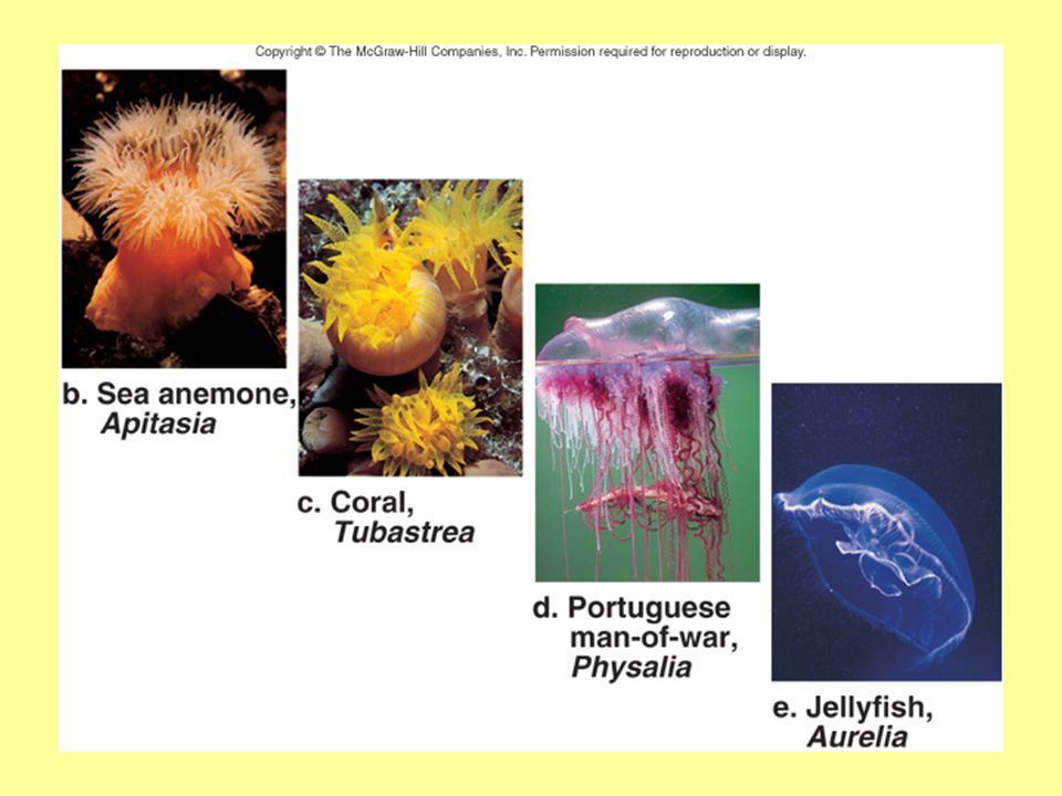
27
Phylum Cnidaria Named for – specialized stinging cells called cnidocytes (contain the stingers called nematocysts – these may contain poison) Symmetry – radial Organization – have endoderm & ectoderm - tissue level Acoelomates – no body cavity (so no organs) Non-segmented; sac body plan Habitat – mostly salt water, hydra found in fresh water
 Symmetry – radial Organization – have endoderm & ectoderm - tissue level Acoelomates – no body cavity (so no organs) Non-segmented; sac body plan Habitat – mostly salt water, hydra found in fresh water")
28
Body forms: may be a polyp or a medusa, or may alternate between the two forms. Polyp – tentacles up, usually sessile Medusa – tentacles down, usually active

30
Examples of animals – Sea anemone – solitary polyp, very colorful Coral – some solitary, most colonial, polyp form, calcium carbonate skeleton, form reefs Portuguese man-of-war – colony of polyp & medusa individuals, each with specialized jobs such as feeding & reproduction Jellyfish – medusa, can live at great depths Hydra – freshwater, polyp form, commonly attached to underwater rocks or plants, less than 1cm in length

32
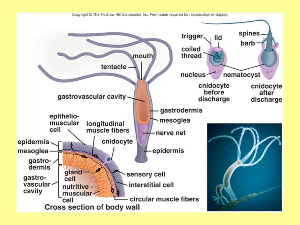
Basal disc Ovary Developing egg Sperm Testis Bud ------------------------GVC – Gastrovascular cavity -------Gastrodermis Epidermis---- Stinging capsule Tentacle Mouth

33
Sac body plan – single opening serves as mouth and anus Outer tissue layer – epidermis Inner tissue layer – gastrodermis Mesoglea – jelly-like material that separates above 2 tissue layers Circular & longitudinal muscle fibers Tentacles surround mouth Large space inside called gastrovascular cavity

34
Life processes Movement: Directional Move by contraction & expansion of body Tentacles can grab prey Response: Nerve net – interconnecting nerve cells communicate with sensory cells throughout body

35
Support Hydrostatic skeleton –fluid-filled closed chambers; internal pressures generated by muscle contractions cause movement as well as maintain the shape of the animals

36
Diet – protists and small animals Feeding – Sting prey with nematocysts Stuff food into mouth using tentacles Don’t chase their prey – but movement of medusa can help drawn food in towards body. Food passes into GVC (gastrovascular cavity) Digestion – cells of gastrodermis take in food through endocytosis and digest the food in their vacuoles.
 Digestion – cells of gastrodermis take in food through endocytosis and digest the food in their vacuoles..")
37
Internal transport: Nutrients and oxygen pass from cell to cell through the process of diffusion Respiration: Diffusion between epidermal cells and exterior watery environment, and between gastrodermal cells and fluid within GVC Excretion: Wastes are excreted through the mouth

38
Reproduction: Usually appear as separate males & females Sexual reproduction – see life cycle diagram given separately Jellyfish – Release eggs & sperm into water Swim as larva, settle as polyps, then divide to become medusa Several young from one fertilized egg Hydra – release egg or sperm from body wall; meet, swim as larva, settle as polyp

39
Medusa Polyp Planula Blastula Egg Sperm

40
Asexual reproduction Budding – cluster of cells form, break off, grow into adults Regeneration – fragments develop into new animals

41
Comb jellies Nematocysts firing Coral feeding Clown fish in anemone

42
Phylum Platyhelminthes Meaning – flatworms Symmetry - bilateral Sac body plan; non-segmented; acoelomates. Organization - 3 germ layers – endoderm, ectoderm, & mesoderm – organ level Have organs for all life processes except respiration and circulation Habitat – fresh or salt water, moist environments, inside host

43
Examples: Marine flatworms Tapeworms Freshwater planarians Flukes

44
Planarians - Lifestyle - free-living Habitat - freshwater Movement – secrete slime, push through with cilia, muscles Excretion – flame cells – interconnecting canals throughout body Response - small brain - ladder of nerves - light-sensitive eyespots - Auricles sensitive to chemicals (all located in the head - cephalization).
.")
45
Planarian

46
Flame cells Excretory pore Flame cells GVC Pharynx Mouth Endoderm Mesoderm Ectoderm GVC Cilia Eyespot Brain Auricle Longitudinal nerve cord Transverse nerve cord

47
Ovary Oviduct Testes Sperm duct Penis Vagina

48
Diet – plankton (small worms or crustaceans) Feeding – wrap around prey - secrete slime - extend pharynx – sucking motion tears up and swallows food Digestion – occurs in GVC
 Feeding – wrap around prey - secrete slime - extend pharynx – sucking motion tears up and swallows food Digestion – occurs in GVC")
49
Respiration and internal transport - Flattened body allows for diffusion of oxygen & nutrients from cell to cell Gender - hermaphrodites Sexual reproduction – cross-fertilization, have sex, exchange sperm, each planaria gets pregnant - 2 sets of young hatch from eggs 2-3 weeks later.

50
Asexual reproduction – capable of regeneration; 1 worm can even grow 2 heads or 2 tails!!

51
Parasitic Flatworms Two classes - flukes and tapeworms Require host to carry on life cycle – see next slide Primary host – infect as adult Secondary host – infect as larval stage Tapeworm – Anterior scolex with hooks and suckers to hold itself inside the gut. No digestive system; absorbs host’s digested food through skin Special enzymes counteract host’s digestive enzymes

52
Body is an assembly line of square sections called proglottids, which contain male and female sex organs. As proglottids mature, they break off, pass with feces, and release eggs. When animals feed in feces-contaminated food, eggs hatch inside, and larva eventually form cysts in muscles of secondary host. When humans eat infected meat, larva hatch from cyst, attach to intestine, and grow to adulthood.

53
1.Hooks 2.Suckers 3.Scolex 4.Neck 5.Proglottids 6.Testes 7.Ovary 8.Eggs 9.Detached proglottid 10.Longitudinal nerve cord 11.Brain (ganglia) 12.Transverse nerve cord
 12.Transverse nerve cord")
54
Life cycle:

55
Contracted by eating undercooked, infected beef, pork, or fish. Transmitted to cats & dogs by fleas that have eaten feces of infected host Primary symptom – weight loss In prolonged infections – worms migrate to eyes, heart, brain, lungs, & liver & form cysts – cause swelling, cramps, diarrhea, anemia, & seizures Diagnosed by fecal exam; treated with medicine

56
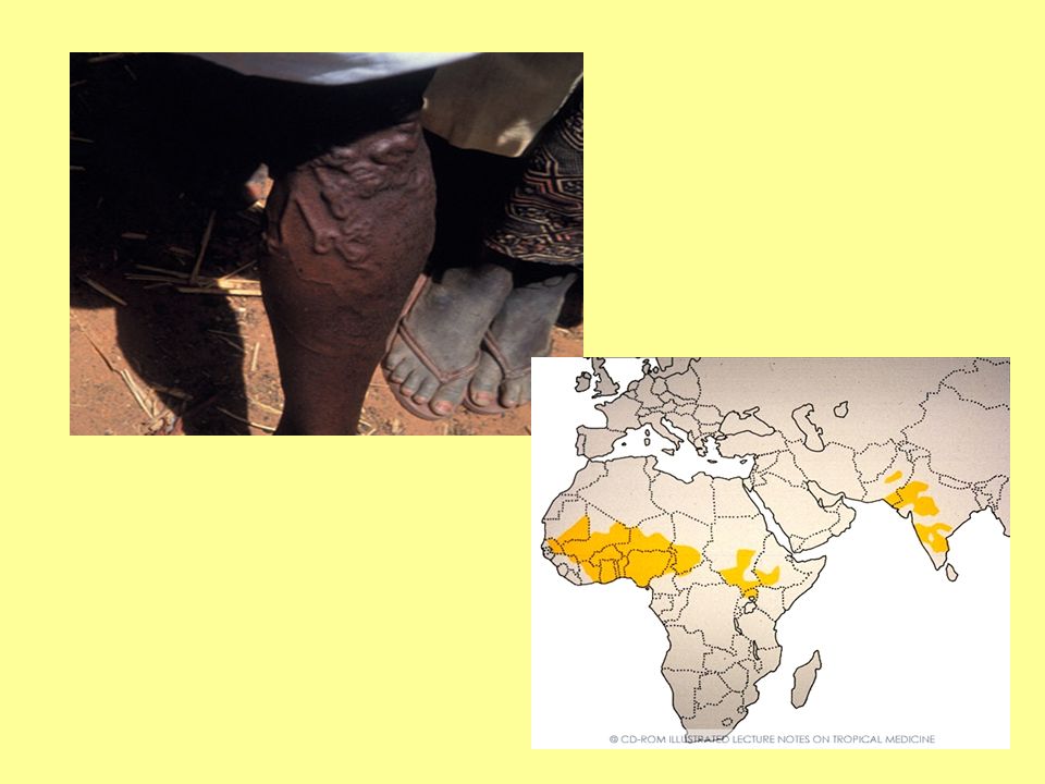
Fluke Have sucker at anterior end to attach to host Various species – can infect digestive tract, bile duct, blood, & lungs. Blood flukes cause schistosomiasis – one of the most common worm infections worldwide (about 200 million in mostly Middle East, Asia, Africa, & S. America) Common in areas with poor sewage treatment Enter through skin when in infected water – see life cycle diagram next slide
 Common in areas with poor sewage treatment Enter through skin when in infected water – see life cycle diagram next slide.")
57
Schistosomiasis

58
Diagosed by fecal exam; treated with medicine Symptoms: nausea, abdominal pain, increased bowel movements, diarrhea, weight loss, fatigue Burrow through host, feed on host’s blood & tissues. Can live for up to 2-3 decades inside host (usually only 5-10 years) Reproduce non-stop – 100 – 300 eggs/day
 Reproduce non-stop – 100 – 300 eggs/day.")
59
Phylum Nematoda (Roundworms) Non-segmented Bilateral symmetry Organization - 3 germ layers – endoderm, ectoderm, & mesoderm – organ level Pseudocoelomates (body cavity) – filled with fluid – space for organs Tube-within-a-tube body plan – complete digestive tract with mouth and anus. Habitat – fresh or salt water, soil, inside host (both plants & animals)
.")
60
Life Processes Internal transport - fluid in pseudocoelom transports nutrients & oxygen Support – fluid in pseudocoelom provides support (hydrostatic skeleton). Respiration - Breathe through their skin - diffusion. Movement - longitudinal muscles – move with whiplike motion – inefficient in water, good in soil & host.

61
Excretion – waste is removed through the anus Feeding & digestion – Feed using muscular pharynx which shoots out from mouth Food is digested in intestine Gender - appear as separate males & females Sexual reproduction – male injects sperm into female, she lays eggs.

62
Roundworm anatomy

63
Types of nematodes: Ascaris Contracted when one eats vegetables contaminated with human waste.

64
Trichina worm: Trichinosis is contracted when eating undercooked pork containing encysted larvae.

65
Filaria worm: Carried by mosquitoes and causes elephantiasis by blocking lymphatic drainage.

66
Pinworms: Common infections in children.

67
Hookworm: Once common in the southern United States. Person becomes infected when walking in soil contaminated with human waste

68
Guinea worm: Person becomes infected when drinking water with water fleas contaminated with guinea worm eggs. Worm emerges from leg once fully grown.

71
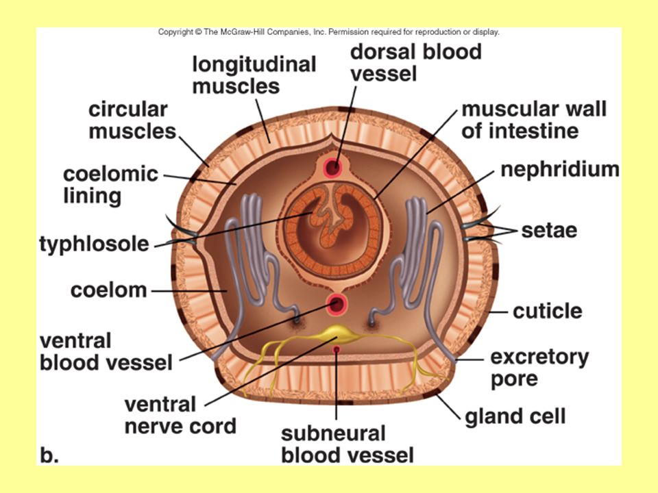
Phylum Annelida (segmented worms) Symmetry - bilateral Organization - 3 germ layers – organ level Coelomates -have a body cavity – more complex organs Tube-within-a-tube body plan – specialized organs in digestive tract Segmented both externally, and internally by partitions called septa. Habitat – fresh or salt water, soil Giant earthworm video clip: http://www.youtube.com/watch?v=DZig6EL5B6A&feature=related

72
Support - a hydrostatic skeleton Movement - move by alternating contraction of longitudinal and circular muscles found in each segment. –Partitioning of the coelom permits each body segment to move independently. Respiration - breathe through their skin. Internal transport - closed circulatory system with blood vessels that run the length of the body and branch to every segment.

73
Excretion - nephridia (tiny tubules found in each segment) remove nitrogen waste through openings in body wall; anus removes waste from digested food. Response - a brain connected to a ventral solid nerve cord with ganglia in each segment. Three classes – Polychaeta – marine worms Oligochaeta – earthworms Hirudinea - leeches

74
Marine Worms Have paddle-like appendages at the side of each segment called parapodia. –Add surface area for respiration Found in bundles on the parapodia - bristles that anchor the worm called setae. Only have functional sex organs during breeding seasons. During breeding seasons, some worms form sex organs in special segments and shed these segment during breeding. Eggs or sperm within float to surface where fertilization takes place – hatch as larva.

75
Lifestyles of these worms vary: Sessile tube worms that filter feed with tentacles Clam worm is a predator with powerful jaws, & a defined head region with eyes & sense organs.

76
Earthworm, Lumbricus

78
1.Mouth 2.Pharynx 3.Esophagus 4.Crop 5.Gizzard 6.Intestine 7.Anus 8.Aortic arches (hearts) 9.Dorsal blood vessel 10.Ventral blood vessel 11.Brain 12.Ventral nerve cord 13.Clitellum 14.Setae
 9.Dorsal blood vessel 10.Ventral blood vessel 11.Brain 12.Ventral nerve cord 13.Clitellum 14.Setae")
79
Clitellum Testis Sperm reservoir Seminal receptacle Ovary

80
Tubule Excretory pore Nephridium

81
H.Intestine I.Coelom J.Muscle K.Muscle L.Epidermis L1. Cuticle M1. Blood vessel M2. Blood vessel N. Nerve cord O. Nephridium P. Setae

82
Earthworms Head is not well-developed; no parapodia. Have pairs of setae in each segment; when muscles contract in each segment, setae anchor in the soil, and aid locomotion. Most scavenge for food in the soil – feed on leaves & decaying matter Digestion: Mouth Pharynx (swallows food) Esophagus (connects pharynx & crop) Crop (stores food) Gizzard (grinds food – contains small stones swallowed by the worm) Intestine (digests food)
 Esophagus (connects pharynx & crop) Crop (stores food) Gizzard (grinds food – contains small stones swallowed by the worm) Intestine (digests food).")
83
Internal transport - Five “hearts” (aortic arches) pump blood and a branch blood vessel reaches each segment. Gender - These worms are hermaphroditic. Reproduction Meet at clitellum, which secretes a ring of mucus Each injects sperm into mucus Tube slides forward, picking up eggs Tube slides off body & is left behind Fertilization occurs within tube Worms hatch in a few weeks – no larval stage

85
Segmentation in earthworms is evidenced by: –Body rings –Coelom divided by septa –Setae on most segments –Ganglia and lateral nerves in each segment –Nephridia in most segments –Longitudinal and circular muscles in each segment –Branch blood vessels in each segment

86
Leeches Most in fresh water, some in soil or salt water. No setae; 2 suckers (1 small anterior, 1 large posterior) to feed. Some are free-living predators; most are fluid feeders that attach themselves to open wounds. Bloodsuckers cut through tissue with 3 saw-like jaws – leaves “Y” – shaped wound. Anesthetic in saliva prevents victim from feeling attack and dilates blood vessels; anticoagulant (hirudin) in their saliva keeps blood from clotting; pouches in crop allows for storage of up to 5 times their weight – long time between feedings.
 to feed. Some are free-living predators; most are fluid feeders that attach themselves to open wounds. Bloodsuckers cut through tissue with 3 saw-like jaws – leaves Y – shaped wound. Anesthetic in saliva prevents victim from feeling attack and dilates blood vessels; anticoagulant (hirudin) in their saliva keeps blood from clotting; pouches in crop allows for storage of up to 5 times their weight – long time between feedings..")
88
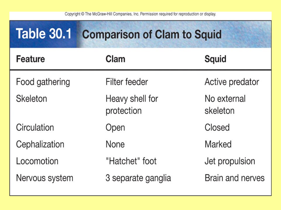
Phylum Mollusca Meaning – soft-body Coelomates (reduced in some) Symmetry – bilateral Organization – organ level Tube-within-a-tube body plan No segmentation Found in fresh or salt water, and on land
 Symmetry – bilateral Organization – organ level Tube-within-a-tube body plan No segmentation Found in fresh or salt water, and on land")
89
Molluscan diversity

90
3 distinct parts to all mollusk bodies: –Visceral mass – soft-bodied part containing organs –Foot – muscular part used for movement –Mantle – membrane covering visceral mass; in some molluscs, it secretes the shell Molluscan groups are distinguished by a modification of the foot.

91
Gastropods – (stomach-foot) the foot is ventrally flattened. Ex: slugs & snails Muscle contractions along foot move the animal Some species have a shell Some species live on land – mantle also functions as a lung by moving air in & out through respiratory pores Radula – tongue with teeth to scrape up food

92
Cephalopods - the foot has evolved into tentacles about the head; ex: squid & octopus Cephalization - brain and nerves; camera-type eye; scent; pigments in skin to change colors Move rapidly by jet propulsion Feed using beak and radula Have a closed circulatory system. Escape predators by secretion from ink gland No shell; hydrostatic skeleton; cartilage around brain Sperm transferred to female in “dart;” she lays and guards eggs – no larval form

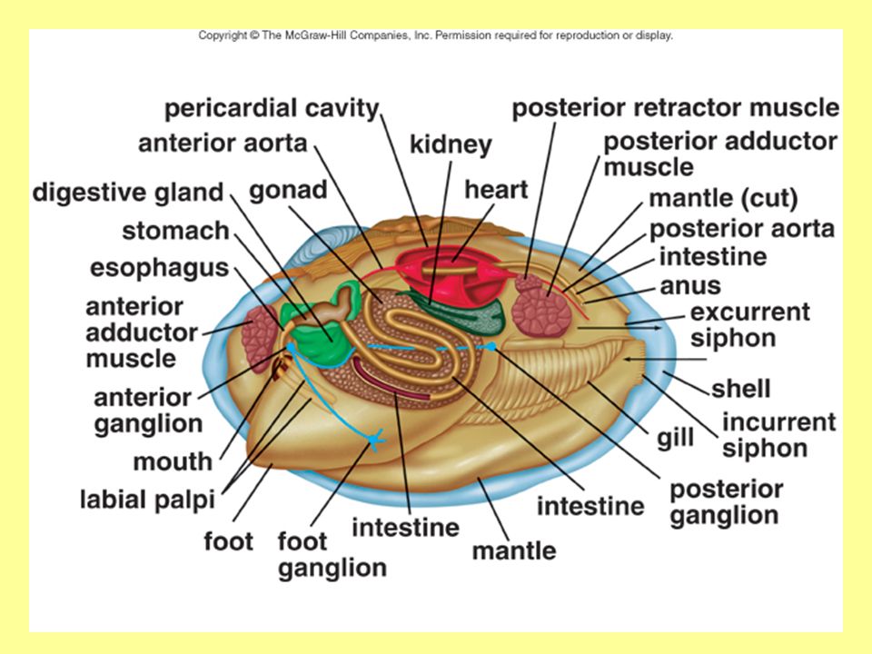
93
Bivalves –Ex: clams, oyster, scallops & mussels –A coelom is present but reduced –Support - Two shells held together by muscles –Respiration - using gills hanging on both sides of visceral mass –Movement - have a hatchet-shaped foot – in some species, this can allow for movement – most are sessile –Response - no cephalization - 3 pairs of ganglia connected by nerves control body.

94
Internal transport – heart pumps blood into an aorta, then through sinuses rather than blood vessels (open circulatory system). Feeding - filter feeding. –Water enters by an incurrent siphon; exits by excurrent siphon. –Food trapped on the gills is swept toward the mouth. Digestion - mouth with labial palps (helps bring food to mouth), an esophagus, a stomach, and an intestine.
, an esophagus, a stomach, and an intestine..")
95
Excretion - 2 kidneys remove waste from cavity around heart; food waste passes through anus, which empties at an excurrent siphon. Reproduction: Sexes are usually separate A single gonad (either ovary or testis) is located around the coils of the intestine. Some clams release both eggs & sperm into water Other species release sperm only into water, enters female, and fertilizes egg at gills.
 is located around the coils of the intestine. Some clams release both eggs & sperm into water Other species release sperm only into water, enters female, and fertilizes egg at gills..")
97
Muscle Mouth Foot Mantle Gills

98
Mouth Esophagus Muscles Stomach Digestive gland Heart Muscles Anus Excurrent siphon Incurrent siphon Mantle Gills Intestine

100
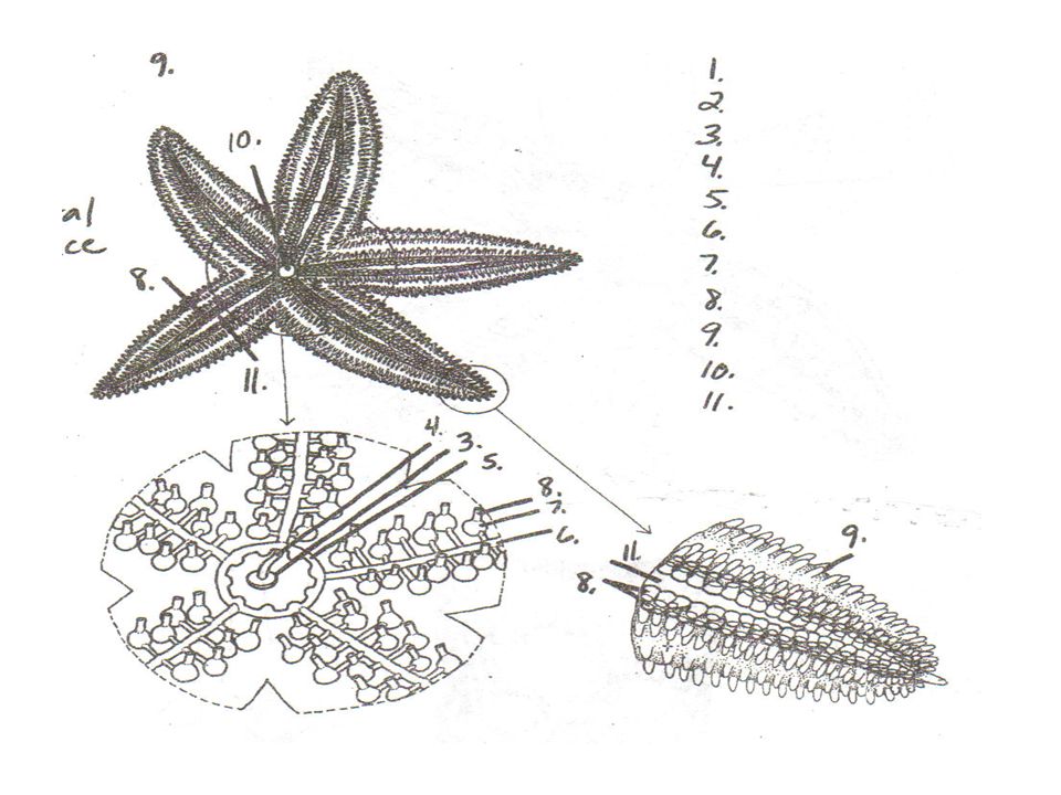
Phylum Echinodermata Habitat – salt water Examples: Sea lilies, feather stars, sea cucumbers, brittle stars, sea urchins, sand dollars, & sea stars Symmetry – radial as adult, larva is a free- swimming filter feeder with bilateral symmetry. Organization – organ level Coelomates; non-segmented Tube-within-a-tube body plans

101
Echinoderm diversity

102
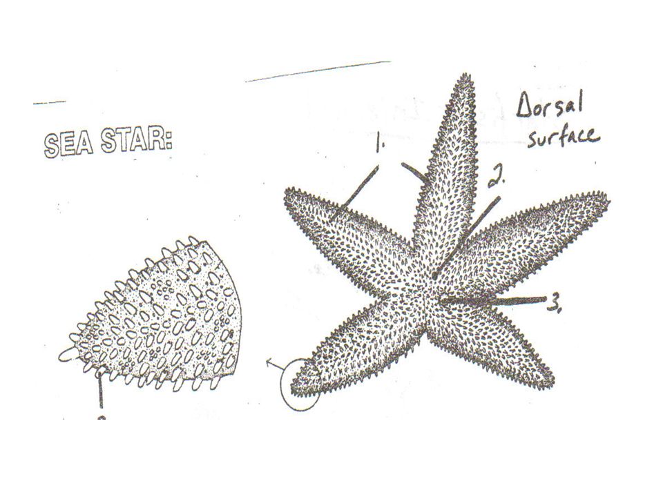
Sea Stars Support - endoskeleton consisting of spine-bearing, calcium-rich plates; spines protrude through skin. Movement - water vascular system –Water enters this system through the sieve plate (aka madreporite), passes into a stone canal, then to the ring canal, into radial canal, then into ampullae, and into tube feet; expansion and contraction of tube feet move the sea star along. Internal transport - fluid in body cavity & water vascular system –Cilia lining the peritoneum move fluid through coelom to transport nutrients & oxygen.
, passes into a stone canal, then to the ring canal, into radial canal, then into ampullae, and into tube feet; expansion and contraction of tube feet move the sea star along. Internal transport - fluid in body cavity & water vascular system –Cilia lining the peritoneum move fluid through coelom to transport nutrients & oxygen..")
103
Respiration – Gas exchange occurs across skin gills and tube feet Excretion - waste diffuses through fluid in coelom & body wall; food waste passes through anus Feeding and digestion – –Eat bivalves (2-shelled mollusks) –Everts its cardiac stomach inside the shells and secretes enzymes; partially digested food is taken in. –Digestion continues in pyloric stomach with enzymes made in digestive glands in each arm

104
Response – Central nerve ring with radial nerves in each arm Sensory tube feet at end of each arm to “taste” the water Photosensitive eyespot at the end of each arm Reproduction – gonads in each arm – open directly to outside Asexually - by regeneration – 1 small piece can grow into a new sea star Sexually - by releasing eggs & sperm into water

105
Sea star anatomy

106
1.Rays/arms 2. Anus 3.Madreporite/ sieve plate 4.Stone canal 5.Ring canal 6.Radial canal 7.Ampulla 8.Tube feet 9.Spine 10.Mouth 11.Ambulacral groove

109
1. Madreporite/ Sieve plate 2. Digestive Gland 3.Sex organs 4.Ampulla 5.Ray/arm 6.Eyespot 7.Radial canal 8.Spines 9.Stomach (2 parts – pyloric And cardiac)
.")
110
Phylum Arthropoda Meaning – “jointed foot” Symmetry – bilateral Coelomates – complex internal organs Tube-within-a-tube body plan Segmented The most varied and numerous of animals (over 1 million identified, probably 30 million really exist!!) Five Classes – Crustaceans, Insects, Arachnids, Millipedes, & Centipedes
 Five Classes – Crustaceans, Insects, Arachnids, Millipedes, & Centipedes")
111
Successful because of: Flexible exoskeleton for support (made of chitin, offers protection, muscles attach to it, locomotion, and retains moisture); shed exoskeleton (molt) during growth Jointed appendages Segmented; specialization of body regions (3 body regions – head, thorax, and abdomen – with specialized appendages in each region) A well-developed nervous system (brain, nerve cord, eyes, & antenna) Small size; high reproductive rate Live in water, land and air
; shed exoskeleton (molt) during growth Jointed appendages Segmented; specialization of body regions (3 body regions – head, thorax, and abdomen – with specialized appendages in each region) A well-developed nervous system (brain, nerve cord, eyes, & antenna) Small size; high reproductive rate Live in water, land and air")
112
Arthropod diversity

113
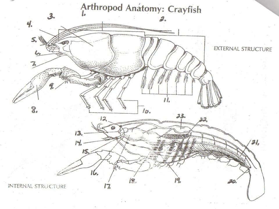
Crustaceans Mostly marine; crayfish, shrimp, crab, lobster Response: compound eyes, 2 pair of antennae, brain, & ventral nerve cord. Feeding: specialized mouth parts. Digestion: stomach – 2 regions – 1 to grind food, 1 to filter coarse particles & prevent them from entering digestive glands where digestion takes place. Locomotion: 5 pairs of walking legs include a first pair of pinching claws (called chelipeds). End of abdomen has uropod & telson used to help crayfish move backwards.
. End of abdomen has uropod & telson used to help crayfish move backwards..")
114
In the crayfish, head and thorax are fused into a cephalothorax which is covered on the top and sides by a carapace. Internal transport - The crayfish has an open circulatory system - heart pumps blood into a hemocoel consisting of sinuses where the hemolymph flows about the organs. Respiration takes place by gills under the hard carapace. Excretion - Green glands – excrete metabolic wastes through duct; food waste passes out through anus

115
Reproduction - the abdominal segments have swimmerets, which are used to hold the eggs in the female. Sexes are separate in the crayfish; can tell male from female by looking at first pair of swimmerets – point upward in male Swimmerets - transfer sperm to female.

116
Male crayfish, Cambarus

118
1.Cephalothorax14. Mouth 2.Abdomen15. Esophagus 3.Carapace16. Ganglion (nerve) 4.Antenna17. Stomach 5.Antennule18. Digestive gland 6.Mandible (jaw)19. Gills 7.Maxilla20. Anus 8.Cheliped (claw)21. Intestine 9.Maxilliped22. Testis 10.Walking legs23. Heart 11.Swimmerets24. Telson 12.Brain 13.Green gland
 4.Antenna17. Stomach 5.Antennule18. Digestive gland 6.Mandible (jaw)19. Gills 7.Maxilla20. Anus 8.Cheliped (claw)21. Intestine 9.Maxilliped22. Testis 10.Walking legs23. Heart 11.Swimmerets24. Telson 12.Brain 13.Green gland.")
119
Insects Many exhibit social behavior, such as bees or ants. Response: in addition to brain & nerve cord, head usually bears a pair of antennae, compound eyes, simple eyes, and in some tympanum for the reception of sound waves. Locomotion: thorax bears three pairs of legs and up to 2 pairs of wings. Excretion - Malpighian tubules collect nitrogen waste, which is added to digestive tract; waste then passes out through anus.

120
Compound eye of insects

121
Insect diversity

122
Internal transport: open circulatory system; heart pumps hemolymph into aorta that leads to a hemocoel (contains sinuses), where it circulates before returning to the heart. Feeding: mouthparts specialized for diet – for ex. chewing vegetation, siphoning nectar. Digestion: mouthparts chew food, crop stores, digestion occurs in stomach & intestine.

123
Some insects, such as grasshoppers, are adapted to a terrestrial life: Respiration - tracheae (small tubules); open to outside by holes called spiracles (on abdomen) Have wings that allow them to evade enemies Third pair of legs is suitable for jumping.
; open to outside by holes called spiracles (on abdomen) Have wings that allow them to evade enemies Third pair of legs is suitable for jumping.")
124
Reproduction: Mate by having sex Female grasshopper has ovipositor at posterior end to dig hole and lay eggs. Grasshoppers undergo gradual metamorphosis from nymph to adult (referred to as incomplete metamorphosis). Butterflies undergo complete metamorphosis, changing from larva to pupa to adult.
. Butterflies undergo complete metamorphosis, changing from larva to pupa to adult..")
125
Female grasshopper

126
1.Head7. Auditory membrane (ears) 2.Thorax8. Wing 3.Abdomen9. Ovipositor (F only) 4.Antennae10. Spiracles 5.Simple eyes (3)11. Jumping leg (2) 6.Compound eye (2) 12. Walking legs (4)
 4.Antennae10. Spiracles 5.Simple eyes (3)11. Jumping leg (2) 6.Compound eye (2) 12. Walking legs (4).")
127
13.Mouth19. Intestine 14.Pharynx20. Anus 15.Esophagus21. Malpighian tubules 16.Crop22. Heart 17.Gizzard23. Aorta 18.Cecum24. Salivary gland

128
Arachnids Include terrestrial spiders, scorpions, ticks, and mites. Ticks and mites are parasitic 2 body segments – cephalothorax & abdomen The cephalothorax bears six pairs of appendages: the chelicerae and the pedipalps, and four pairs of walking legs. Chelicerae contain fangs to deliver poison Pedipalps sense or hold the prey

129
Spiders are well-adapted to life on land: Excretion - have Malphigian tubules – they secrete uric acid, helping to conserve water. Respiration - using a “book lung” – inner body wall folds inward to look like pages of book. Spiders spin silk used in various ways. Where spiders spin webs, the type of web is a feature that demonstrates the evolutionary relationship among spiders. Response - 8 simple eyes and no antennae

130
Arachnid diversity

131
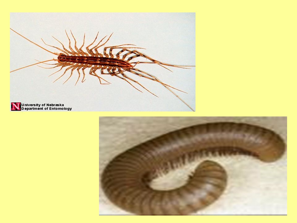
Centipedes Head and numerous body segments 1 pair of legs per segment (about 30 legs) Breathe with trachea Carnivores; nocturnal; move quickly Poison claws to paralyze prey Millipedes Head and numerous body segments 2 pairs of legs per segment (about 70 legs) Breathe with trachea Herbivores, move slowly, harmless to humans
 Breathe with trachea Carnivores; nocturnal; move quickly Poison claws to paralyze prey Millipedes Head and numerous body segments 2 pairs of legs per segment (about 70 legs) Breathe with trachea Herbivores, move slowly, harmless to humans")
Similar presentations