Download presentation
Presentation is loading. Please wait.

1
صدق الله العظيم الاسراء اية 58

2
By Dr. Abdel Aziz M. Hussein Lecturer of Physiology Member of American Society of Physiology

9
Two types of cochlear potentials can be recorded in the cochlea; a) Endocochlear potential:a) Endocochlear potential: b) Cochlear microphonic potential:b) Cochlear microphonic potential:
 Endocochlear potential:a) Endocochlear potential: b) Cochlear microphonic potential:b) Cochlear microphonic potential:")
10
A resting electrical potential of about +80 mV ( ) endolymph in scala media and perilymph in scala vestibuli and scala tympani It is due to difference in chemical composition ( ) endolymph and perilymph It is maintained by a K + pump (in stria vascularis) that transports K + from the perilymph to the endolymph
 endolymph in scala media and perilymph in scala vestibuli and scala tympani It is due to difference in chemical composition ( ) endolymph and perilymph It is maintained by a K + pump (in stria vascularis) that transports K + from the perilymph to the endolymph")
11
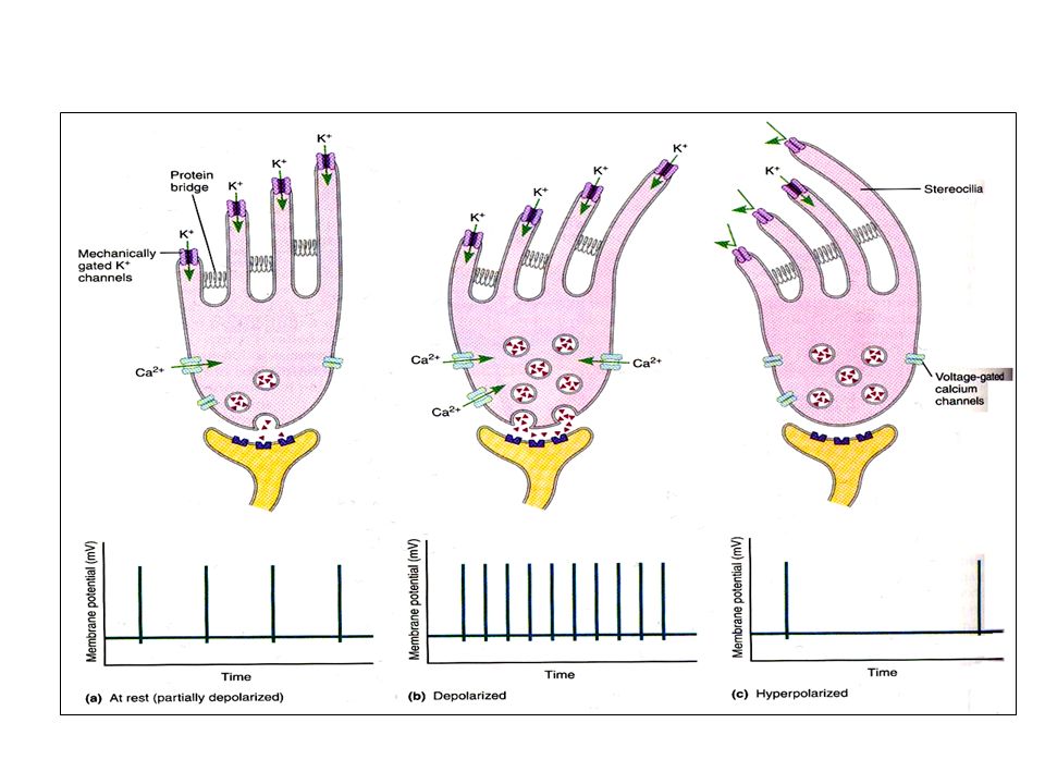
Tight junctions between hair cells and the adjacent supporting cells which prevent the endolymph to reach the bases of hair cells. So, hair cells have –ve intracellular potential of -60 mV with respect to the perilymph, but -140 mV with respect to the endolymph, [-60-(+80)] = [-140] mV surrounding its upper surfaces. This high electrical potential at the tips of the stereocilia greatly sensitizes the cell, thereby increasing its ability to respond to the slightest sound stimulus
] = [-140] mV surrounding its upper surfaces. This high electrical potential at the tips of the stereocilia greatly sensitizes the cell, thereby increasing its ability to respond to the slightest sound stimulus.")
12
Hair cells Endolymph Perilymph Basilar Membrane - 140 mv - 60 mv Tight Junctions Organ of Corti + 80 mv + 0 mv

13
These are receptor potentials that can be recorded from most parts of the cochlea when the ear is exposed to sound. They are recorded from an electrode placed at or near the round window. They represent the sum of potentials generated by a large population of hair cells mainly that produced by lateral hair cells. So, its detection in surface recordings has been considered a distinctive sign of outer hair cell integrity.

14
Its magnitude is dependent on the proximity of the recording electrodes to the hair cells and proportional with the intensity of sound, hence the degree of displacement of basilar membrane. Cochlear microphonic potential is maintained so long the basilar membrane is vibrating and it corresponds closely to the sound stimulus regarding: the frequency, the wave form and the amplitude They have the same characteristics of receptor potentials i.e. can summate, proportional with the intensity of sound (graded), and not obey all or none rule.
, and not obey all or none rule..")
17
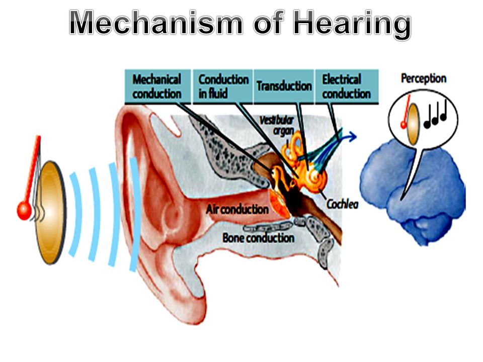
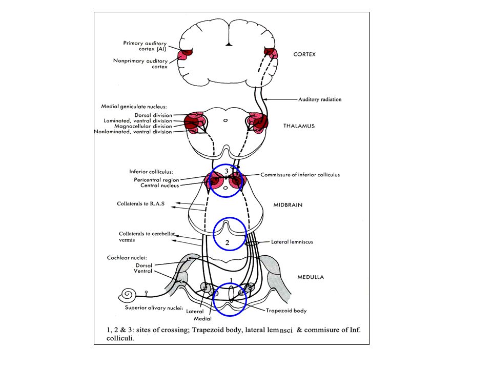
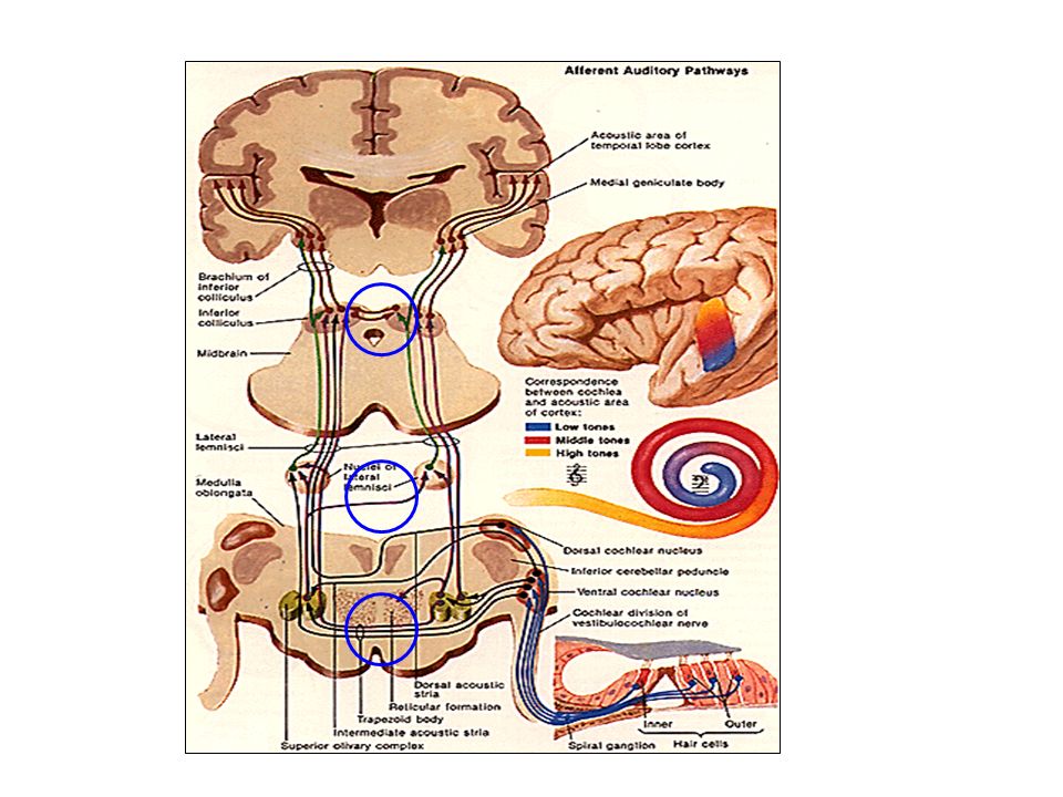
The auditor pathway from the cochlea to the cerebral cortex consists of at least 4 neurons and may increase to 6 neurons

19
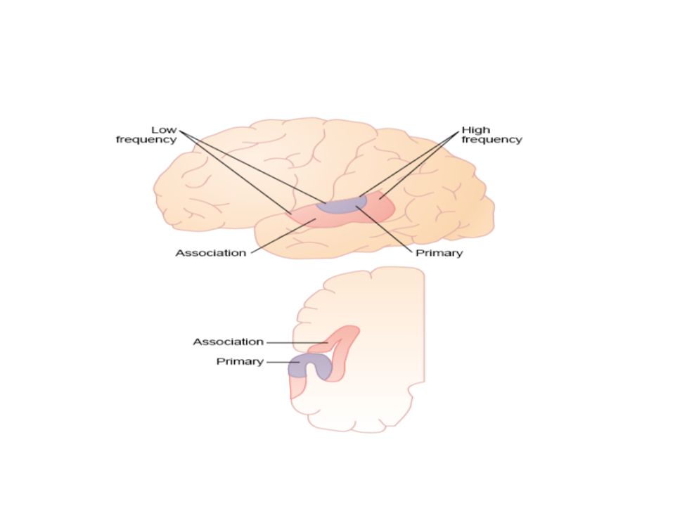
formed of 1ry and 2ry areas; A) Primary auditory area (areas 41 & 42): located in the upper part of temporal lobe B) Secondary (associated) auditory area (area 22): Surrounds the primary area and covers the insular cortex
 Primary auditory area (areas 41 & 42): located in the upper part of temporal lobe B) Secondary (associated) auditory area (area 22): Surrounds the primary area and covers the insular cortex")
20
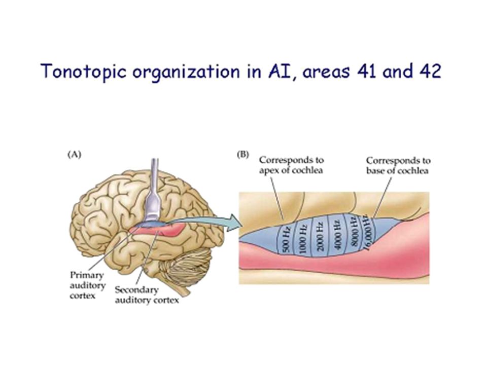
Receive auditory impulses from both ears from ipsilateral MGB. Anterolateral part receives impulses from the apex of the cochlea (low pitched sounds) Posteromedial part receives impulses from the base of the cochlea (high pitched sounds)
 Posteromedial part receives impulses from the base of the cochlea (high pitched sounds).")
24
Functions: 1.Conscious perception of pitch, amplitude, and sound pattern without understanding its meaning. 2.Perception of the source of the sound.Lesion: 1.Bilateral damage of the primary auditory areas greatly reduces the person's capacity for hearing 2.Unilateral damage slightly reduces hearing of the opposite ear ?

25
It receives impulses from the primary auditory area.Functions: Its function is to associate sound information with afferent information from other sensory areas of the cortex for interpreting and understanding the meaning of sounds.Lesion: The person will be unable to interpret the meaning of the heard sound (auditory aphasia or word deafness).
.")
26
1.Signals from one ear are transmitted to both sides of the brain. 2.At least 3 crossing-over occur between the right and left pathways in the brain stem: 3.Many fibers from the auditory tracts pass directly into the RAS of brain stem which projects upward to cerebral cortex and downward to spinal cord → activates the entire nervous system in response to a loud sound. 4.Some fibers also go to the vermis of the cerebellum which is also activated in response to sudden noise. 5.Inferior colliculi represent the center of spinal reflexes of hearing. 6.A high degree of spatial arrangement is maintained in the fiber tracts from the cochlea all the way to the cortex.

30
Human ear can recognize frequencies from 20-20.000 Hz with maximum sensitivity of ear occur at frequencies from 1,000 to 4,000 HZ. Discrimination of sound pitch can be explained by 2 theories; a) The place principle or theory:a) The place principle or theory: b) The frequency principle or theory:b) The frequency principle or theory:
 The place principle or theory:a) The place principle or theory: b) The frequency principle or theory:b) The frequency principle or theory:.")
31
It is the most accepted theory. Basilar membrane fibers are short thick stiff fibers at the base → maximally activated by the high frequency sounds and long thin lax fibers at the apex → maximally activated by low frequency sounds So each frequency causes vibration of its own particular "place" on the basilar membrane. So, the basilar membrane serves as a frequency analyzer and have tonotopic map There is a 2 nd tonotopic map in cochlear nuclei However, the place principal cannot explain discrimination of frequencies - from 200-20 HZ occurring at the apex of the cochlea.

34
It was the first theory suggested to explain frequency discrimination. It postulates that, for low frequency sounds, the basilar membrane vibrates in the same frequency and the auditory nerve fibers can fire at the same frequency of the sound. While at high frequency sounds the nerve cannot discharge at the same rate due to the absolute refractory period (the nerve fibers cannot transmit impulses at rate greater than 1000 impulses/sec).
..")
35
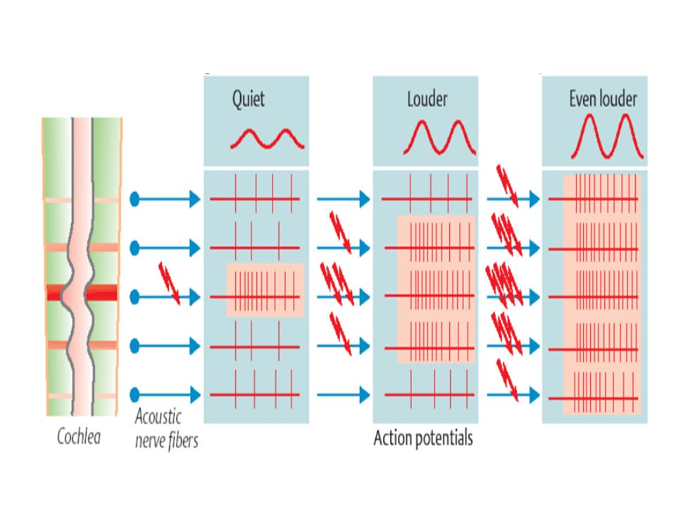
It is determined by the number of impulses discharged from the hair cells to the auditory cortex. This occurs by means of 3 different ways: 1. Temporal summation: ↑ sound intensity →↑ movement the basilar membrane →↑ firing of hair cells 2. Spatial summation: ↑ sound intensity →↑ movement the basilar membrane →↑ number of hair cells stimulated →↑ neurons stimulated in auditory cortex. 3. Certain hair cells do not become stimulated until the vibration of the basilar membrane reaches a relatively high intensity (in very loud sounds).
..")
37
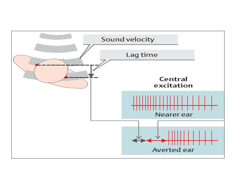
It depends on the binaural hearing. Superior olivary nuclei and auditory cortex have a role Sound localization depends on: 1.Time-lag ( ) entries of sound in both ears. 2.Difference ( ) sound intensities in the two ears. Ears are roughly 20 cm apart and the speed of sound is 342 m/sec, so the time delay between the arrival of sound wave to one ear and the opposite side is about 0.06 msec. SON divided into 2 parts: 1.Medial superior olivary nucleus →detect time-lag. 2.Lateral superior olivary nucleus → detect intensity differences
 entries of sound in both ears. 2.Difference ( ) sound intensities in the two ears. Ears are roughly 20 cm apart and the speed of sound is 342 m/sec, so the time delay between the arrival of sound wave to one ear and the opposite side is about 0.06 msec. SON divided into 2 parts: 1.Medial superior olivary nucleus →detect time-lag. 2.Lateral superior olivary nucleus → detect intensity differences.")
40
The folds and bulges of the ear pinna produce different reflections of sounds based on their angle of entry along the vertical plane. Role of auditory cortex is proved to be important because destruction of auditory cortex on both sides of the brain causes loss of the ability to detect the direction from which the sound comes

41
THANKS

Similar presentations


 Physical properties of sound>")


