Download presentation
Presentation is loading. Please wait.

4
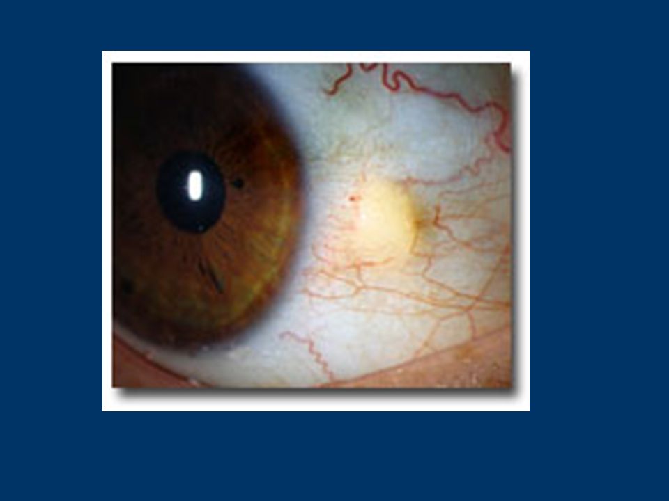
PINGUECULAE A pinguecula is a type of conjunctival degeneration in the eye. It is extremely common and is seen as a yellow-white deposit on the conjunctiva adjacent to the limbus (the junction between the cornea and sclera). It is to be distinguished from pterygium clinically, which is a wedge shaped area of fibrosis, that appears to grow into the cornea. It is most prevalent in tropical climates and is in direct correlation with UV exposure. Histologically it shows degeneration of the collagen fibres of the conjunctival stroma with thinning of the overlying epithelium and occasional calcification. They may enlarge slowly but is a benign condition requiring no treatment.
. It is to be distinguished from pterygium clinically, which is a wedge shaped area of fibrosis, that appears to grow into the cornea. It is most prevalent in tropical climates and is in direct correlation with UV exposure. Histologically it shows degeneration of the collagen fibres of the conjunctival stroma with thinning of the overlying epithelium and occasional calcification. They may enlarge slowly but is a benign condition requiring no treatment..")
6
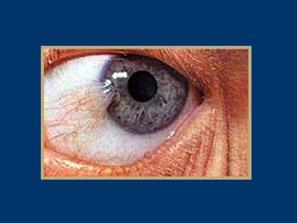
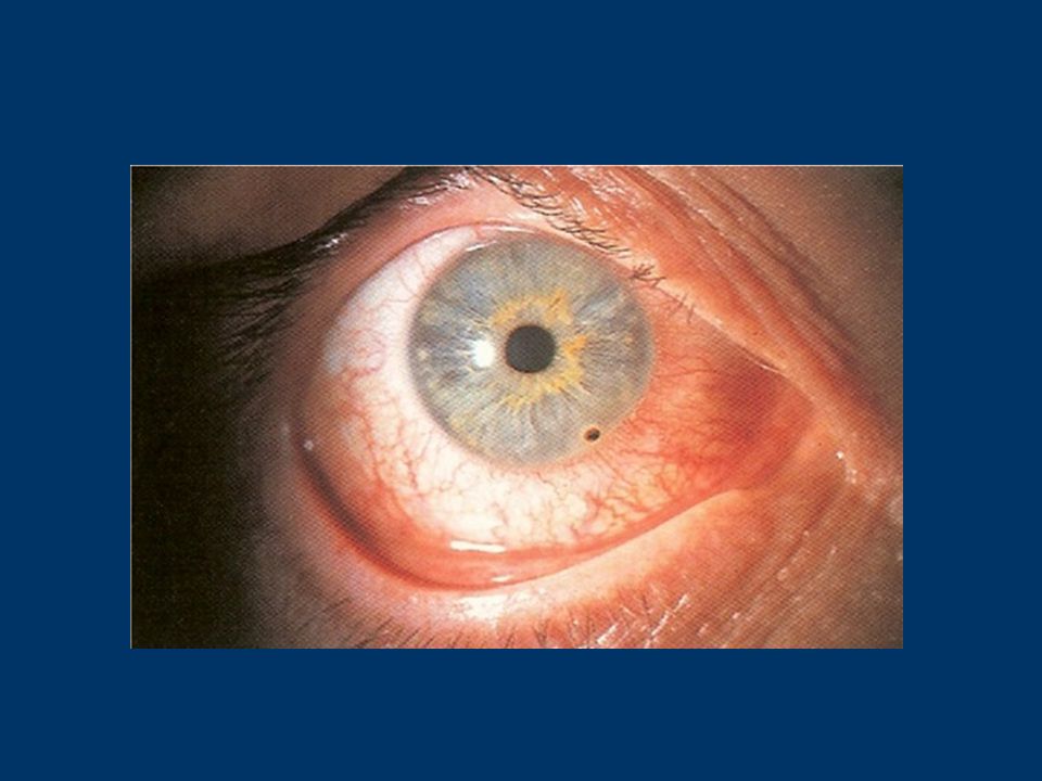
Pterygium: In ophthalmology, it is a degenerative condition of the eye affecting the conjunctiva and the cornea. It it thought to be an irritative phenomenon associated with exposure to ultraviolet light and is found particularly in people who work outdoors in hot, dusty climates. Globally there is a relationship between decreased incidence in the upper latitudes and relatively increased incidence in lower latitudes - this is thought to be because of elevated levels of ultraviolet light exposure in the lower latitudes. are seen as wedge-shaped lesions spreading onto the cornea almost always from the nasal side (1) pathologically, it is a degeneration of Bowman's membrane of the cornea which extends into the conjunctival epithelium. The process usually begins at the medial and lateral borders of the cornea. It progresses towards the centre taking with it a continuation of the conjunctival epithelium the condition is usually asymptomatic. The highest prevalence is in patients over the age of 40 years, while patients aged 20-40 years are reported to have the highest incidence of pterygia (1). It is uncommon for patients to present with pterygia prior to age 20 years. visual disturbances may result from encroachment on the pupillary area. it may be removed surgically but recurrence is common. Protective glasses for outdoor work are advised.
 pathologically, it is a degeneration of Bowman s membrane of the cornea which extends into the conjunctival epithelium. The process usually begins at the medial and lateral borders of the cornea. It progresses towards the centre taking with it a continuation of the conjunctival epithelium the condition is usually asymptomatic. The highest prevalence is in patients over the age of 40 years, while patients aged years are reported to have the highest incidence of pterygia (1). It is uncommon for patients to present with pterygia prior to age 20 years. visual disturbances may result from encroachment on the pupillary area. it may be removed surgically but recurrence is common. Protective glasses for outdoor work are advised..")
9
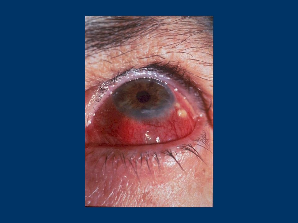
Episcleritis and Scleritis

11
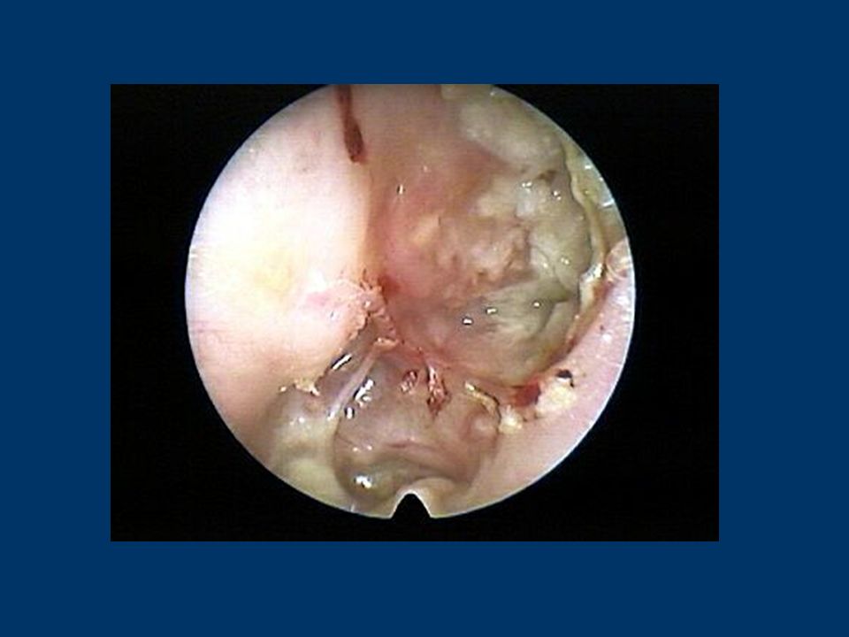
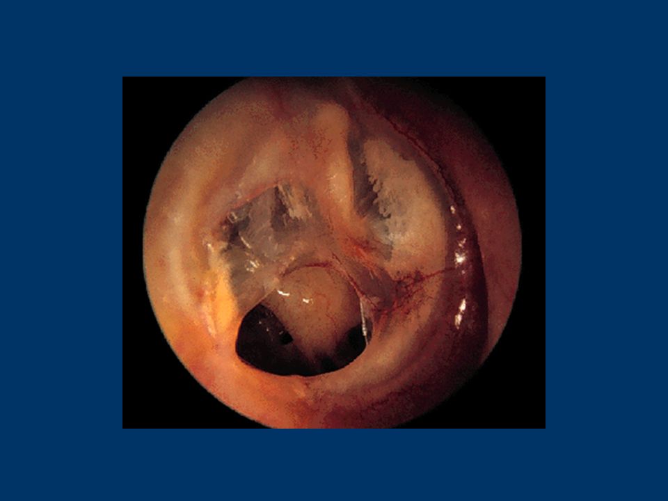
Cholesteatoma: Are skin or stratified keratinising squamous epithelium growing in the middle ear they are a greasy-looking mass or accumulation of debris that is seen in a retraction pocket or perforation. They often take the form of a cyst or pouch that sheds layers of old skin Divided into two types: congenital - presents as a pearly white mass located behind an intact tympanic membrane acquired - results from a retracted or perforated tympanic membrane with an ingrowth of epithelium Aetiology : the cause is unknown. It may result from blockage of the Eustachian tube producing a chronic negative pressure in the middle ear which would cause the tympanic membrane to be sucked inwards as a retraction pocket Usually, the pars flaccida is indrawn but any thin part of the pars tensa may be involved. The pockets gradually expand as the skin desquamates. Invariably, they become infected and smelly A cholesteatoma is potentially very dangerous because local expansion may result in damage to adjacent vital structures such as dura, lateral sinus, facial nerve and the semicircular canal.

13
Nasal Polyps

15
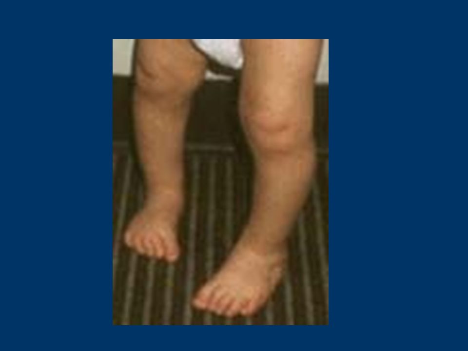
Tibial Torsion: Extremely common and provided it is symmetrical and the child is developmentally normal, it will usually correct by age 4. This condition generally presents with intoeing in toddlers aged between one and three years With child sitting, lateral malleolus is in front of medial. Also normally anterior superior iliac spine, patella and hallux in line More commonly affects the left tibia than the right. Most cases resolve spontaneously In a tiny proportion of cases a derotational osteotomy may be indicated

16
Define ‘Orthopaedics’ (etymologically)
")
17
LINK

19
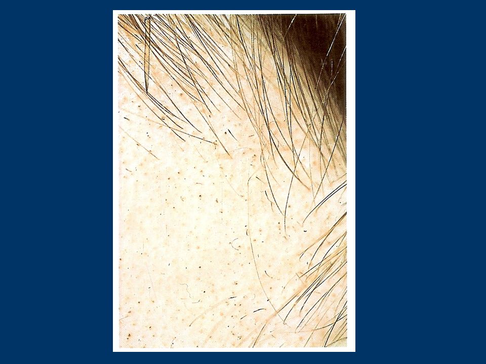
Alopecia Areata: Presentation is typically with localised, round bald patches developing suddenly over one or two weeks, without any preceding symptoms. At the edge of the patch, there may be small, broken hairs with a tapering shaft - 'exclamation mark' hairs. The scalp shows no sign of inflammation, scaling or scarring. The finger nails may be pitted and ridged. The patches may spread to involve the entire scalp - alopecia totalis - or body - alopecia universalis. The condition usually resolves in a period of three to six months but repeated episodes of hair loss are not uncommon. Hair regrowth is initially white.

21
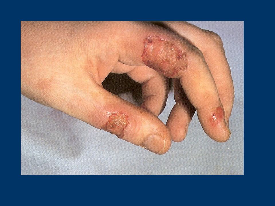
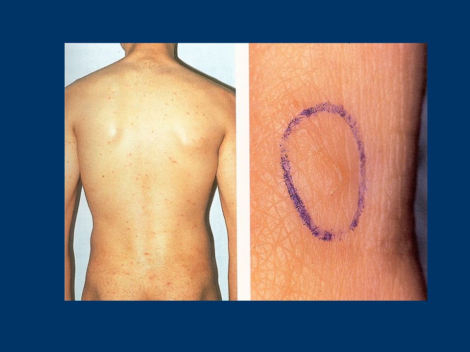
Dermatitis Artefacta

23
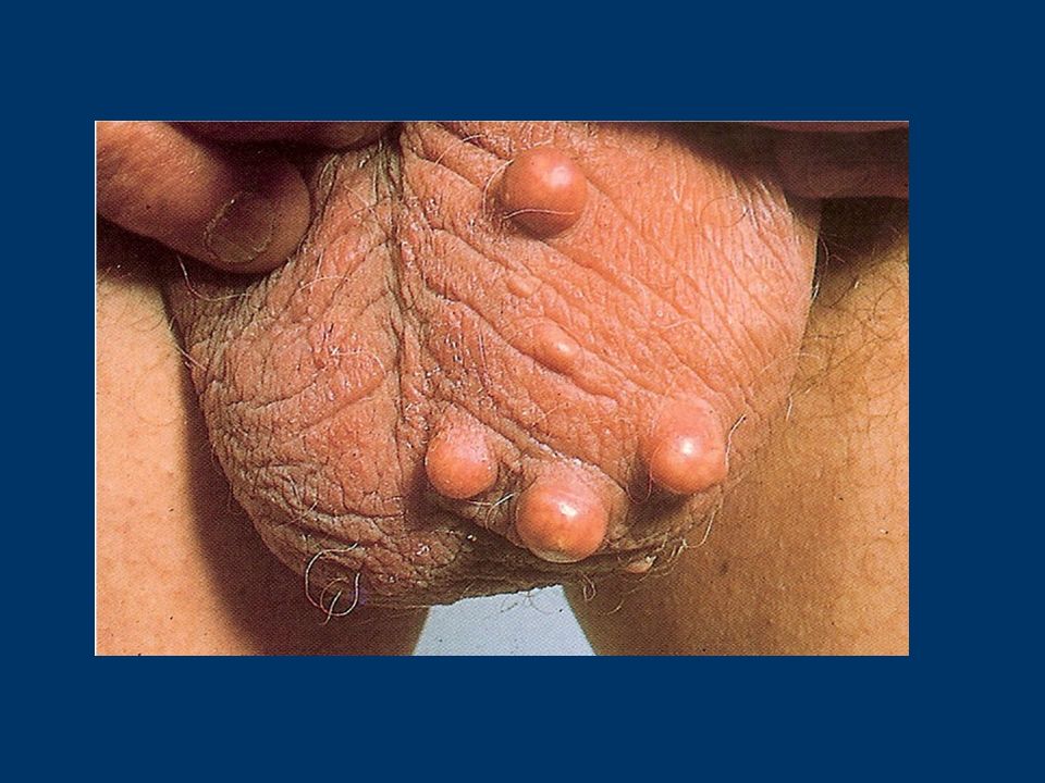
Epidermoid Cysts

25
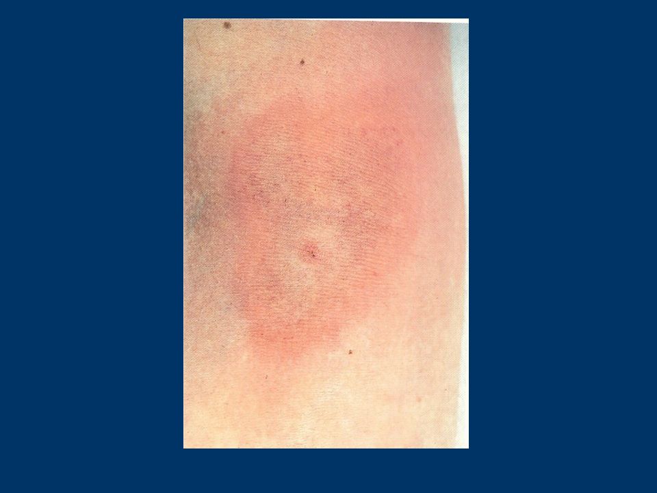
Lyme Disease (Erythema Migrans)
")
27
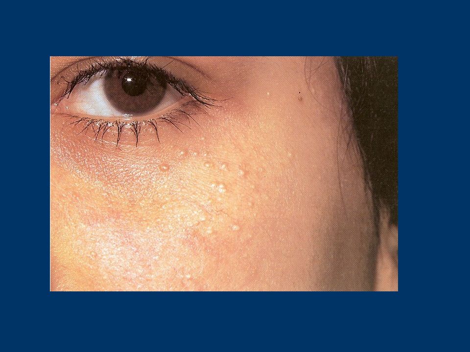
Milia

29
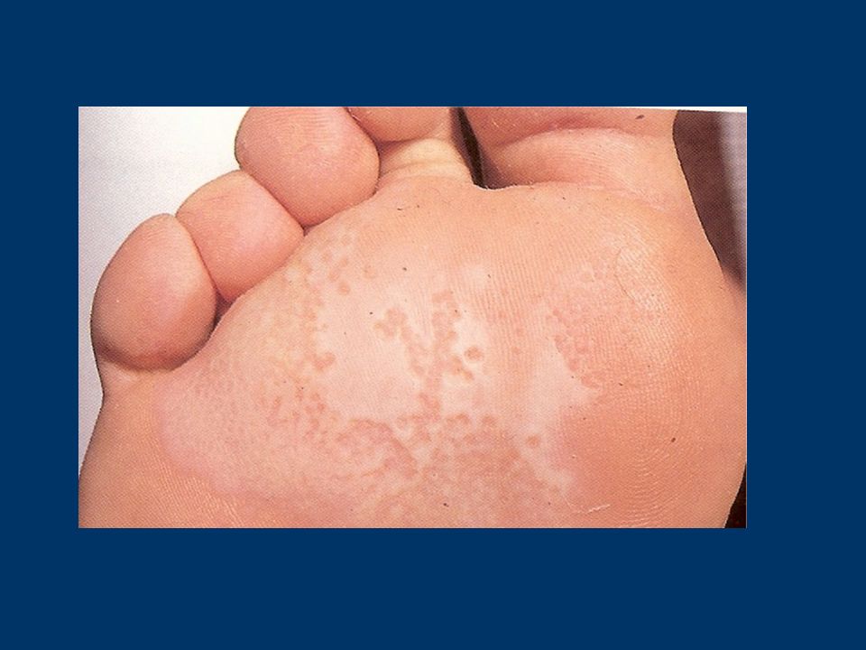
Pitted Keratolysis

31
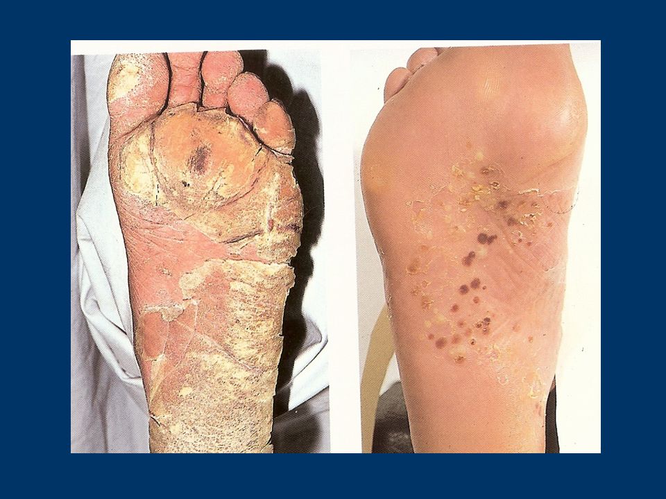
Psoriasis And Pustular Psoriasis

33
CSOM Otorrhoea > 3 months Recurrent infections Hearing loss Safe tubo-tympanic (central perforation) Unsafe attico-antral (attic disease) Surgery in children
 Unsafe attico-antral (attic disease) Surgery in children")
36
Scabies

38
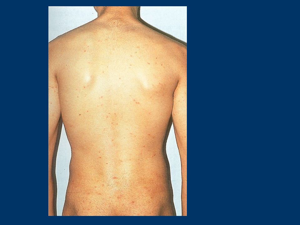
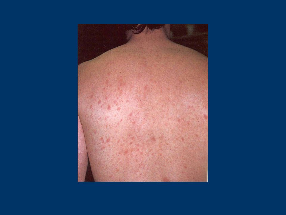
Toxic Erythema

40
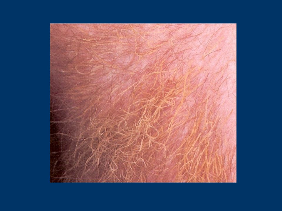
Trichomycosis Axillaris

42
Corneal Foreign Body

45
Pityriasis Rosea

47
Split nails (Lamellar due to water and detergents)
")
49
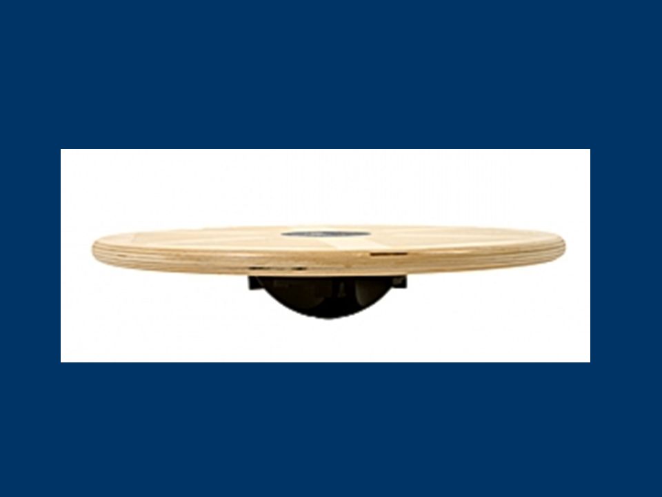
Wobble board (how many senses?)
")
Similar presentations