Download presentation
Presentation is loading. Please wait.

1
Lab skills

2
Safety Issues Follow instructions, use all instruments correctly. Wear safety goggles Report injuries and defective equipment Tie back loose hair and clothing. Beware of hot glassware,

3
Do not heat stoppered bottles, Never point a test tube in you partners direction Never taste or directly inhale anything, use a wafting motion. Do not pour back into reagent bottles. Clean area before and after activity.

4
Triple Beam Balance

5
Make sure zero mark- all weight at zero Start moving 100’s first. If too much go back a notch. move tens and then move ones if necessary Add all sliders up for total mass of object.

6
When using a paper or some sort of dish be sure to deduct the mass of the dish from the mass of the dish with the substance. Or zero the scale out with the dish on it.

7
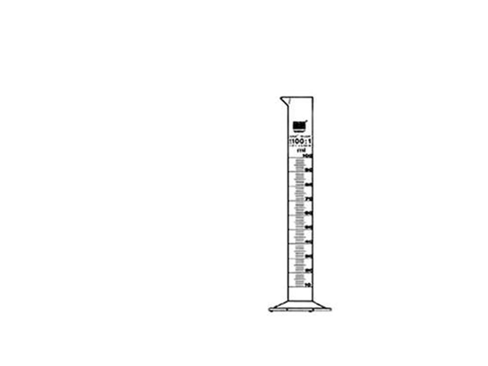
Graduated Cylinder Mensicus: the curved surface of the liquid that is where the reading is taken.

9
Metric ruler

11
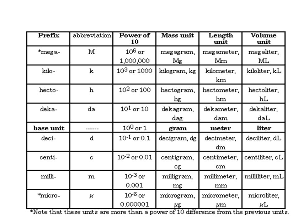
Meters = Length or distance Liters = Volume Grams = Mass

12
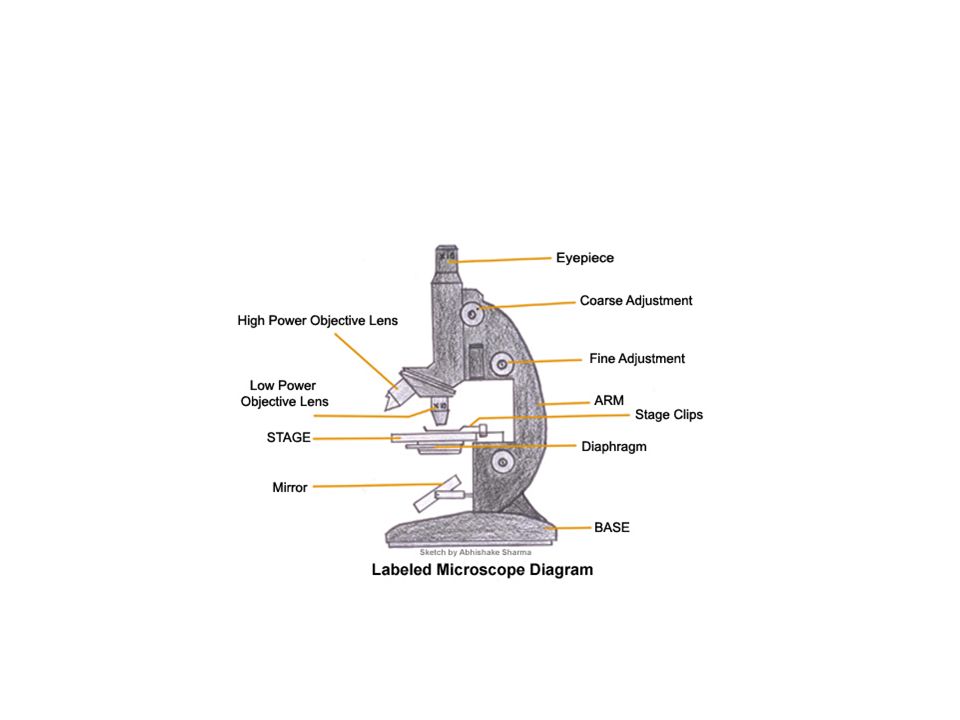
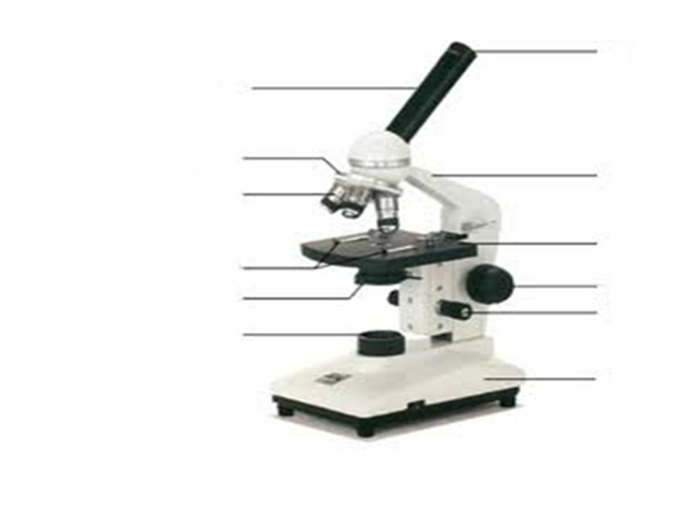
Compound Light Microscope: Consists of 2 lenses the eyepiece (ocular) and the objective (low power and High power) parts-label and define.
 and the objective (low power and High power) parts-label and define.")
15
Eyepiece: The lens the viewer looks through to see the specimen. The eyepiece usually contains a 10X or 15X power lens. Body tube : The body tube connects the eyepiece to the objective lenses.

16
Arm: The arm connects the body tube to the base of the microscope. Coarse adjustment: Brings the specimen into general focus. Used with Low power objective Fine adjustment: Fine tunes the focus and increases the detail of the specimen. Used with the high power objectives

17
Nosepiece: A rotating turret that houses the objective lenses. The viewer spins the nosepiece to select different objective lenses.

18
Objective lenses: One of the most important parts of a compound microscope, as they are the lenses closest to the specimen. A standard microscope has three, four, or five objective lenses that range in power from 4X to 100X. When focusing the microscope, be careful that the objective lens doesn’t touch the slide, as it could break the slide and destroy the specimen.

19
Specimen or slide: The specimen is the object being examined. Most specimens are mounted on slides, flat rectangles of thin glass. The specimen is placed on the glass and a cover slip is placed over the specimen. This allows the slide to be easily inserted or removed from the microscope. It also allows the specimen to be labeled, transported, and stored without damage.

20
Stage: The flat platform where the slide is placed. Stage clips: Metal clips that hold the slide in place. Aperture: The hole in the middle of the stage that allows light from the illuminator to reach the specimen.

21
On/off switch: This switch on the base of the microscope turns the illuminator off and on. Illumination: The light source for a microscope. Older microscopes used mirrors to reflect light from an external source up through the bottom of the stage; however, most microscopes now use a low-voltage bulb.

22
Iris diaphragm: Adjusts the amount of light that reaches the specimen.. Condenser: Gathers and focuses light from the illuminator onto the specimen being viewed. Base: The base supports the microscope and it’s where illuminator is located.

23
Find object using low power (see large amount with less detail). Use Coarse adjusting knob (larger knob). Brighter image Flip to high power (zoom) (see less of image but more detail). Use Fine adjusting knob (smaller knob). Darker image Be careful not to disrupt the specimen of crack the slide or lens.
. Brighter image Flip to high power (zoom) (see less of image but more detail). Use Fine adjusting knob (smaller knob). Darker image Be careful not to disrupt the specimen of crack the slide or lens..")
24
Lighting system: consists of a mirror, diaphragm(controls the amount of light reaching the specimen) Disc diaphragm: flat plate with a number of different sized openings. Iris diaphragm is made of overlapping plates and the size of the opening may be adjusted and substage illuminator.

25
Total magnification = eyepiece x objective

26
Images are upside down and backwards. Microscopes are used to make small details more visible. Resolution: the ability of the microscope to show two points close together. Controls the sharpness of the image.

27
Optical Microscope: uses light to produced enlarged images( light microscope) Lenses are pieces of curved glass that bend the light and make the image appear larger.
 Lenses are pieces of curved glass that bend the light and make the image appear larger.")
28
Simple Microscope: magnifying glass. Stereomicroscope: two oculars.

29
Electron Microscope-Excellent magnification and provides a very detailed picture. Good for examination of nonliving specimens

30
Dissecting Microscope- images are magnified but seen as they are. Two oculars. Three dimensional image. Good for viewing external features

31
Phase Contrast Microscope: makes unstained elements visible.

32
Prep of wet mount. Staining object makes them more visible. No air bubbles this interferes with the item you are trying to view. Certain stains bind to specific proteins therefore some stains are used to make specific items visible

33
A vital stain is used on living tissue only other stains will kill the tissue cells. Apply the stain to one side of the wet mount and use a paper towel to draw the water and stain across the slide and under the coverslip. This leaves the slide in tact.

34
Prep of a wet mount

35
Staining a slide

36
Types of Stains- Methylene Blue Lugol’s Iodine

37
Indicators- Chemicals when added in small amounts will indicate or show the presence of a specific substance.

38
Benedicts (Fehling’s) solution. (glucose solution) Blue----(heating)---yellow(+).
 solution. (glucose solution) Blue----(heating)---yellow(+).")
39
Lugol’s solution (Iodine). Test for starch. Orange ------ Purple(+)
. Test for starch. Orange Purple(+)")
40
Bromthymol Blue (CO 2 ). Blue (O 2 ) --------- yellow (CO 2 )(+).
. Blue (O 2 ) yellow (CO 2 )(+).")
41
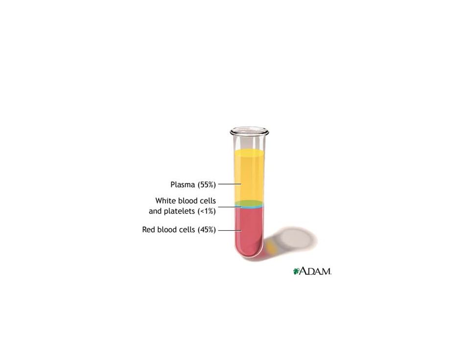
Centrifugation: The instrument used is the centrifuge. It spins the specimen in a circular motion and separates materials according to their densities. Most Dense (Heavy) bottom, least dense (lightest) top.
 bottom, least dense (lightest) top..")
43
Ultracentrifuge: same principle only stronger spinning action so there is more separation. Microcentrifuge: same principle only separating a small sample

44
Dissection: the separation of a large organism into its parts. Micro-dissection: dissection on a very small object with small instruments being used.

45
Tissue Culture: a live tissue sample that is used for research.

46
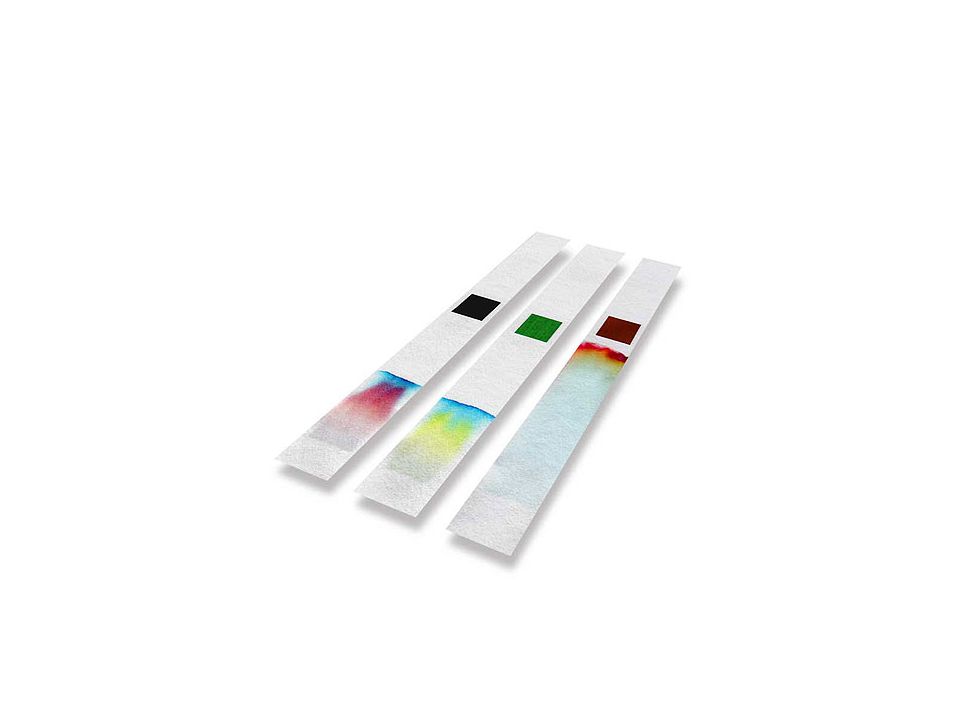
Chromatography: Is used to separate and analyze complex mixtures. The mixture is placed on a paper. A solvent is placed in a container.

47
. The paper is placed in the container with the liquid touching the very bottom. The mixtures particles will separate according to their size.

48
By comparing the known materials to the unknown materials we can identify the components in a mixture. The particles moving are in the mobile phase and the paper is the stationary phase (not moving)
.")
50
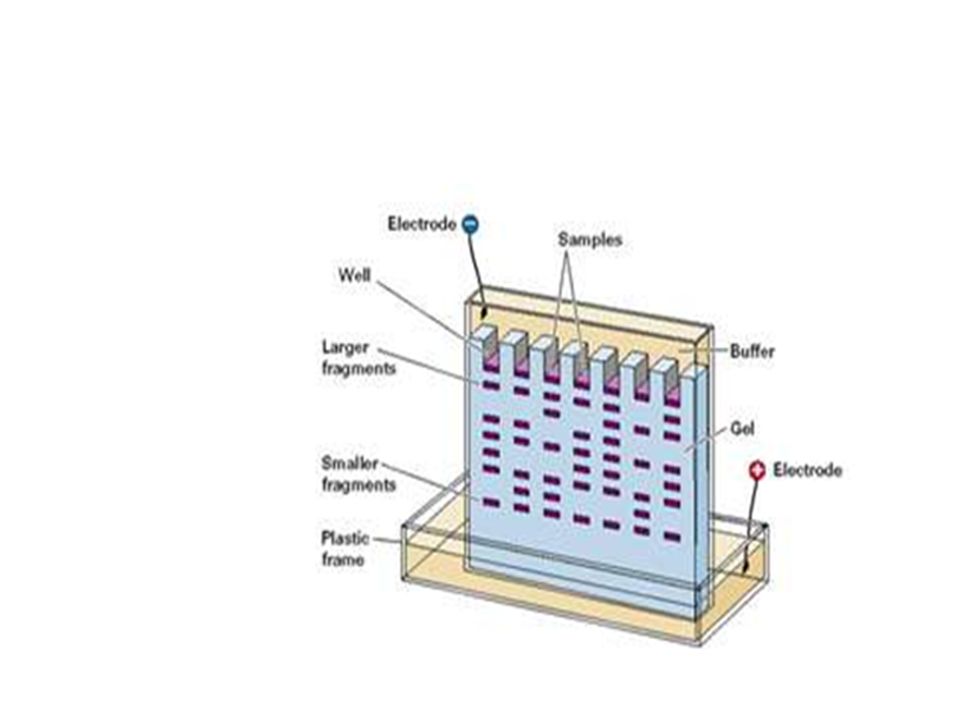
Electrophoresis: the particles are separated thru the use of an electric current. This method can both identify and measure the amount of a substance in a test solution.

51
The electric current causes the particles to migrate (move) based on their size and charge. The test solution results are compared to known results and the particles of a mixture are identified

53
Always stop both procedures before the liquid runs off the paper. If that happens the test results are invalid and need to be repeated (provided there is enough test sample left over)
.")
Similar presentations


 Electron (up to X 200,000) Scanning Electron Microscope (SEM) Transmission Electron Microscope (TEM)>")


>")


