Download presentation
Presentation is loading. Please wait.

1
Histology lab 1 basic tissues
Dr Abubakr H Mossa Anatomy Sr. instructor 8/10/2012

2
Objectives: Define the tissue Identify the basic types of tissues
Identify the general features of the epithelium Identify the different types of epithelium Identify the functions of epithelium Define the connective tissue Identify the elements of the CT Identify the types of CT Identify the structure of bone & cartilage and their types

3
Histology is the study of the microscopical structure of body tissues.
A tissue is defined as a group of similar cells and their related intercellular substance grouped together to perform a common function. The basic types of tissues are: Epithelium Connective tissue Muscle Nervous tissue

4
Epithelium Epithelium is a tissue with the following features:
It consists of one or more than one layer of cell sheets. Little intercellular space. Rests on a basement membrane: supports the epithelium and separates it from the underlying connective tissue. Avascular. Epithelium contains no blood or lymph vessels. It derives its nutrition by diffusion of tissue fluid from the vessels in the underlying connective tissue.

5
Classification of epithelium:
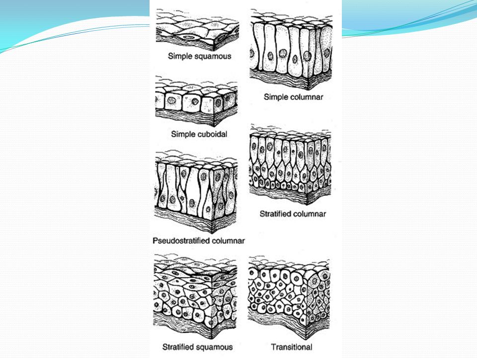
Criteria far classifying epithelium 1. Number of cells (thickness). a. Simple epithelium. consists of only one layer of cells. b. Stratified epithelium. Two or more layers of cells. 2. Shape (when viewed in profile). a. Squamous epithelium. Cells appear flattened or scale-like. b. Cuboidal epithelium. Cell height is same as cell width. c. Columnar epithelium. Cell height is greater than cell width. 3. Free surface. a. Ciliated epithelium. Cilia are present at the cell surface. b. Moist or non-keratinized epithelium. The cells at free surface are not cornified. c. Dry or keratinized epithelium. Cells at free surface are cornified.
. a. Simple epithelium. consists of only one layer of cells. b. Stratified epithelium. Two or more layers of cells. 2. Shape (when viewed in profile). a. Squamous epithelium. Cells appear flattened or scale-like. b. Cuboidal epithelium. Cell height is same as cell width. c. Columnar epithelium. Cell height is greater than cell width. 3. Free surface. a. Ciliated epithelium. Cilia are present at the cell surface. b. Moist or non-keratinized epithelium. The cells at free surface are not cornified. c. Dry or keratinized epithelium. Cells at free surface are cornified.")
7
Simple squamous: One layer of cells Flat cells with flat nuclei
Found in the Lung alveoli Bowman’s capsule (Kidney) Endothelium?: lining of blood vessels Bowman’s capsule
 Endothelium : lining of blood vessels. Bowman’s capsule.")
8
Lung alveoli: Simple squamous

9
Simple cuboidal: One layer of cells Cells are equal in width & height
Nucleus is round & central Found in: Renal tubules Thyroid gland follicles

10
Simple columnar: One layer of cells Cells are tall
Nucleus is basal & oval Goblet cells are seen (mucus secreting cells) Found in: Lining of stomach, intestine & gall bladder
 Found in: Lining of stomach, intestine & gall bladder.")
11
Stratified squamous non keratinized:
Multiple layers of cells of different shapes The superficial layer has cells of squamous type Moist, it is not covered by the dead layer of keratine Examples: Tongue covering Esophagus lining Vaginal lining

12
Stratified squamous keratinized:
Many layers of different shapes The superficial layers have squamous cells Dry, a dead layer of keratine is covering the tissue Example: skin

13
Special types of epithelium

14
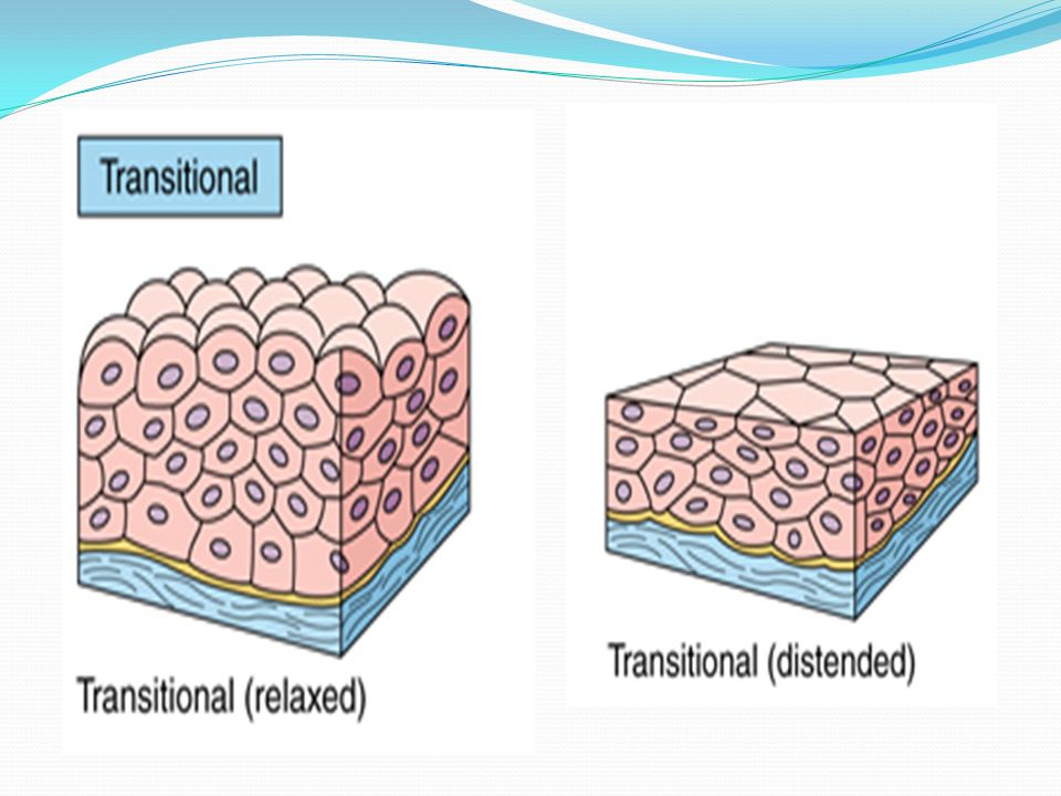
Transitional epithelium:
Many layers of cells (4-6 layers) The superficial layer has cells with changing shape, in relaxing state they are dome shape while they are flat in distended state. Moist surface Example: Urothelium: epithelium lining ureter & urinary bladder.
 The superficial layer has cells with changing shape, in relaxing state they are dome shape while they are flat in distended state. Moist surface. Example: Urothelium: epithelium lining ureter & urinary bladder.")
16
Pseudostratified epithelium:
All cells are in contact with the basement membrane, but not all of them reach the surface of the epithelium. The nuclei of cells are located at different heights within the epithelium and give the epithelium a stratified appearance. The epithelium looks stratified but it is not - hence its name "pseudostratified". Examples: the trachea .

17
Functions of epithelium:
1. Absorption. Example: lining of intestine. 2. Excretion: Example: secretory portion of sweat gland. 3. Secretion: Example: acinar cells of pancreas. 4. Protection. Example: epidermis of skin. 5. Lubrication. Example: goblet cells of small intestine and colon. 6. Sensation: Example: taste buds of tongue or olfactory cells in olfactory epithelium of superior nasal concha. 7. Reproduction. Example: germinal epithelial cells in seminiferous tubules of testis. 8. Transport: Example: ciliated epithelial cells in trachea which move the mucus and particular matter toward the pharynx for expectoration or swallowing.

18
Connective tissue

19
Connective tissue: It formed by cells separated by varying amounts of extracellular substance “matrix”. This tissue is important to support the body structure, protect the organs & help in providing nutrition to other tissues. So the contents of CT are: Cells: Fibroblasts & Fibrocytes & adipocytes (fixed cells) Fibers: Collagen, elastic & reticular fibers Ground substance:
 Fibers: Collagen, elastic & reticular fibers. Ground substance:")
20
CT:

21
Adipose tissue: Adipose tissue is essentially loose connective tissue
contains large numbers of adipocytes. Fat storage tissue to provide energy for body activities.

22
Special types of connective tissue: cartilage & bone

23
Cartilage: Functions: Composition: Weight bearing Helps in growth
Supports developing organs Withstanding of bending & torsional forces. Cells… Ground substance Fibers..

24
Composition of cartilage:
Cells: Matrix: (GS + fibers) called CHONDROCYTES Found in spaces of the matrix called LACUNAE Either single or in groups called “cell nests” Chondrocytes are round or “D” shaped cells. CHONDROBLASTS are the immature cartilage cells. They are found around the cartilage (perichondrium) Fibers are collagen or elastic Ground substance is stiff & composed of water & glycoprotein. Cartilage is avascular.
 called CHONDROCYTES. Found in spaces of the matrix called LACUNAE. Either single or in groups called cell nests Chondrocytes are round or D shaped cells. CHONDROBLASTS are the immature cartilage cells. They are found around the cartilage (perichondrium) Fibers are collagen or elastic. Ground substance is stiff & composed of water & glycoprotein. Cartilage is avascular.")
25
Types of cartilage: 3 types 1. HYALINE
Eg: Nose, trachea, costal cartilages, ends of long bones 2. ELASTIC Eg: External ear, epiglottis 3. FIBROCARTILAGE Eg: Intervertebral discs, pubic symphysis, knee joint

26
Hyaline cartilage: Features: Chondrocytes are found in cell nests.
The matrix appears homogenous. Fibers are not seen. (they have the same refractive index of the GS. Location:… Perichondrium is seen.

27
Elastic cartilage: Features: Cells are more & larger.
Cells are present either singly or in small groups. Fibers are seen ,branching & anastomosing, between the cells. Perichondrium is present. Location: …..

28
White fibrocartilage:
Features: Less no. of cells. Cells are seen singly in rows between the fibers. Fibers are more which are arranged in the form of thick bundles. NO perichondrium. Location:….

29
Bone: Functions of bone: Composition:
support - for muscles, organs, and soft tissues leverage and movement - the synovial joints protection - for critical organs calcium phosphate storage - mineral balance hemopoiesis - formation of blood cells Cells. Ground substance fibers

30
Composition of bone: Cells: Matrix: Fibers: collagen fibers
GS: protein polysaccharides & hyaluronic acid with calcium hydroxy-apatite (which gives rigidity to bone) Osteoblasts - bone forming cells - produce unmeniralized matrix “the osteoid”- calcify the matrix Osteocytes - mature bone cells- occupy lacunae- connected to other cells by processes through canaliculi Osteoclasts – phagocytic cells- multinucleated cells
 Osteoblasts - bone forming cells - produce unmeniralized matrix the osteoid - calcify the matrix. Osteocytes - mature bone cells- occupy lacunae- connected to other cells by processes through canaliculi. Osteoclasts – phagocytic cells- multinucleated cells.")
31
Composition of bone:

32
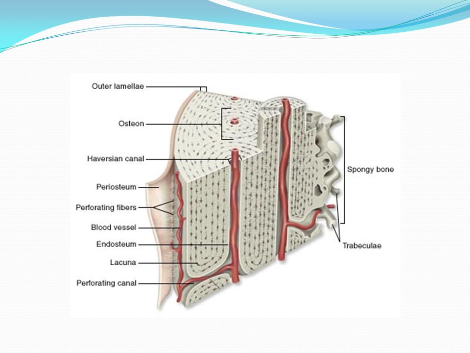
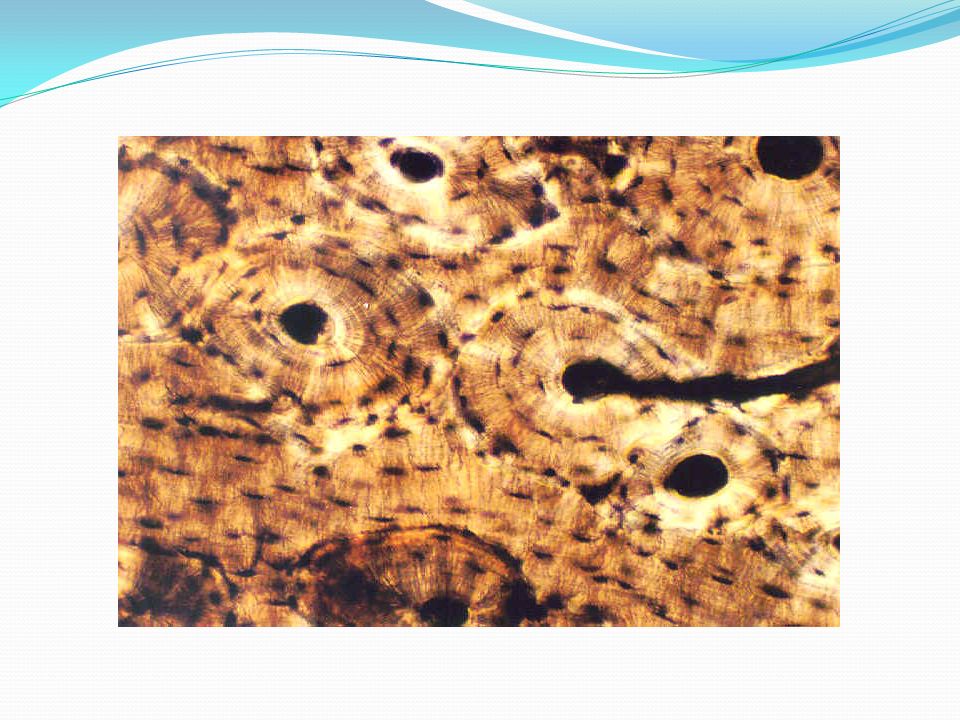
Osteon or Haversian system:
Osteons, or Haversian systems, are the units of structure in mature bone. They are tightly arranged running generally parallel to the long axis of the bone. At the center of each is an Haversian canal which carries blood vessels and nerves.

33
Osteon: Canaliculi (small canals) connect the Haversian canals with lacunae containing the osteocytes. Osteocytes extend processes into the canaliculi and receive nutrients and get rid of wastes through the canaliculi. Around the lacunae and canaliculi, lamellae or layers of two types can be identified: concentric lamellae form circular rings around each Haversian canal, and interstitial lamellae, fill in the spaces between existing osteons. Volkmann’s canals – channels lying at right angles to the central canal, connecting blood and nerve supply of the periosteum to that of the Haversian canal

36
Types of bone: Compact Bone: Cancellous (Spongy) Bone
a very dense tissue forming the outer layer of all bones and diaphyses of long bones. always contains numerous osteons or Haversian Systems. Haversian systems are only found in compact bone. Of the most obvious features is the absence of Haversian Systems or Osteons. During bone formation, the first bone to form is always cancellous. The bone tissue is arranged in thin plates or trabeculae.

37
Thanks

Similar presentations