Download presentation
Presentation is loading. Please wait.

1
BMS 231: 2015/2016 Anatomy of the Urinary System DR SOBIA IKRAM DR AQEELA BANO DR SADIA FARHAN

2
Table of Contents 1. Objectives for this lesson 2. Functions and Organisation of the Urinary system 3. Location and External anatomy of the kidney. 4. Internal Gross anatomy and parts of the kidney 5. Parts of the nephron and general functioning of the nephron.. 6. Definition,function and location of the Urinary Bladder, ureters and urethra.

3
Objectives When you finish this lesson, you should be able to Describe the Functions and Organisation of the Urinary system. Understand the Location and External Anatomy of the kidney. Identify the Internal gross Anatomy and parts of the kidney. Understand the parts of the nephron and their general functioning. Define and explain the location and functions of the urinary bladder, ureters and urethra.

4
Functions of the urinary system 1.Remove nitrogenous wastes (urine) 2.Maintain electrolyte, acid-base, and fluid balance of blood 3.Homeostatic organ 4.Acts as blood filter 5.Release hormones: calcitriol & erythropoietin
 2.Maintain electrolyte, acid-base, and fluid balance of blood 3.Homeostatic organ 4.Acts as blood filter 5.Release hormones: calcitriol & erythropoietin")
5
Organs of the Urinary System 5 Kidneys Ureters Urinary bladder Urethra

6
kidneys The Kidneys are the blood filtering organs of the urinary system. The kidneys are fist-sized, bean shaped structures that remove nitrogenous wastes (urine). The kidneys regulate the amount of water, salts and other substances in the blood.
. The kidneys regulate the amount of water, salts and other substances in the blood..")
7
Location and External Anatomy Located Retroperitonealy. Lateral to T 12 – L 3 Vertebra L3

8
HILUM Is a depression present On the medial surface of each kidney. Vessels and nerves enter and exit from the hilum. RENAL CAPSULE is the membrane that surrounds the kidney. AN AVERAGE KIDNEY is about 12 cm tall, 6 cm wide, 3 cm thick Location and External Anatomy

9
External anatomy of the Kidney 9 12 cm 6cm 3cm

10
10

11
Internal Gross Anatomy of the Kidney Sagittal Section through the Kidney shows 1.Renal cortex 2.Renal Medulla Renal pyramid 3.Renal pelvis Major calices Minor calices

12
renal capsule renal cortex renal medulla renal pelvis renal pyramids ureter Internal Gross Anatomy of the Kidney

13
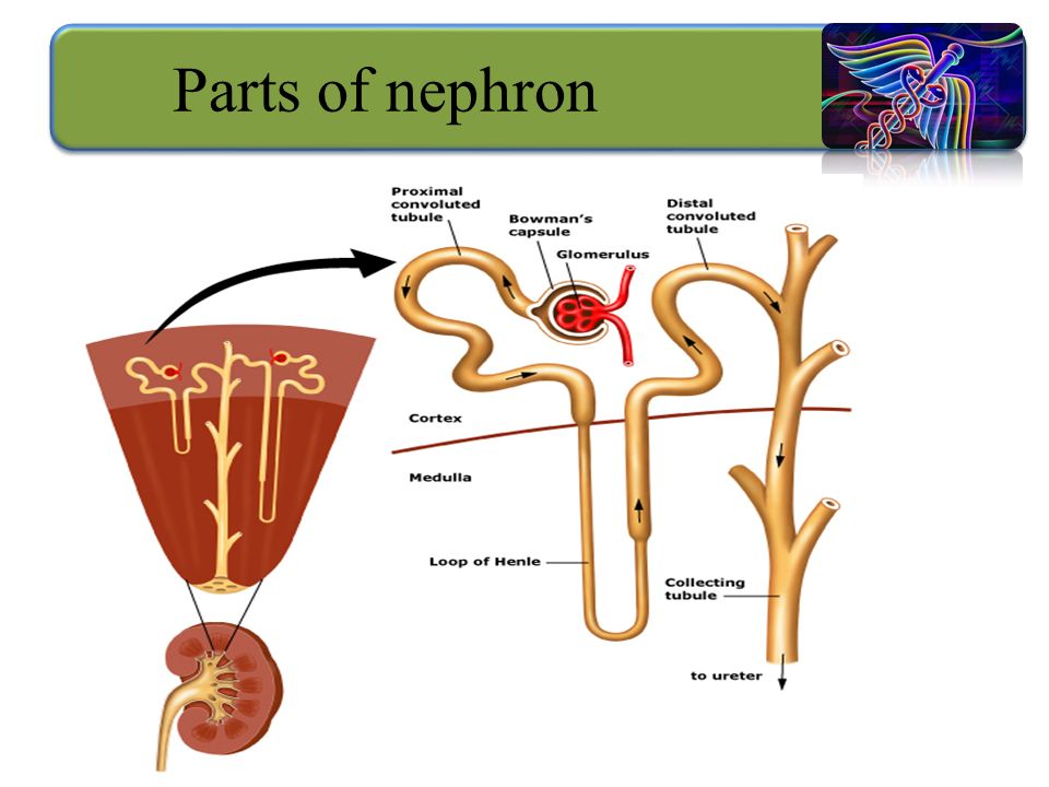
Nephrons They are the functioning unit of the kidney About 1 million nephrons are in a kidney.

14
1.Bowman’s capsule 2.Glomerulus 3.Proximal convoluted tubule 4.Loop of Henle 5.Distal convoluted tubule 6.Collecting tubule Parts of nephron

16
The ureters are tubes made of smooth muscles that carry urine from the kidneys to the urinary bladder. Ureter

17
Superiorly Continuous with the renal pelvis Inferiorly Pass through the abdominal cavity, behind the peritoneum, into the pelvic cavity where they enter the posterior wall of the bladder 25-30 cm in length 17 Ureter

18
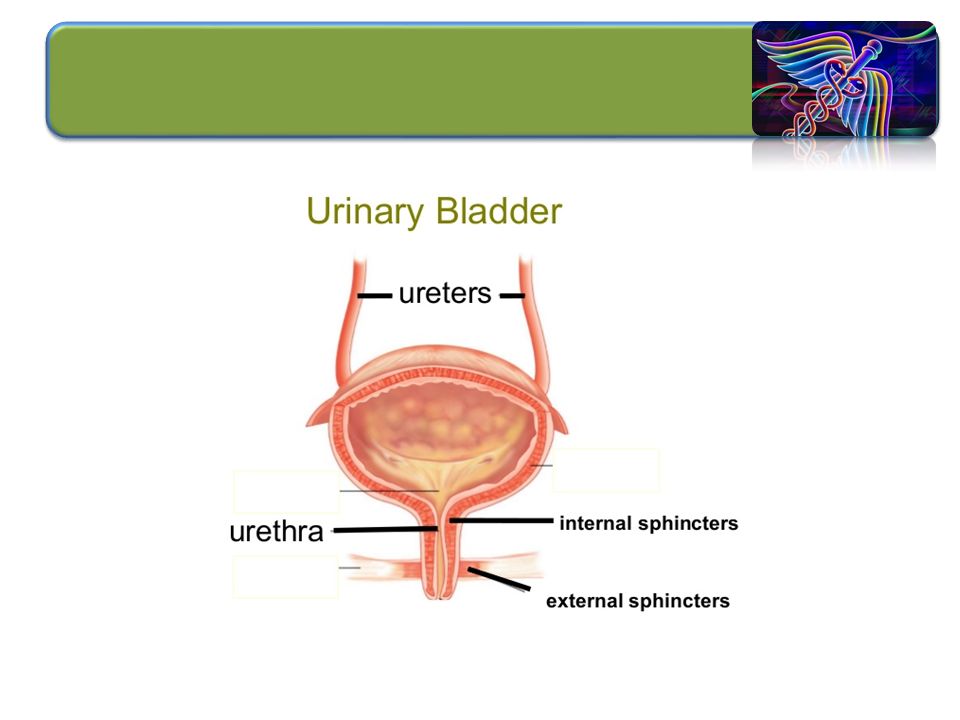
Urinary bladder

19
Sagittal section of male and female bladder

20
Urinary Bladder Wall of bladder –Mucosa ( inner) –Muscular layer (middle ) Detrusor –Adventitia (outer)
 –Muscular layer (middle ) Detrusor –Adventitia (outer)")
21
Internal urethral sphincter: Smooth muscle Involuntary control More superiorly located External Urethral sphincter: Skeletal muscle Voluntary control Posteriorly located Sphincters of the Bladder

23
IN FEMALES –Length of 3–4 cm IN MALES – 20 cm in length – three regions anatomically –Prostatic urethra Passes through the prostate gland –Membranous urethra Through the urogenital diaphragm –Spongy (penile) urethra Passes through the length of the penis Urethra
 urethra Passes through the length of the penis Urethra")
24
Sagittal section of Urethra

25
Frontal view of male urethra

26
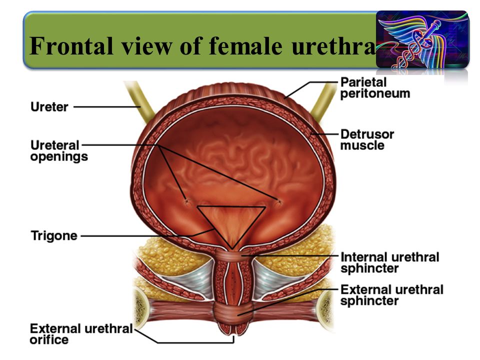
26 Frontal view of female urethra

Similar presentations


 Ureters (2) Urinary bladder Urethra.>")


