Download presentation
Presentation is loading. Please wait.

1
Organ of vision

2
The Five Senses Touch Hearing SIGHT Taste Smell

3
“Special” senses Special senses monitor vision, hearing, olfaction, gustation, and equilibrium through specialized sense organs These sense organs are highly specialized

4
External Anatomy Sensory Organ for vision -Situated in bony, orbital cavity for protection –Eyelids= shades that add protection form injury, strong light, dust –Eyelashes= hairs to filter dust & dirt

5
External Anatomy

6
Sight The eye is the organ that captures light.

7
Sight

8
Color Blindness

9
All sensory receptors send info to the CNS via an action potential… At the CNS, info is routed according to the stimulus and its location The stronger the stimulus, the higher the frequency of action potentials Some receptors adapt, that is their sensitivity to a stimulus is reduced if the stimulus is continually applied (smell) –The RAS can heighten or reduce awareness of sensory information
 –The RAS can heighten or reduce awareness of sensory information")
10
A complex sensory organ: the eye. is surrounded by accessory structures that act to protect, lubricate, and support it is a light, compact, durable, and highly specialized hollow organ that weighs about 8 oz and measures 1 inch in diameter. is divided into anterior (aqueous) & posterior (vitreous) cavities. its walls are made of 3 “tunics”
 & posterior (vitreous) cavities. its walls are made of 3 tunics .")
11
Accessory structures of the eye… eyelids (palpebrae) eyelashes & brows exocrine glands lacrimal apparatus Conjunctiva 6 extrinsic occulomotor muscles: – the inferior, superior, lateral and medial rectus muscles –the superior and inferior oblique muscles
 eyelashes & brows exocrine glands lacrimal apparatus Conjunctiva 6 extrinsic occulomotor muscles: – the inferior, superior, lateral and medial rectus muscles –the superior and inferior oblique muscles")
12
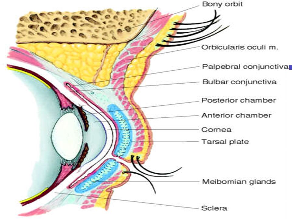
Within the upper eyelid –Tarsal plates, connective tissue gives upper lid shape –Meibomian glands, in the plates, lubricate the lids, stops overflow of tears, airtight seal when lids closed

13
Cornea – clear, covers & protects iris & pupil

15
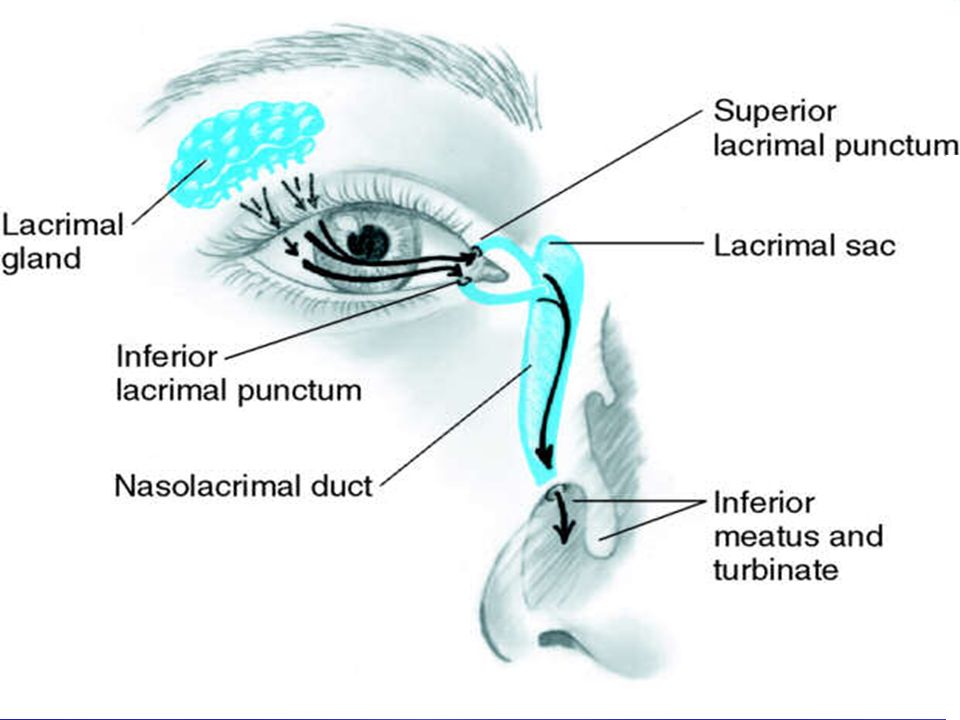
Lacrimal apparatus – irrigates conjunctiva & cornea –3 parts A.Lacrimal gland, upper, outer corner of eye = tears B.Puncta= inner canthus, tear drainage C.Nasolacrimal duct= allows tears to drain from puncta to nasolacrimal sac. Tears then empty into the inferior meatus of the nose

17
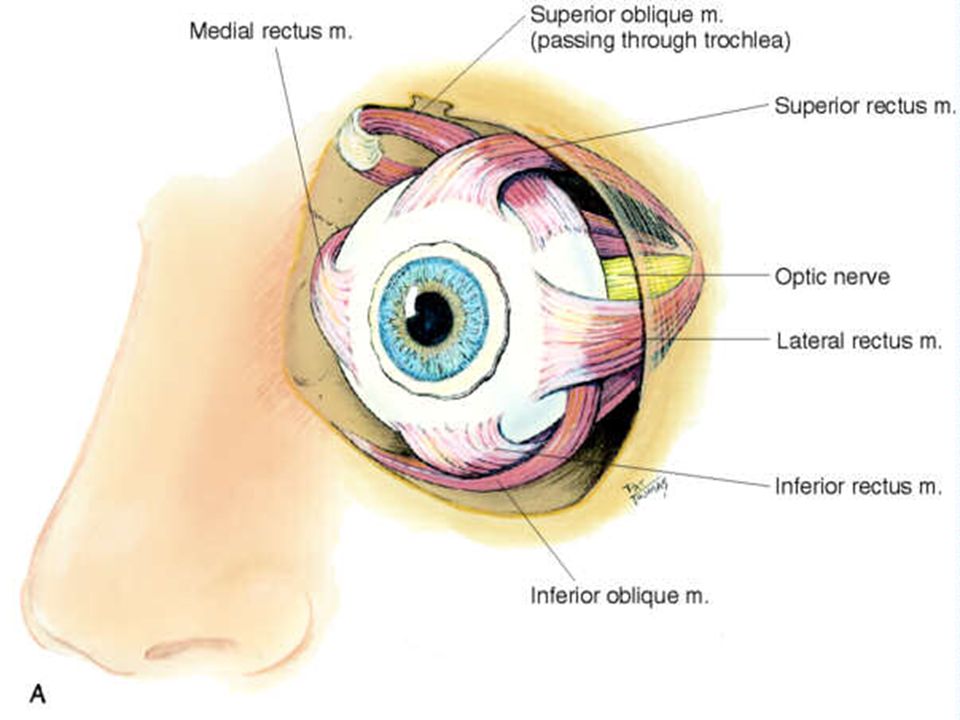
Extraoccular muscles 6 muscles –Attach eyeball to orbit –Straight and rotary movement –Four straight muscles 1.Superior rectus 2.Inferior rectus 3.Lateral rectus 4.Medial rectus

18
Two slanting/ oblique muscles 5.Superior 6.Inferior Humans have a Binocular, single – image visual system – Eyes normally move as a pair

21
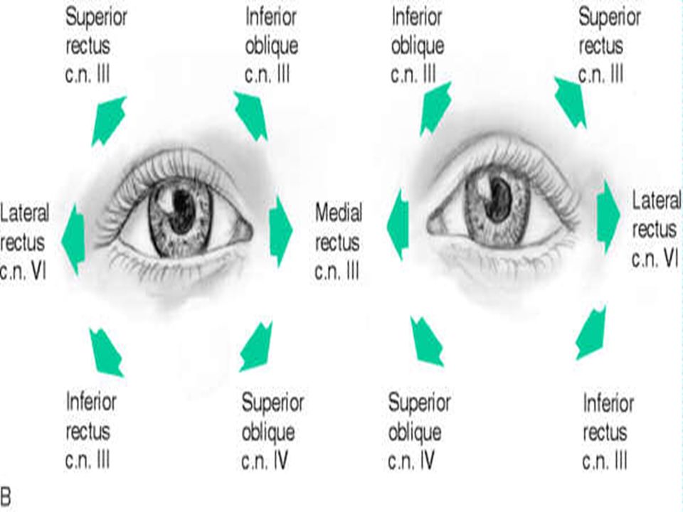
Eye movement stimulated by Cranial NervesEye movement stimulated by Cranial Nerves III OculomotorIII Oculomotor IV TrochlearIV Trochlear VI AbducensVI Abducens

22
Eye anatomy….. http://www.macula.org/ anatomy/eyeframe.htm l http://www.macula.org/ anatomy/eyeframe.htm l http://www.macula.org/ anatomy/eyeframe.htm l The hollow eye is divided into 2 cavities: An anterior cavity which contains aqueous humor A posterior cavity which holds vitreous humor Humors act to stabilize eye shape and provide nutrients

23
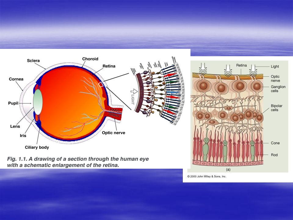
The Tunics of the eye… Fibrous - the sclera & anterior cornea Vascular – contains blood vessels, lymphatics, choroid & intrinsic muscles of the iris &ciliary bodies (they support the lens) Neural – the retina, it contains the rods and cones (photoreceptor cells), bipolar &ganglion cells
 Neural – the retina, it contains the rods and cones (photoreceptor cells), bipolar &ganglion cells")
24
Exposed part of the eye –Conjunctiva, folded envelope b/t eyelids & eyeball thin mucous membrane, transparent protective covering of the exposed part of the eye. Palpebral conjunctiva lines the lids, is clear but has sm.bld. Vessels Bulbar conjunctiva is over eyeball, white sclera show through, merges at limbus with cornea

25
Retinal organization … The retina is made of several cell layers: –Photoreceptor cells – rods lie along the periphery & cones lie at the back of the retina –Bipolar cells synapse with the rods and cones –Ganglion cells synapse with the bipolar cells –The axons of the ganglion cells form the optic nerve –http://www.macula.org/anatomy/retinaframe.html http://www.macula.org/anatomy/anatomy.html http://www.macula.org/anatomy/retinaframe.html http://www.macula.org/anatomy/anatomy.htmlhttp://www.macula.org/anatomy/retinaframe.html http://www.macula.org/anatomy/anatomy.html

26
Macula lutea – area on the retina where the visual image forms, it contains only cones with the greatest numbers at the fovea centralis Optic Disc or “blind spot” is the area where the ganglion cell axons exit the eye to form the optic nerve

28
Other structures of the eye: Lens – held in place by suspensory ligaments, it functions to focus the visual image onto the retina Cornea – clear portion of the fibrous tunic it is contiguous with the sclera Iris – part of the vascular tunic, it contains blood vessels, pigment, and 2 smooth muscle layers to control the width of the pupil. Ciliary body – a thick region of the choroid that encircles the lens and supports the suspensory ligments of the lens

29
Accommodation- focusing an image on the retina by changing lens shape light bends/refracts as it passes from 1 medium to another. In the eye it goes through the cornea, a. humor, lens, & v. humor. The refraction of light is constant through all but the lens The lens changes shape to keep the image focused on the retina for greatest visual acquity Accommodation occurs with response to light and to the distance of the object being viewed http://www.kscience.co.uk/animations/eye.s wf http://www.kscience.co.uk/animations/eye.s wf http://www.kscience.co.uk/animations/eye.s wf

30
The Physiology of Vision… How is it that we see? Photoreceptors respond to visible light: Rods – sensitive to photons (energy) but not their wavelength (color) allow for vision at night/dim light The 3 types of Cones (green, red, blue) need bright light & are responsive to wavelength: they allow us to “see” color Lack of functional cones = colorblindness
 but not their wavelength (color) allow for vision at night/dim light The 3 types of Cones (green, red, blue) need bright light & are responsive to wavelength: they allow us to see color Lack of functional cones = colorblindness.")
31
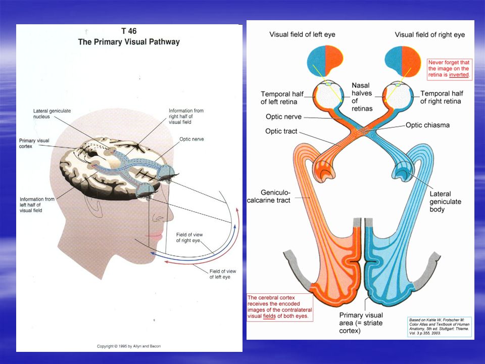
The visual pathway…. Photoreceptor stimulation Bipolar cell activation Stimulation of ganglion cell’s whose axons form the… Optic nerve that cross at the diencephalon and goes to the thalamus that routes info to the visual cortex of occipital lobe and the reflex centers of brain stem At the optic chiasm, a partial crossover of nerve fibers occurs http://www.sumanasinc.com/webcontent/anisampl es/neurobiology/visualpathways.html http://www.sumanasinc.com/webcontent/anisampl es/neurobiology/visualpathways.html http://www.sumanasinc.com/webcontent/anisampl es/neurobiology/visualpathways.html

Similar presentations


 separated by the palpebral fissue Eyelashes Tarsal glands Lacrimal apparatus Vision Accessory structures.>")


