Download presentation
Presentation is loading. Please wait.

1
Human Physiology: Unit-1
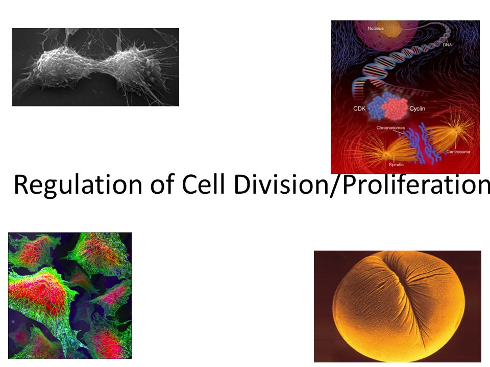
Regulation of Cell Division/Proliferation BY DR BOOMINATHAN Ph.D. M.Sc.,(Med. Bio, JIPMER), M.Sc.,(FGSWI, Israel), Ph.D (NUS, SINGAPORE), PDF (USA) PONDICHERRY UNIVERSITY Sixth Lecture 21/August/2012 Source: Collected from different sources on the internet and modified by Dr Boominathan Ph.D.
, M.Sc.,(FGSWI, Israel), Ph.D (NUS, SINGAPORE), PDF (USA) PONDICHERRY UNIVERSITY. Sixth Lecture. 21/August/2012. Source: Collected from different sources on the internet and modified by Dr Boominathan Ph.D.")
2
Regulation of Cell Division/Proliferation
3
Learning objectives Cell Division Cell cycle
Cyclin-CDKs that control cell cycle progression Checkpoints p53/RB Control of cell cycle/division/proliferation Cancer

4
THINK: Of how many cells are you composed?
When an organism grows bigger do you get more cells or just bigger cells or both? When do your cells divide the fastest? Slowest? Do cells ever stop dividing? Are all cells capable of division and replacement?

5
Cell division

6
How the cell cycle works
works.html Mitosis and Cytokinesis: chapter2/animation__mitosis_and_cytokinesis.html

7
Comparision of mitosis and meiosis
Control of cell cycle

8
Coordination of cell division
A multicellular organism needs to coordinate cell division across different tissues & organs critical for normal growth, development & maintenance coordinate timing of cell division coordinate rates of cell division not all cells can have the same cell cycle

9
Frequency of cell division
Frequency of cell division varies by cell type embryo cell cycle < 20 minute skin cells divide frequently throughout life 12-24 hours cycle liver cells retain ability to divide, but keep it in reserve divide once every year or two mature nerve cells & muscle cells do not divide at all after maturity permanently in G0 G2 S G1 M metaphase prophase anaphase telophase interphase (G1, S, G2 phases) mitosis (M) cytokinesis (C) C
 mitosis (M) cytokinesis (C) C.")
10
Control of the Cell Cycle /Cell proliferation

11
Interphase Interphase ~ 90% of the time.
Ø G1: Little new cell absorbs nutrients and grows larger. Does protein synthesis, its job. Ø S phase: Synthesis of new DNA (DNA replication) for daughter cells in preparation for mitosis. Ø G2: Cell continues to grow, do protein synthesis, do its job. Gets too large, needs to divide.
 for daughter cells in preparation for mitosis. Ø G2: Cell continues to grow, do protein synthesis, do its job. Gets too large, needs to divide.")
12
Cyclin & Cyclin-dependent kinases
CDKs & cyclin drive cell from one phase to next in cell cycle proper regulation of cell cycle is so key to life that the genes for these regulatory proteins have been highly conserved through evolution the genes are basically the same in yeast, insects, plants & animals (including humans) CDK and cyclin together form an enzyme that activates other proteins by chemical modification (phosphorylation). The amount of CDK molecules is constant during the cell cycle, but their activities vary because of the regulatory function of the cyclins. CDK can be compared with an engine and cyclin with a gear box controlling whether the engine will run in the idling state or drive the cell forward in the cell cycle.
 CDK and cyclin together form an enzyme that activates other proteins by chemical modification (phosphorylation). The amount of CDK molecules is constant during the cell cycle, but their activities vary because of the regulatory function of the cyclins. CDK can be compared with an engine and cyclin with a gear box controlling whether the engine will run in the idling state or drive the cell forward in the cell cycle.")
13
The cell cycle engines Cyclin Dependent Kinases (CDKs) CDK cyclin
substrate ATP P product + ADP

14
Cyclin D-CDK4 Cyclin E-CDK2 Cyclin A-CDK2 Cyclin B-CDC2

15
CDK activity

16
Mitogens stimulate the onset of the cell cycle

17
Mitogens control cyclin D expression

18
Mitogens control cyclin D expression
- Mitogens act by activating the D-Cdk4/6 complexes - Mitogens act by inhibiting CKIs - Mitogen signaling is correlated with growth, answering the question: “have I grown enough?”

19
We actually have: 3 D-type cyclins 2 E-type cyclins 2 A-type cyclins
3 B-type cyclins

20
cyclin D and growth w/o growth signals, sub-threshold levels of enzymes will lead to quiescence (G0) START/Restriction point

21
- Activated D-Cdk4/6 initiates transcription of cyclin E and activation of E-Cdk2
- Activated E-Cdk2 allows progression through START - From here on, it’s a cell cycle clock game

23
APC = anaphase-promoting complex
The different cyclins are degraded by two different E3 ligases e.g. cyclin B, the G2/M cyclin is degraded by APC = anaphase-promoting complex

24
cyclins must be removed for mitosis to be completed
Remember? cyclins must be removed for mitosis to be completed Protein Level Time cyclin A cyclin B M

25
CDKs are positively regulated by cyclins
For example, Cyclin-X promotes synthesis of the next cyclin that in turn, promotes destruction of the Cyclin-X These regulatory activities are indirect

26
Mechanisms of CDKs regulation
1. Abundance of cyclins 2. CDK phosphorylation 3. Binding to CKIs (inhibitory proteins) CDK Cyclin active + inactive CDKI CDKI
 CDK. Cyclin. active. + inactive. CDKI. CDKI.")
27
Activating phosphorylation is catalyzed by Cdk-Activating Kinases (CAKs). However, they are abundant and not regulated 1 3 Cdk Phosporylation of Thr by CAK 2 4 Cyclin Substrate binding to the kinase

28
Mechanisms of CDKs regulation
1. Abundance of cyclins 2. CDK phosphorylation 3. Binding to CKIs (inhibitory proteins) CDK Cyclin active CDKI + inactive CDKI
 CDK. Cyclin. active. CDKI. + inactive. CDKI.")
29
Cdk inhibitor proteins (CKIs)
- Discovered by asking : “what binds to CDKs”? - The INK4 family proteins bind to CDK4/6, blocking cyclin D binding - The Cip/Kip family proteins bind to blocking active site of multiple CDKs - CKIs normally regulate entry into S phase

30
CKIs Regulate the G1-S Transition

31
Overview of Cell Cycle Control
There’s no turning back, now! Overview of Cell Cycle Control Two irreversible points in cell cycle replication of genetic material separation of sister chromatids Checkpoints process is assessed & possibly halted centromere sister chromatids single-stranded chromosomes double-stranded

32
Checkpoint control system
Checkpoints cell cycle controlled by STOP & GO chemical signals at critical points signals indicate if key cellular processes have been completed correctly

33
Checkpoint control system
3 major checkpoints: G1/S can DNA synthesis begin? G2/M has DNA synthesis been completed correctly? commitment to mitosis spindle checkpoint are all chromosomes attached to spindle? can sister chromatids separate correctly?

34
G1/S checkpoint G1/S checkpoint is most critical
primary decision point “restriction point” if cell receives “GO” signal, it divides internal signals: cell growth (size), cell nutrition external signals: “growth factors” if cell does not receive signal, it exits cycle & switches to G0 phase non-dividing, working state
, cell nutrition. external signals: growth factors if cell does not receive signal, it exits cycle & switches to G0 phase. non-dividing, working state.")
35
Progression of the cell cycle is regulated by feedback from intracellular events

36
G0 phase G0 phase non-dividing, differentiated state
most human cells in G0 phase liver cells in G0, but can be “called back” to cell cycle by external cues nerve & muscle cells highly specialized arrested in G0 & can never divide

37
Activation of cell division
How do cells know when to divide? cell communication signals chemical signals in cytoplasm give cue signals usually mean proteins activators inhibitors experimental evidence: Can you explain this?

38
“Go-ahead” signals Protein signals that promote cell growth & division
internal signals “promoting factors” external signals “growth factors” Primary mechanism of control phosphorylation kinase enzymes either activates or inactivates cell signals We still don’t fully understanding the regulation of the cell cycle. We only have “snapshots” of what happens in specific cases.

39
Cell cycle signals Cell cycle controls cyclins Cdk’s
inactivated Cdk Cell cycle controls cyclins regulatory proteins levels cycle in the cell Cdk’s cyclin-dependent kinases phosphorylates cellular proteins activates or inactivates proteins Cdk-cyclin complex triggers passage through different stages of cell cycle activated Cdk

40
Cyclins & Cdks Interaction of Cdk’s & different cyclins triggers the stages of the cell cycle There are multiple cyclins, each with a specific role. Cyclins are unstable. Some are triggered for destruction by phosphorylation. Others are inherently unstable and are synthesized discontinuously during the cell cycle. Oscillations in the activities of cyclin-dependent kinases (CDKs) dictate orderly progression through the cell division cycle. In the simplest case of yeast, a progressive rise in the activity of a single cyclin-CDK complex can initiate DNA synthesis and then mitosis, and the subsequent fall in CDK activity resets the system for the next cell cycle. In most organisms, however, the cell cycle machinery relies on multiple cyclin- CDKs, whose individual but coordinated activities are each thought to be responsible for just a subset of cell cycle events. Leland H. Hartwell Checkpoints Tim Hunt Cdks P. Nurse Cyclins
 dictate orderly progression through the cell division cycle. In the simplest case of yeast, a progressive rise in the activity of a single cyclin-CDK complex can initiate DNA synthesis and then mitosis, and the subsequent fall in CDK activity resets the system for the next cell cycle. In most organisms, however, the cell cycle machinery relies on multiple cyclin- CDKs, whose individual but coordinated activities are each thought to be responsible for just a subset of cell cycle events. Leland H. Hartwell. Checkpoints. Tim Hunt. Cdks. P. Nurse. Cyclins.")
41
Nobel Prize Leland H. Hartwell Checkpoints Tim Hunt Cdks P Nurse Cyclins They won the Nobel Prize in Physiology or Medicine for their discoveries of protein molecules that control the division (duplication) of cells
 of cells.")
42
Spindle checkpoint G2 / M checkpoint M cytokinesis C G2 mitosis G1 S
Chromosomes attached at metaphase plate Replication completed DNA integrity Inactive Active Active Inactive Cdk / G2 cyclin (MPF) M APC cytokinesis C G2 mitosis G1 S Cdk / G1 cyclin Inactive MPF = Mitosis Promoting Factor APC = Anaphase Promoting Complex Active G1 / S checkpoint Growth factors Nutritional state of cell Size of cell
 M. APC. cytokinesis. C. G2. mitosis. G1. S. Cdk / G1 cyclin. Inactive. MPF = Mitosis Promoting Factor. APC = Anaphase Promoting Complex. Active. G1 / S checkpoint. Growth factors. Nutritional state of cell. Size of cell.")
43
Summary - The cell cycle is controlled by Cdks, activated by cyclins and CDK activating Kinases, and inhibited by CKIs - Cyclins are positively and negatively regulated by cyclin-Cdks complexes - Any process in the cell cycle is dependent on the previous one - The cell cycle progresses in the right order

44
70kg human ~ 1013 cells

45
Growth factor signals growth factor cell division cell surface
nuclear pore nuclear membrane P P cell division cell surface receptor Cdk protein kinase cascade P E2F P chromosome Rb P E2F cytoplasm Rb nucleus

46
Example of a Growth Factor
Platelet Derived Growth Factor (PDGF) made by platelets in blood clots binding of PDGF to cell receptors stimulates cell division in fibroblast (connective tissue) heal wounds Erythropoietin (EPO): A hormone produced by the kidney that promotes the formation of red blood cells in the bone marrow. EPO is a glycoprotein (a protein with a sugar attached to it). The kidney cells that make EPO are specialized and are sensitive to low oxygen levels in the blood. These cells release EPO when the oxygen level is low in the kidney. EPO then stimulates the bone marrow to produce more red cells and thereby increase the oxygen-carrying capacity of the blood. EPO is the prime regulator of red blood cell production. Its major functions are to promote the differentiation and development of red blood cells and to initiate the production of hemoglobin, the molecule within red cells that transports oxygen. EPO has been much misused as a performance-enhancing drug (“blood doping”) in endurance athletes including some cyclists (in the Tour de France), long-distance runners, speed skaters, and Nordic (cross-country) skiers. When misused in such situations, EPO is thought to be especially dangerous (perhaps because dehydration can further increase the viscosity of the blood, increasing the risk for heart attacks and strokes. EPO has been banned by the Tour de France, the Olympics, and other sports organizations.
 made by platelets in blood clots. binding of PDGF to cell receptors stimulates cell division in fibroblast (connective tissue) heal wounds. Erythropoietin (EPO): A hormone produced by the kidney that promotes the formation of red blood cells in the bone marrow. EPO is a glycoprotein (a protein with a sugar attached to it). The kidney cells that make EPO are specialized and are sensitive to low oxygen levels in the blood. These cells release EPO when the oxygen level is low in the kidney. EPO then stimulates the bone marrow to produce more red cells and thereby increase the oxygen-carrying capacity of the blood. EPO is the prime regulator of red blood cell production. Its major functions are to promote the differentiation and development of red blood cells and to initiate the production of hemoglobin, the molecule within red cells that transports oxygen. EPO has been much misused as a performance-enhancing drug ( blood doping ) in endurance athletes including some cyclists (in the Tour de France), long-distance runners, speed skaters, and Nordic (cross-country) skiers. When misused in such situations, EPO is thought to be especially dangerous (perhaps because dehydration can further increase the viscosity of the blood, increasing the risk for heart attacks and strokes. EPO has been banned by the Tour de France, the Olympics, and other sports organizations.")
47
Growth Factors and Cancer
Growth factors can create cancers proto-oncogenes (normal) normal growth factor genes that become oncogenes (cancer-causing) when mutated stimulates cell growth if switched “ON” can cause cancer Eg., Myc (activates cyclins) tumor-suppressor genes inhibits cell division if switched “OFF” can cause cancer Eg., : p53, RB
 normal growth factor genes that become oncogenes (cancer-causing) when mutated. stimulates cell growth. if switched ON can cause cancer. Eg., Myc (activates cyclins) tumor-suppressor genes. inhibits cell division. if switched OFF can cause cancer. Eg., : p53, RB.")
48
Cancer & Cell Growth Cancer is essentially a failure of cell division control unrestrained, uncontrolled cell growth What control is lost? lose checkpoint stops gene p53 plays a key role in G1/S restriction point p53 protein halts cell division if it detects damaged DNA options: stimulates repair enzymes to fix DNA forces cell into G0 resting stage keeps cell in G1 arrest causes apoptosis of damaged cell ALL cancers have to shut down p53 activity p53 is the Cell Cycle Enforcer p53 was discovered mainly by Drs. Arnold Levine & David Lane Bert Vogelstein, Moshe Oren, Varda Rotter…

49
p53 — master regulator gene
NORMAL p53 p53 allows cells with repaired DNA to divide. p53 protein DNA repair enzyme p53 protein Step 1 Step 2 Step 3 DNA damage is caused by heat, radiation, or chemicals. Cell division stops, and p53 triggers enzymes to repair damaged region. p53 triggers the destruction of cells damaged beyond repair. ABNORMAL p53 abnormal p53 protein cancer cell Step 1 Step 2 DNA damage is caused by heat, radiation, or chemicals. The p53 protein fails to stop cell division and repair DNA. Cell divides without repair to damaged DNA. Step 3 Damaged cells continue to divide. If other damage accumulates, the cell can turn cancerous.

50
Rb inhibits G1 cell cycle progression
Rb and G1 cell cycle progression. Rb exists in different states of phosphorylation: unphosphorylated, hypophosphorylated, and hyperphosphorylated. The hypophosphorylated state occurs after phosphorylation by cyclin D–CDK4/6, and this leaves Rb associated with E2F family transcription factors such that E2F is unable to activate transcription. Rb becomes hyperphosphorylated after the activation of cyclin E–CDK2. At this point, E2F dissociates from E2F and can initiate the transcription of genes required for progression into S phase. One of the genes activated by E2F is cyclin E generating a positive feedback loop to aid in the progression through G1-ps. Foster D A et al. Genes & Cancer 2011;1: Copyright © by SAGE Publications

51
Positive Feedback Loop

52
Restriction point control and the G1-S transition.
Restriction point control and the G1-S transition. As cells enter the division cycle from quiescence, the assembly of cyclin D-dependent kinases in response to mitogenic signals requires Cip/Kip proteins, which are incorporated into catalytically active holoenzyme complexes. The cyclin D-dependent kinases initiate Rb phosphorylation, releasing E2F from negative constraints and facilitating activation of a series of E2F-responsive genes, the products of which are necessary for S-phase entry. Activation of cyclin E by E2F enables formation of the cyclin E-cdk2 complex. This is accelerated by the continued sequestration of Cip/Kip proteins into complexes with assembling cyclin D-cdk complexes. Cyclin E-cdk2 completes the phosphorylation of Rb, further enabling activation of E2F-responsive genes, including cyclin A. Cyclin E-cdk2 also phosphorylates p27Kip1, targeting it for ubiquitination and proteasomal degradation. The initiation of the self-reinforcing E2F transcriptional program together with degradation of p27Kip1 alleviates mitogen dependency at the restriction point and correlates with the commitment of cells to enter S phase. In subsequent cycles, cyclin D-dependent kinases remain active as long as mitogens are present, and levels of p27Kip1 remain low. All p27Kip1 in cycling cells is complexed with cyclin D-cdk complexes. Mitogen withdrawal results in cyclin D degradation, liberating p27Kip1 from this latent pool. The resulting inhibition of cyclin D- and E-dependent kinases leads to cell cycle arrest, usually within a single cycle. Sherr C J Cancer Res 2000;60: ©2000 by American Association for Cancer Research

53
How tumor suppressor genes block cell division

54
Development of Cancer Cancer develops only after a cell experiences ~6 key mutations (“hits”) unlimited growth turn on growth promoter genes ignore checkpoints turn off tumor suppressor genes (p53) escape apoptosis turn off apoptotic genes immortality = unlimited divisions turn on chromosome maintenance genes promotes blood vessel growth turn on blood vessel growth genes overcome anchor & density dependence turn off touch-sensor gene
 escape apoptosis. turn off apoptotic genes. immortality = unlimited divisions. turn on chromosome maintenance genes. promotes blood vessel growth. turn on blood vessel growth genes. overcome anchor & density dependence. turn off touch-sensor gene.")
55
What causes these “hits”?
Mutations in cells can be triggered by UV radiation chemical exposure radiation exposure heat cigarette smoke pollution age genetics

56
Tumors Mass of abnormal cells Benign tumor Malignant tumors
abnormal cells remain at original site as a lump p53 has halted cell divisions most do not cause serious problems & can be removed by surgery Malignant tumors cells leave original site lose attachment to nearby cells carried by blood & lymph system to other tissues start more tumors = metastasis impair functions of organs throughout body

57
Traditional treatments for cancers
Treatments target rapidly dividing cells high-energy radiation Inhibits/apoptose rapidly dividing cells chemotherapy stop DNA replication stop mitosis & cytokinesis stop blood vessel growth

58
Resources Mitosis CANCER
How Cancer grows Mitosis: Cell Cycle & Cancer Animations: Cell Biology & Cancer Animations: Mitosis & Meiosis Interactive Exercise: Mitosis Animations: Plant Cell Mitosis:

59
Questions??

Similar presentations


 Coordination of cell division A multicellular organism needs to coordinate cell division across different tissues.>")


