Download presentation
Presentation is loading. Please wait.

1
Introduction to Dermatology
Clinical Pathology

2
Function of the Skin Mechanical protection Environmental protection
Water light Thermoregulation Sensory functions Pigmentation Prevents solar damage Metabolic/immunologic functions Secretion Excretion Vitamin D production Antimicrobrial action

3
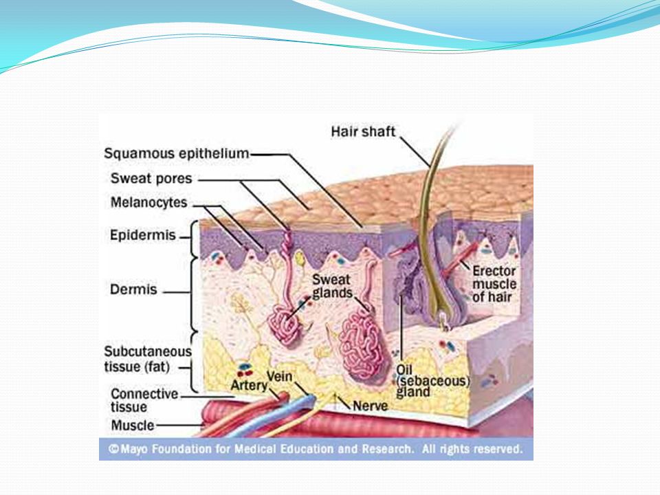
Structure of the Skin Epidermis Dermis Subcutaneous layer
Squamous keratinized epithelium (5 layers) sits on basement membrane Dermis Collagen fibers, blood, lymphatic vessels, nerves, fibroblasts, ground substance. Subcutaneous layer Hair follicles Epidermal invaginations into the dermis.
 sits on basement membrane. Dermis. Collagen fibers, blood, lymphatic vessels, nerves, fibroblasts, ground substance. Subcutaneous layer. Hair follicles. Epidermal invaginations into the dermis.")
6
Dermatologic Diagnostic Tools
Signalment Breed, sex, age, color History Diet Environment Date of onset Acute vs. chronic Initial distribution of lesions Pruritic Physical Exam General PE Identify lesion Location of lesions/distribution Pruritis

8
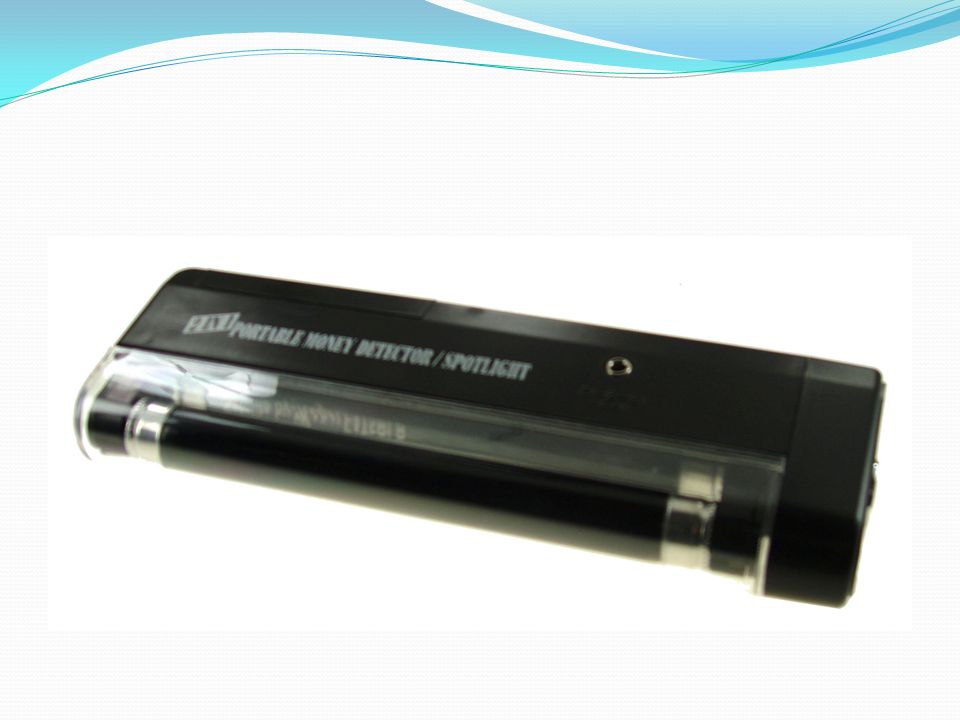
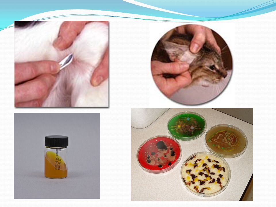
Diagnostic Tests Wood’s lamp Skin scraping Tape strip test
Direct smear/impression smears Fungal cultures/ microscopic exams Bacterial culture Biopsy Fine needle aspirate Swab

11
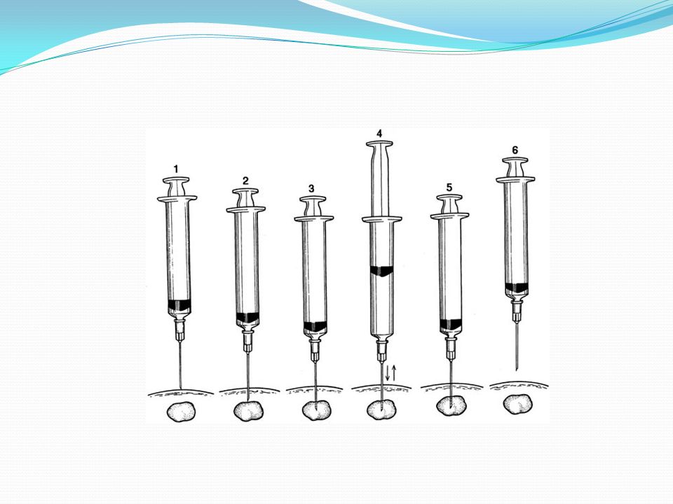
Skin Biopsy Punch

13
Dermatologic Terms for Lesions
Primary Lesion: Develop spontaneously as a direct reaction of the underlying disease. Secondary Lesion: Evolve from primary lesions.

14
Primary Lesion: Macule
Circumscribed flat spot having color change. Petechia are hemorrhagic types of macules.

15
Primary Lesion: Papule
Small, solid elevation of the skin Many are pink or reddish swellings Plaque: A large flat top elevation formed by a coalition of papules.

16
Primary Lesion: Pustule
Small, circumscribed elevation of the epidermis filled with pus.

17
Primary Lesion: Wheal A sharply circumscribed, raised, lesion consisting of edema. Hives

18
Primary Lesion: Vesicle
Elevation of the epidermis with clear fluid Seen in viral or autoimmune dermatoses.

19
Primary Lesion: Tumor Neoplastic enlargement of subcutaneous tissues.

20
Secondary Lesion: Scales
Accumulation of loose fragments of the keratin from the horny layer of the skin. (like dandruff) Epidermal collarettes: A special type of scale arranged into a circular rim. Remnants of the “roof” of a vesicle or pustule.
 Epidermal collarettes: A special type of scale arranged into a circular rim. Remnants of the roof of a vesicle or pustule.")
21
Secondary Lesion: Crusts
Form from dried exudate, serum, pus, cells, and scales. In pyodermas, crusts are yellowish-green.

22
Secondary Lesion: Excoriation
Superficial removal of epidermis Usually self-induced due to pruritis Abrasion that is self-induced

23
Secondary Lesion: Ulcers
A defect in the epidermis and exposing underlying dermis.

24
Secondary Lesion: Lichenification
Thickening/ hardening of the skin Due to chronic friction or trauma Can see in elbow pads

25
Secondary Lesion: Hyperpigmentation
Abnormal pigment of the skin

26
Dermatophytosis/Ringworm
Infection of the hair shafts and skin. Microsporum canis most common cause In rare instances- Microsporum gyseum and Trichophyton spp. Infective spores in soil, by direct contact, and by environmental fomites, ventilation. Trauma to skin may promote infection Other risk factors: Age Immune competence Lesions may be circular, irregular, crusts, scales, hair thinning.

28
Classification of Dermatophytes
Anthropophilic: Inhabit people only Zoophilic: Inhabit both animals and people Geophilic: Free-living saprophytes in soil. May be contaminant in cultures. Microsporum gypseum only species that causes lesions in animals.

29
Diagnosis: Woods Lamp: Fungal Culture: Microscopic Exam of the colony:
50% of Microsporum canis strains will fluoresce under a woods lamp. Looking for an “apple green” fluorescence Fungal Culture: Saboraud’s medium or Dermatophyte Test Medium (DTM) specifically designed for ringworm diagnosis. Color change before 10 days Microscopic Exam of the colony:
 specifically designed for ringworm diagnosis. Color change before 10 days. Microscopic Exam of the colony:")
31
DTM Procedure Pluck samples from suspicious lesions using a sterile hemostat. For asymptomatic carriers, use sterile toothbrush to comb cat fur. Place gently on DTM culture. Close lid of bottle, but do not tighten down. Store in darkened area. Results in 5-12 days. Positive result: growth and color change at the same time (day). Check every other day after Day 3.
. Check every other day after Day 3.")
32
Special DTM notes: Pigs
Often have contamination from geophilic/saprphytic fungi. Swab lesion with alcohol, let dry, then collect sample.

34
Dermatophyte Identification using Colony Morphology
Microsporum canis: Surface is white and woolly. Reverse side is yellow. Microsporum gypseum: Surface is coarsely powdery, light tan to cinnamon brown. Reverse is brownish yellow. Trichophyton mentagrophytes: Surface is cream colored and powdery. Reverse is yellowish to brown.

35
Microsporum canis Confirm with microscopic exam
Macroconidia have thick walls, spindle shaped 8-15 cells and possess a terminal knob.

36
Microsporum gypseum Spindle shaped but broader with no terminal knobs. Less than 6 cells on macroconidia.

37
Trichophyton Mentagrophytes
Few macroconidia, slender and cigar shaped with thin walls. Microconidia are numerous and arranged in grape-like clusters.

38
Direct Microscopic examination of Ringworm
Select a few hairs or skin scrape. May be suspended in mineral oil, through direct tape method or placed in a drop of 20% KOH (if use this method, gently heat and let stand for minutes). Examine under low and high power for fungal spores. If looking at colonies, tease out a little colony material and place on slide. Gently touch 2 cm strip of clear tape to surface of colony and then stain with new methylene blue or lactophenol cotton blue stains.
. Examine under low and high power for fungal spores. If looking at colonies, tease out a little colony material and place on slide. Gently touch 2 cm strip of clear tape to surface of colony and then stain with new methylene blue or lactophenol cotton blue stains.")
39
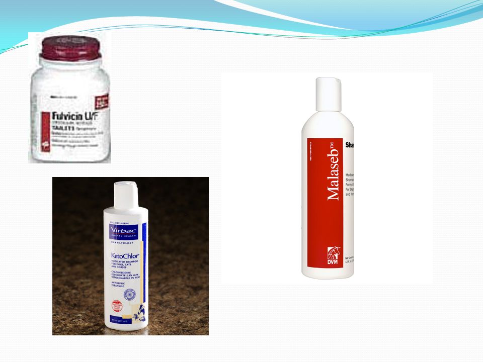
Dermatophytosis treatment
Systemic antifungals: Griseofulvicin (expensive and hard to get) Clip hair/shave down Program (Lufeneron): Off label use Topical antifungals: Miconazole, Chlorhexidine (malaseb shampoo and wipes), Ketoconazole (ketochlor shampoo), also topical lotions and creams. Solution of Lime-sulfur dip
 Clip hair/shave down. Program (Lufeneron): Off label use. Topical antifungals: Miconazole, Chlorhexidine (malaseb shampoo and wipes), Ketoconazole (ketochlor shampoo), also topical lotions and creams. Solution of Lime-sulfur dip.")
Similar presentations


>")


>")


