Download presentation
Presentation is loading. Please wait.

1
Paediatric Nephrology

2
Teaching website

5
UTI – cumulative incidence
Boys Girls By 2 years 2.2% 2.1% By 7 years 2.8% 8.2% By 16 years 3.6% 11.3%

6
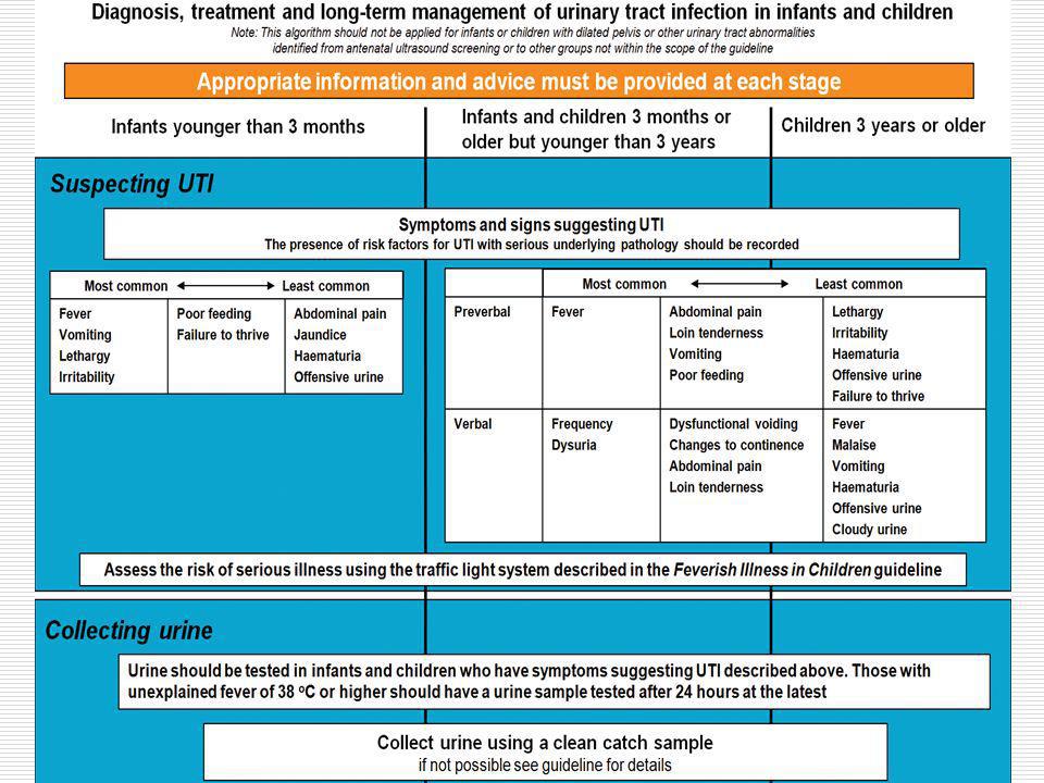
When to suspect a UTI Infants Pyrexia (>38.5oC) Poor feeding
Vomiting Abdominal discomfort Febrile seizure

7
When to suspect a UTI (2) Older children Frequency Dysuria Wetting
Abdominal pain Pyrexia

8
Diagnosis Collect a urine specimen MSU Clean catch specimen
Bag specimen Catheter specimen Suprapubic aspirate Pad specimen

10
Leucocyte esterase Identifies presence of white blood cells
High sensitivity for UTI but low specificity

11
Nitrite test Reliable sign of infection when positive
BUT high false negative rate Urine has to have been in the bladder for at least an hour. This lowers the false negative rate.

12
Use of both nitrite & leucocyte esterase tests
Has not replaced urine culture in patients suspected of having a UTI.

13
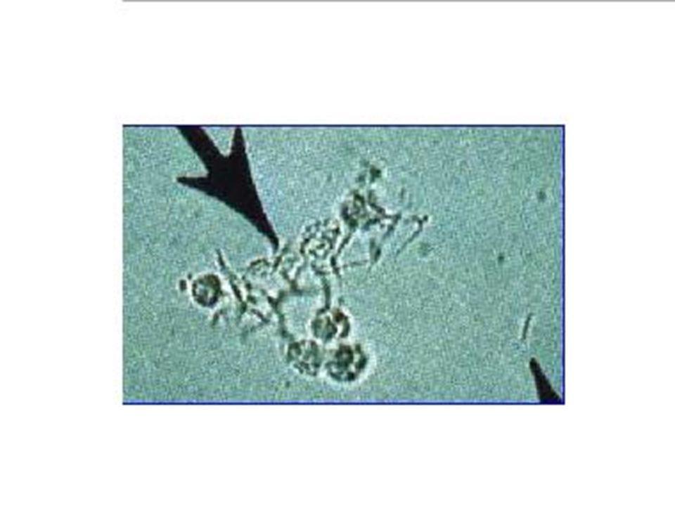
What do you do with the urine?

14
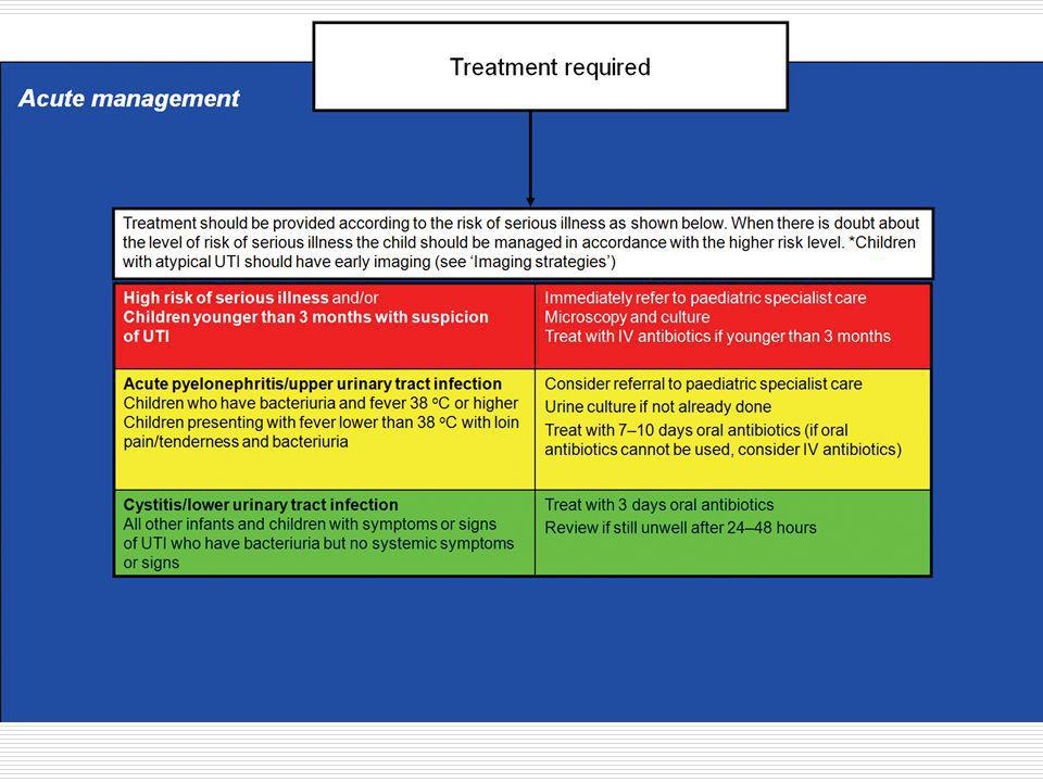
Aims of treatment Prevention of renal scarring
Achieved through prompt initiation of antibiotic therapy, particularly in those groups at highest risk Infants Children with vesicoureteric reflux

16
Ultrasound - Normal

17
Ultrasound - Hydronephrosis

18
Ultrasound - Scarring

19
DMSA scan - normal

20
DMSA - scarring

21
DMSA - scarring

22
MCUG - normal

23
MCUG - normal

24
MCUG – R sided grade II VUR

25
MCUG – Bilateral VUR

26
MCUG – Bilateral VUR

27
Management of VUR Antibiotic prophylaxis until 4 -5 years old
Surgery if continue to get UTIs Reimplantation Injection of Deflux

33
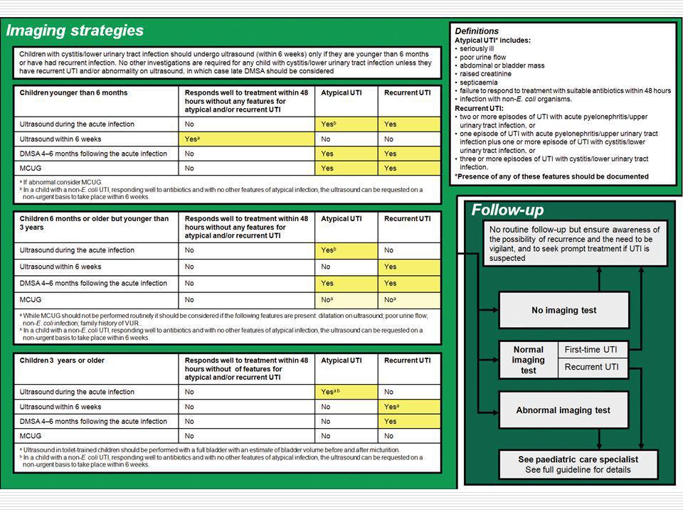
IMPORTANT MESSAGE Only do an investigation if the result will potentially alter your management of the patient.

34
Any questions?

36
Nephrotic syndrome Triad of: Heavy proteinuria Hypoalbuminaemia Oedema

39
Normal glomerulus (em)
")
40
EM showing foot process fusion

41
Interstitial fluid Ph - Hydrostatic pressure Po - Oncotic pressure

42
Mechanisms of oedema formation (2)
Increased hydrostatic pressure Hypervolaemia Increased venous pressure Reduced oncotic pressure Hypoalbuminaemia Increased capillary permeability Sepsis

43
Complications Oedema Hypovolaemia Infection Cool peripheries
Prolonged capillary refill time Abdominal pain Increased blood pressure Infection Hypogammaglobulinaemia

44
Complications (2) Hypercoaguable state Raised haematocrit
Loss of anti-thrombin III

45
VQ scan in nephrotic patient

46
Complications (2) Hypercoaguable state Hyperlipidaemia Hypothyroidism
Raised haematocrit Loss of anti-thrombin III Hyperlipidaemia Hypothyroidism

47
Treatment Lots of steroids

48
Any questions?

49
Red cell cast

50
Tubules filled with red blood cells – source of red cell casts

52
Dysmorphic red blood cells – Indicates they have had to squeeze through the glomerular basement membrane

53
Causative organism - Streptococcus

54
Resultant infections Impetigo Tonsillitis

55
Complement

56
Normal glomerulus

57
Glomerulus showing a proliferative nephritis – note the increased number of nuclei seen

58
Higher magnification

59
Immunofluorescent staining for C3

60
By electron microscopy, the immune deposits of post-infectious glomerulonephritis are predominantly subepithelial, as seen below, with electron dense subepithelial "humps" above the basement membrane and below the epithelial cell. The capillary lumen is filled with a leukocyte demonstrating cytoplasmic granules. By electron microscopy, the immune deposits of post-infectious glomerulonephritis are predominantly subepithelial, as seen above with electron dense subepithelial "humps" above the basement membrane and below the epithelial cell. The capillary lumen is filled with a leukocyte demonstrating cytoplasmic granules. Another example is shown below illustrating the subepithelial deposits.

62
Features of acute nephritis
Haematuria Proteinuria Oliguria Hypertension Oedema Renal impairment

63
Assessment of renal function
Glomerular filtration rate (GFR) mls/min Number of mls of blood cleared of a freely filtered substance each minute. Correct for body surface area – mls/min/1.73m2 Creatinine clearance GFR 1 / serum creatinine
 mls/min. Number of mls of blood cleared of a freely filtered substance each minute. Correct for body surface area. – mls/min/1.73m2. Creatinine clearance. GFR 1 / serum creatinine.")
64
Creatinine clearance Fact If serum [Cr] = 100 µmol/l and
If serum [creatinine] is constant, the rate of production of creatinine must equal its excretion. If serum [Cr] = 100 µmol/l and Urine [Cr] = 10 mmol/l and Urine production = 60 ml/hr What is the rate of creatinine production? What is the creatinine clearance?
![Creatinine clearance Fact If serum [Cr] = 100 µmol/l and](http://slideplayer.com/slide/800079/3/images/64/Creatinine+clearance+Fact+If+serum+%5BCr%5D+%3D+100+%C2%B5mol%2Fl+and.jpg "If serum [creatinine] is constant, the rate of production of creatinine must equal its excretion. If serum [Cr] = 100 µmol/l and. Urine [Cr] = 10 mmol/l and. Urine production = 60 ml/hr. What is the rate of creatinine production What is the creatinine clearance")
65
Creatinine production
Urine [Cr] = 10 mmol/l Urine production = 60 ml/hr Creatinine excretion =

66
Creatinine production
Urine [Cr] = 10 mmol/l Urine production = 60 ml/hr Creatinine excretion = 10 x 0.06 = 0.6 mmol/h = 600 µmol/h = 10 µmol/min

67
Creatinine clearance Serum [Cr] = 100 µmol/l Creatinine excretion = 10 µmol/min Creatinine clearance =
![Creatinine clearance Serum [Cr] = 100 µmol/l Creatinine excretion = 10 µmol/min Creatinine clearance =](http://slideplayer.com/slide/800079/3/images/67/Creatinine+clearance+Serum+%5BCr%5D+%3D+100+%C2%B5mol%2Fl+Creatinine+excretion+%3D+10+%C2%B5mol%2Fmin+Creatinine+clearance+%3D.jpg "Creatinine clearance Serum [Cr] = 100 µmol/l Creatinine excretion = 10 µmol/min Creatinine clearance =")
68
Creatinine clearance Serum [Cr] = 100 µmol/l Creatinine excretion = 10 µmol/min Creatinine clearance = 10 ÷ 100 = 0.1 l/min = 100 ml/min
![Creatinine clearance Serum [Cr] = 100 µmol/l Creatinine excretion = 10 µmol/min Creatinine clearance = 10 ÷ 100 = 0.1 l/min = 100 ml/min](http://slideplayer.com/slide/800079/3/images/68/Creatinine+clearance+Serum+%5BCr%5D+%3D+100+%C2%B5mol%2Fl+Creatinine+excretion+%3D+10+%C2%B5mol%2Fmin+Creatinine+clearance+%3D+10+%C3%B7+100+%3D+0.1+l%2Fmin+%3D+100+ml%2Fmin.jpg "Creatinine clearance Serum [Cr] = 100 µmol/l Creatinine excretion = 10 µmol/min Creatinine clearance = 10 ÷ 100 = 0.1 l/min = 100 ml/min")
69
Renal impairment What is the creatinine clearance if the serum [Cr] rises to 200 µmol/l?
![Renal impairment What is the creatinine clearance if the serum [Cr] rises to 200 µmol/l](http://slideplayer.com/slide/800079/3/images/69/Renal+impairment+What+is+the+creatinine+clearance+if+the+serum+%5BCr%5D+rises+to+200+%C2%B5mol%2Fl.jpg "Renal impairment What is the creatinine clearance if the serum [Cr] rises to 200 µmol/l")
70
Renal impairment Serum [Cr] = 200 µmol/l Creatinine excretion =
![Renal impairment Serum [Cr] = 200 µmol/l Creatinine excretion =](http://slideplayer.com/slide/800079/3/images/70/Renal+impairment+Serum+%5BCr%5D+%3D+200+%C2%B5mol%2Fl+Creatinine+excretion+%3D.jpg "Renal impairment Serum [Cr] = 200 µmol/l Creatinine excretion =")
71
Renal impairment Serum [Cr] = 200 µmol/l Creatinine excretion = 10 µmol/min Creatinine clearance =
![Renal impairment Serum [Cr] = 200 µmol/l Creatinine excretion = 10 µmol/min Creatinine clearance =](http://slideplayer.com/slide/800079/3/images/71/Renal+impairment+Serum+%5BCr%5D+%3D+200+%C2%B5mol%2Fl+Creatinine+excretion+%3D+10+%C2%B5mol%2Fmin+Creatinine+clearance+%3D.jpg "Renal impairment Serum [Cr] = 200 µmol/l Creatinine excretion = 10 µmol/min Creatinine clearance =")
72
Renal impairment Serum [Cr] = 200 µmol/l Creatinine excretion = 10 µmol/min Creatinine clearance = 10 ÷ 200 = 0.05 l/min = 50 ml/min
![Renal impairment Serum [Cr] = 200 µmol/l Creatinine excretion = 10 µmol/min Creatinine clearance = 10 ÷ 200 = 0.05 l/min = 50 ml/min](http://slideplayer.com/slide/800079/3/images/72/Renal+impairment+Serum+%5BCr%5D+%3D+200+%C2%B5mol%2Fl+Creatinine+excretion+%3D+10+%C2%B5mol%2Fmin+Creatinine+clearance+%3D+10+%C3%B7+200+%3D+0.05+l%2Fmin+%3D+50+ml%2Fmin.jpg "Renal impairment Serum [Cr] = 200 µmol/l Creatinine excretion = 10 µmol/min Creatinine clearance = 10 ÷ 200 = 0.05 l/min = 50 ml/min")
Similar presentations


 Acute GN Definition (Hricik et al, 1998) Syndrome characterized by the abrupt onset of macroscopic hematuria; oliguria; acute renal failure; manifested.>")


