Download presentation
Presentation is loading. Please wait.

1
Immune system Chapter 43

2
Figure 43.1

3
Pathogen: Infectious agent Innate immunity: Nonspecific All animals Acquired immunity: Specific Previous exposure

4
Acquired Immunity Active Disease Vaccine Transfer of lymphocytes from a donor (bone marrow transplant) Passive Maternal antibodies Immunoglobulins (gamma globulins)
 Passive Maternal antibodies Immunoglobulins (gamma globulins)")
5
Defense First line –Skin Second line –Cell counterattack Third line –Immune response (antibodies) –Specific
 –Specific")
6
First-line Skin 1. Impenetrable barrier 2. Oil & sweat glands Skin pH--3-5 3. Sweat contains lysozyme Enzyme digests bacterial walls 4. Prevents water loss

7
Skin

8
First line Lysozyme in saliva Stomach acid Digestive enzymes in gut Airway mucous Cilia in airways Acidic urine

9
First line

10
Trachea

11
Second line Invaders Lymphatic system Defense cells Adenoids, tonsils Thymus, spleen Lymph nodes, lymph capillaries & ducts

12
Lymphatic system

13
Thymus Peyer’s patches (in small intestine) Appendix (cecum) Lymph nodes Spleen Lymphatic vessels Tonsils Adenoid Tissue cells Lymphatic vessel Lymphatic vessel Lymph node Masses of defensive cells Blood capillary Interstitial fluid
 Appendix (cecum) Lymph nodes Spleen Lymphatic vessels Tonsils Adenoid Tissue cells Lymphatic vessel Lymphatic vessel Lymph node Masses of defensive cells Blood capillary Interstitial fluid")
14
Second line WBC Antimicrobial proteins Inflammatory response

15
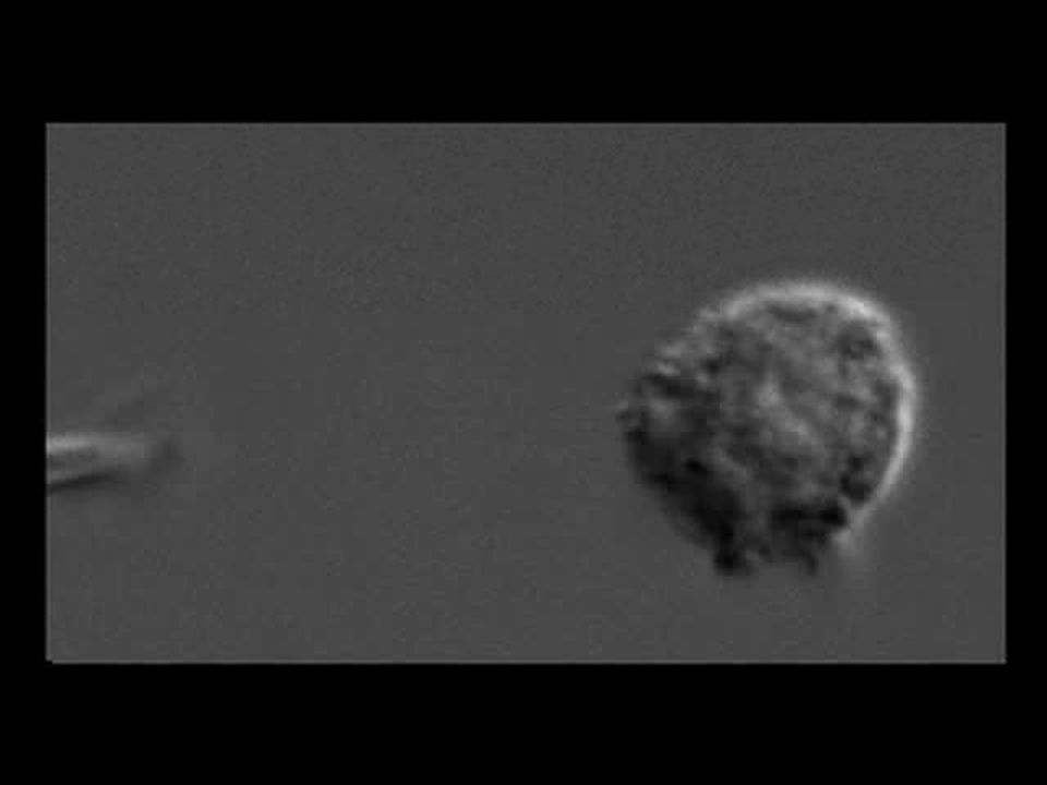
Second line Leukocytes (WBC) Circulating in body 1. Macrophages (monocytes) Kill invaders by ingesting them Phagocytosis Ingestion
 Kill invaders by ingesting them Phagocytosis Ingestion.")
16
Fig. 43-3 Microbes PHAGOCYTIC CELL Vacuole Lysosome containing enzymes

17
Second line 2. Neutrophils Most abundant WBC Phagocytosis Release chemicals--kill bacteria Also kills other neutrophils

19
Second line 3. Eosinophils WBC Low phagocytic activity Parasite defense

20
Second line Antimicrobial proteins 1. Complement system Proteins found in plasma Attack bacterial or fungal cell walls Cause cells lysis Signals other defense responses

21
Second line 2.Interferons Paracrine polypeptide Protect cells in area of virus Prevent viral replication Cancer defense

22
Second line 3. Natural killer cells Kills cells infected by a virus Help fight cancer cells

23
Second line Inflammatory response Local, non-specific Histamines & prostaglandins Vasodilation Increased blood flow to area Edema or swelling WBC (phagocytic), pus formation Shock, systemic
, pus formation Shock, systemic")
24
Second line

25
Inflammatory response Elevated temperature (fever) Interleukin-1 Released by Macrophages Directs hypothalamus to increase temp Helps stimulate defense response
 Interleukin-1 Released by Macrophages Directs hypothalamus to increase temp Helps stimulate defense response")
26
Third line Specific response Lymphocytes T-cells---cellular response B-cell---humoral response Antibodies

27
Third line Antigen Foreign molecule Epitope Antigen determinant Located on surface Causes a specific immune response

28
Third line Self-versus-nonself recognition Genes code for specific proteins Major Histocompatibility Complex proteins MHC proteins Cell recognition Glycoproteins on surface of cells

29
Third line B-cell lymphocytes Made & develops in bone marrow Becomes a plasma cell Produce antibodies in response to specific antigens Immunoglobulins (Ig) Antibodies Humoral immunity
 Antibodies Humoral immunity")
30
Role of B Cells

31
Third line B-lymphocyte structure Antigen receptor Specific Plasma membrane

32
Third line Light chains 2 short polypeptides Heavy chains 2 identical long polypeptides 4 chains held together by disulfide bond Forms Y-shaped molecules

33
Fig. 43-9a Antigen- binding site Antigen- binding site Disulfide bridge Variable regions Constant regions Transmembrane region Plasma membrane Light chain Heavy chains Cytoplasm of B cell (a) B cell receptor B cell V V C C V V CC
 B cell receptor B cell V V C C V V CC.")
34
Fig. 43-10 Antigen-binding sites Antigen- binding sites Epitopes (antigenic determinants) Antigen Antibody B Antibody C Antibody A CC C V V V V C
 Antigen Antibody B Antibody C Antibody A CC C V V V V C.")
35
Antibodies Plasma cells release antigen receptor Specific for antigens Arms of the Y shaped molecule Have different aa sequences

36
Antibodies IgM first response Aggregation of complement proteins IgG major form, second response Stimulates phagocytosis by macrophages IgD receptors for antigens on B cells IgA Present in breast milk, mucous, saliva Provide protection to newborns

37
Antibodies IgE Release histamines Bind to mast cells Insert heavy chain into mast cells Initiate inflammatory response Presence of antigens Vasodilation

38
Antibodies

40
Third line T-cell lymphocytes Made in bone marrow Processed in thymus gland Regulate immune responses Attack cells with specific antigens Cell-mediated immunity

41
Fig. 43-9b Antigen- binding site Variable regions Constant regions Transmembrane region Plasma membrane T cell chain chain Disulfide bridge Cytoplasm of T cell (b) T cell receptor C C V V
 T cell receptor C C V V.")
42
Third line T-cells 1. Helper T cells (CD4) Initiate response based on antigens 2. Memory T cells Remember previous antigens

43
Third line 3. Cytotoxic T cell (CD8) Lyse cells infected by virus 4. Suppressor T cells Turn off immune response

44
Helper T Cells

45
Cytotoxic T Cells

46
Third line response Antigen Macrophage process antigen Secrete cytokines (interleukins or interferons) Stimulates T helper cells
 Stimulates T helper cells")
47
Third line response Recognize antigens Antigen receptor on T-cells Bind to antigens Triggers T-cytotoxic cells, T-memory cells Cytotoxic cells destroy infected cells Stimulates B cells

48
Third line response Antigen receptor on B-cell Binds foreign antigen Triggers formation of a clone of plasma cells Clones produce antibodies Antibodies bind invading antigen Prevent affects of antigen Destruction or blocks effect

50
Immune response Macrophage ⇓ Helper T-cell ⇙ ⇘ B-cell Cytotoxic T-cell ⇙ ⇘ ⇙ ⇘ Plasma Memory Memory Cytotoxic cells cells cells T-cells ⇓ Antibodies

51
T-cells Cytotoxic T-cells Attack transplants (skin grafts) Considered foreign tissue Destroy cancer cells Interferon (lymphomas, renal Ca, melanoma, Kaposi’s sarcoma and Breast Ca) Interleukin (tx cancer)
 Considered foreign tissue Destroy cancer cells Interferon (lymphomas, renal Ca, melanoma, Kaposi’s sarcoma and Breast Ca) Interleukin (tx cancer)")
52
B-cells Primary immune response First exposure Lasts about 2 weeks Memory cells are also produced during the first exposure Secondary immune response Activates memory cells Response faster & lasts longer

54
Immune System Summary First line Skin, cilia, enzymes, pH of skin Second line WBC (macrophages, neutrophils, natural killer cells, eosinophils) Antimicrobial proteins Inflammatory response
 Antimicrobial proteins Inflammatory response")
55
Immune System Summary Third line Lymphocytes (B & T) Antibodies (immunoglobulins) IgM, IgG, IgA, IgE, IgD
 Antibodies (immunoglobulins) IgM, IgG, IgA, IgE, IgD")
56
Monoclonal antibodies Antibodies specific for one antigen Cell cultures produce large quantities Use in lab tests Pregnancy tests Antibody to HCG (human chorionic gonadotropin)
")
57
HIV Human immunodeficiency virus Attacks + destroys CD4+ T-cells T-cells secrete a suppressing factor Blocks other T-cells Infects macrophages & brain cells

58
Autoimmune diseases Systemic Lupus Rheumatoid arthritis Hashimoto thyroiditis

60
Allergy Allergens (antigens) Release IgE Binds mast cells & basophils Stimulates release of chemicals Histamine Drop in BP
 Release IgE Binds mast cells & basophils Stimulates release of chemicals Histamine Drop in BP")
61
Allergy Anaphylactic shock Widespread histamine response Death Bee stings or peanuts or penicillin Contact dermatitis Delayed response Poison ivy, poison oak

63
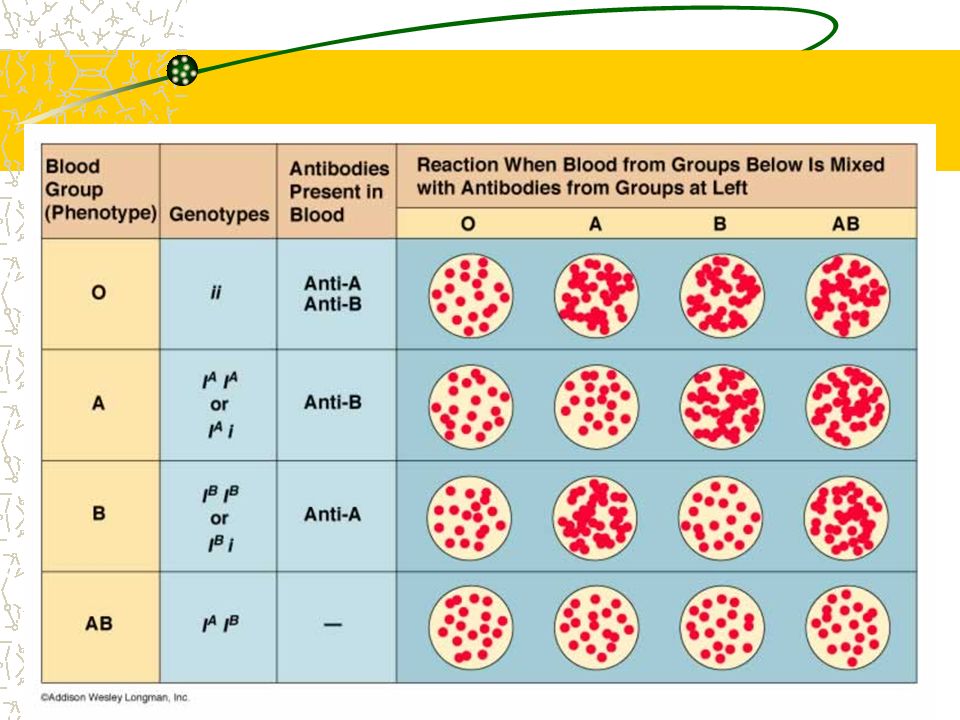
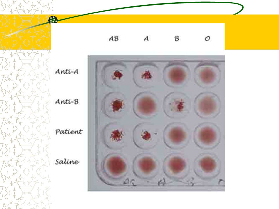
Blood types (ABO) Blood type Surface antigensAntibodies A A anti B B B anti A AB none O anti A & B
 Blood type Surface antigensAntibodies A A anti B B B anti A AB none O anti A & B")
64
ABO

67
Rh factor

Similar presentations


 INNATE IMMUNITY (all animals) Rapid response Recognition of traits shared by broad ranges of pathogens,>")


 INNATE IMMUNITY (all animals) Rapid response Recognition.>")
 Immune system.>")


 engulfs a yeast cell (pathogen)>")
>")
