Download presentation
Presentation is loading. Please wait.

2
中華牙醫學會第三十二屆學術研討會中華民國口腔顎顏面放射線學會九十八年度學術研討會 27th November, 2009 陳玉昆, Y-K Chen Department of Oral Pathology, School of Dentistry, College of Dental Medicine, Kaohsiung Medical University, Kaohsiung, TAIWAN 虛擬顯微鏡於口腔顎顏面 病理學實驗課程之應用 An Application of Virtual Microscopy to the Teaching of Oral & Maxillofacial Pathology Laboratory Course

3
參 考 資 料參 考 資 料 1.Evans AJ, et al. Primary frozen section diagnosis by robotic microscopy and virtual slide telepathology: the University Health Network experience. Human Pathology 2009;40:1070-81. 2.Dee FR. Virtual microscopy in pathology education Human Pathol 2009;40: 1112-21. 3.Weinstein RS, et al. Overview of telepathology, virtual microscopy, and whole slide imaging: prospects for the future. Human Pathol 2009;40:1057-69. 4.Kaohsiung Medical University, Department of Oral Pathology. 5.Marchevsky AM, et al. Self-instructional “Virtual Pathology” laboratories using web-based technology enhance medical school teaching of pathology. Human Pathol 2003;34:423-9. 6.Chen YK, et al. An application of virtual microscopy to the teaching of oral and maxillofacial pathology laboratory course. Oral Surg Oral Med Oral Pathol Oral Radiol Endod 2008;105:342-7. 7.Hanks CT, et al. Internet- and DVD-based instruction in oral pathology for dental students. Oral Surg Oral Med Oral Pathol Oral Radiol Endod 2004;98:195. 8.Farah CS, et al. The e-evolution of microscopy in dental education. J Dental Edu 2009;73:942-9. 9.Dotslide, Olympus company catalogue

4
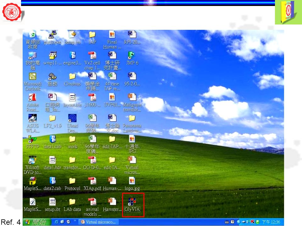
Virtual microscopy: emulate traditional light microscopy using digital image files (virtual slides) manipulated on a computer screen using microscope emulator software Refs. 4, 9 Whole slide imaging: create digital images of the entire area of a glass slide Hard Copy Soft Copy 顯微影像數位掃瞄系統 Automate scan process - 50 slides

5
Virtual microscopy: use computer mouse to perform the control functions ordinarily handled with a traditional light microscope (position the objective lens) Ref. 4

6
OlyVIA 閱片 Dot slide 掃瞄、閱片 Procedures of whole slide scanning (1) Dot slide
 Dot slide")
7
Ref. 4 New slide Procedures of whole slide scanning (2) Dot slide
 Dot slide")
8
Ref. 4 Overview image Procedures of whole slide scanning (3) Dot slide
 Dot slide")
9
Ref. 4 Procedures of whole slide scanning (4) Dot slide
 Dot slide")
10
Ref. 4 Edit scan area Procedures of whole slide scanning (5) Dot slide
 Dot slide")
11
Ref. 4 Procedures of whole slide scanning (6) Dot slide New area
 Dot slide New area")
12
Ref. 4 Live (active) Procedures of whole slide scanning (7) Dot slide
 Procedures of whole slide scanning (7) Dot slide")
13
Ref. 4 Live (active) Procedures of whole slide scanning (8) Dot slide Autofocus (AF)
 Procedures of whole slide scanning (8) Dot slide Autofocus (AF)")
14
Ref. 4 Start Procedures of whole slide scanning (9) Dot slide
 Dot slide")
15
Ref. 4 Procedures of whole slide scanning (10) Dot slide
 Dot slide")
16
Ref. 4 Live (active) Procedures of whole slide scanning (11) Dot slide
 Procedures of whole slide scanning (11) Dot slide")
17
Ref. 4 Procedures of whole slide scanning (12) Dot slide
 Dot slide")
18
Ref. 4 Save file Extended Focal Imaging (EFI) Procedures of whole slide scanning (13) Dot slide
 Procedures of whole slide scanning (13) Dot slide")
19
Ref. 4 Procedures of whole slide scanning (14) Dot slide Extended Focal Imaging (EFI)
 Dot slide Extended Focal Imaging (EFI)")
20
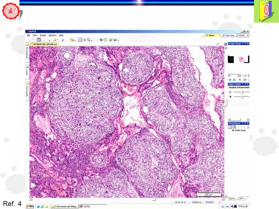
Virtual slide viewer: use iconic representations of 4×, 10×, 20×, and 40× objective lenses and select the magnification of choice by clicking on one of these icons with a computer mouse Ref. 4 OlyVIA

21
Refs. 4, 9 Net Image Server/Web Server, Dotslide and OlyVIA Dot slide/ OlyVIA Dot slide/ OlyVIA Lan/Internet Dot slide/ OlyVIA Dot slide/ OlyVIA Net Image Server Dot slide Web Server WorkstationClients

22
Telepathology: practice of pathology at a long distance, has advanced continuously since 1986 Robotic control of microscopy Ref. 1 Frozen Section Telepathology UHN Toronto Ontario Canada

23
Virtual microscopy or telepathology - attractive to educators due to: (1) nearly perfectly simulates the pan & zoom characteristics of conventional microscopy (2) versatility of computer-assisted education Refs. 3, 8

24
Medical schools in the US: entirely digital for pathology courses, discarding light microscopes, and building virtual slide laboratories UCLA School of Medicine’s Center Ref. 5

25
UCLA School of Medicine’s Center Ref. 5 Table 1. Mean Ratings of Attributes of Virtual Pathology Based on a Likert Scale (1, Not at All Useful to 5 Very Useful), n = 72 Attributes of the Virtual Pathology ModuleMean Standard Deviation Quality of images4.40.69 Image description4.10.87 Case histories framing the slides4.10.88 Pretest3.51.05 Posttest3.70.98 Feedback on posttest3.21.33 Overall format of presentation40.89 Ease of navigation4.60.63 Overall gain in learning4.00.81 Presence of experts to assist me4.20.75
, n = 72 Attributes of the Virtual Pathology ModuleMean Standard Deviation Quality of images Image description Case histories framing the slides Pretest Posttest Feedback on posttest Overall format of presentation40.89 Ease of navigation Overall gain in learning Presence of experts to assist me")
26
UCLA School of Medicine’s Center Ref. 5 Table 2. Mean Ratings of Student Perceptions of Virtual Pathology Based on a Likert Scale (1, Not at All Useful to 5 Very Useful), n = 72 Criteria for Student PerceptionsMean Standard Deviation I enjoyed working on the web- based slide presentation3.40.62 This format was an effective way to learn the given content3.40.62 I was able to complete all of the sections within the time limit3.70.46 The petest helped me in focusing on what I did not know3.00.85 I prefer this interactive format to a lecture presentation3.20.76
, n = 72 Criteria for Student PerceptionsMean Standard Deviation I enjoyed working on the web- based slide presentation This format was an effective way to learn the given content I was able to complete all of the sections within the time limit The petest helped me in focusing on what I did not know I prefer this interactive format to a lecture presentation")
27
Oral Pathology Laboratory course for dental students at University of Michigan - converted from a microscope-based course to an internet-accessible virtual histopathology ‘‘laboratory.’’ (2003) Ref. 7 University of Queensland, School of Dentistry (Brisbane, Australia) implement VM technology (Aperio Systems’ ImageScope) into its courses of Oral Biology (2006) & Oral Pathology (2008) Ref. 8
 implement VM technology (Aperio Systems’ ImageScope) into its courses of Oral Biology (2006) & Oral Pathology (2008) Ref. 8.")
28
口腔病理學 ( 含實驗 ) - KMUOP 1. 屬於基礎範圍,為提昇同學學習之興趣,於 2006 年開 始應用網路及多媒體教學,架構口腔病理科教學網頁 2. 假設虛擬顯微鏡,將口腔病理教學玻璃切片轉換成高解 析度的數位影像檔,置放於本科網站中。下載 OlyVA 軟體, 使用寬頻網路閱覽口病教學切片。 Refs. 4, 6

29
Ref. 4

33
Remote log in Ref. 4

34
………. Log in User name Password administrator Ref. 4

35
Open remote file Ref. 4

36
Search now Ref. 4

38
Open Ref. 4

42
Refs. 4, 6 高醫牙科口腔病理學實驗切片線上教學 小組線上教學 發表文章: An application of virtual microscopy in the teaching of an oral and maxillofacial pathology laboratory course. Oral Surg Oral Med Oral Pathol Oral Radiol Endod 2008;105:342-7 線上考試

43
口病實驗期末考 ( 請務必寫上姓名與學號 ) 姓名 學號 內容: 1. 寫出口腔病理診斷 2. 標示口腔病理特徵 ( 考試時間為 50 分鐘 ) Ref. 4
 姓名 學號 內容: 1. 寫出口腔病理診斷 2. 標示口腔病理特徵 ( 考試時間為 50 分鐘 ) Ref. 4")
44
注意事項 點選 OlyVIA 後,工具列中點選 remote login , 鍵入個人帳號、個人密碼及 IP Address:163.15.173.75 , max.Hits: 改為 500 、 下載考試題目 考試當天,於口病網頁: oralpathol.dlearn.kmu.edu.tw 下載考試用 ppt 檔於 D 槽,務必更改檔名為自己的姓名與學 號,如 ’ 陳大明 9512345’ 。 ( 完成下載考試用 ppt 檔於 D 槽後,請務必關閉口病網頁,否 則視同作弊 ) 作答完成後,檢查無誤後,請將個人 ppt 檔 儲存至老師提供之 USB ,最後請登錄 USB 之 號碼於點名簿上,方可離開電腦教室 ( 請務必詳細閱讀 ) Ref. 4

45
請寫下題目 1-8 之病理診斷 請以英文作答,錯字扣 1 分 題目病理診斷得分 1 Squamous cell carcinoma (M to P-D) 2 AAA 3 BBB 4 CCC 5 DDD 6 EEE 7 FFF 8 Nevus (intramucosal) 得分 (A) Ref. 4

46
請標示下列病理特徵 題目病理特徵得分 Aaaa Bbbb C Nevus cells ( 示範從 OlyVIA 下載圖片之方法與練習用 ) Dddd Eeee Ffff Gggg H Abnormal mitosis( 練習用 ) Iiii Jjjj 得分 (B) 總得分 = A + B = Ref. 4

47
從 OlyVIA 下載圖片之方法 (1) 點選 Copy Display to Clipboard ,複製適合的圖片 Ref. 4
 點選 Copy Display to Clipboard ,複製適合的圖片 Ref. 4")
48
從 OlyVIA 下載圖片之方法 (2) Back Nevus cells Ref. 4
 Back Nevus cells Ref. 4")
49
C. 請從題目 1-8 中找出並標示 Nevus cells 之病理特徵 從 OlyVIA 下載圖片之方法 (3) 貼上所選擇之圖片後, 插上標誌,如星號 ( 可插上多個星號 ) 貼上所選擇之圖片後, 插上標誌,如箭號 Ref. 4
 貼上所選擇之圖片後, 插上標誌,如星號 ( 可插上多個星號 ) 貼上所選擇之圖片後, 插上標誌,如箭號 Ref. 4.")
50
H. 請從題目 1-8 中找出並標示 Abnormal mitosis (009) 之病理特徵 ( 請自我練習 ) 請勿複製新投影片 Ref. 4
 之病理特徵 ( 請自我練習 ) 請勿複製新投影片 Ref. 4")
51
作答完成 請務必更改 ppt 檔名為自己的姓名與學號, 如 ’ 陳大明 9512345’ 再將此 ppt 檔儲存至老師提供之 USB 最後請登錄 USB 之號碼於點名簿上,方可離 開電腦教室 Ref. 4

52
Advantages of virtual microscopy vs traditional microscopy Ref. 2 Accessibility - Access can be anywhere anytime there is computer (and the Web) available - One slide can be viewed by many or duplicated and shared Multiple recuts are not needed - One-of-a-kind slides that cannot be recut can be duplicated into an unlimited number of copies and shared with others, e.g. fortuitous cut in following case
 available - One slide can be viewed by many or duplicated and shared Multiple recuts are not needed - One-of-a-kind slides that cannot be recut can be duplicated into an unlimited number of copies and shared with others, e.g. fortuitous cut in following case.")
53
Advantages of virtual microscopy vs traditional microscopy Refs. 1, 2 Efficiency - Focus, proper condenser adjustment, and lighting are not required -Technical competence in viewing is easier to achieve and less frustrating for trainees who do not have an aptitude for traditional microscopy - There is rapid access to the next slide in the slide box

54
Advantages of virtual microscopy vs traditional microscopy Refs. 2, 4 Pedagogic - Very-low-power over (<<x4) allows to better visualize relationship of pathologic to normal tissue - A thumbnail & location box allows trainee to remain oriented to the whole slide while viewing a high magnification
 allows to better visualize relationship of pathologic to normal tissue - A thumbnail & location box allows trainee to remain oriented to the whole slide while viewing a high magnification.")
55
Advantages of virtual microscopy vs traditional microscopy Ref. 2 Pedagogic - Marking x-y & magnification coordinates of multiple key areas & movement among these areas at the click of the mouse is possible

56
Advantages of virtual microscopy vs traditional microscopy Ref. 2 Pedagogic - Side-by-side viewing, annotation overlays, & integration with descriptions, case scenarios, gross & radiological images or digital photomicrographs is possible

57
Advantages of virtual microscopy vs traditional microscopy Ref. 2 Pedagogic -Group discussion is enhanced as each computer screen can serve as a multihead microscope Conference

58
Disadvantages of virtual microscopy vs traditional microscopy Ref. 2 Pedagogic -Trainees do not learn how to use a traditional microscope if virtual microscopy is used exclusively Technology - Low magnification has less resolution when viewed on a standard computer screen - Refractile objects do not refract well - Original glass slide tissue artifact and imperfections are difficult to scan - Virtual focus acquisition & viewing is inefficient, especially over the Web

59
Ref. 2 Venues for virtual microscopy implementation Education in Pathology 1.Medical student education - Pathology small group teaching - Histopathology laboratories/large groups - Integrated and problem-based curricula - Repositories for sharing among institutions 2. Cytology 3. Hematology 4. House staff education and evaluation 5. Continuing medical education 6. Veterinary pathology and comparative pathology 7. Histology

60
Although the production of virtual microscopic slides remains a time- consuming task and the cost of archiving the images digitally is still considerably higher than archiving on glass slides, virtual microscopy still has many advantages over traditional glass microscopy in oral and maxillofacial pathology education Ref. 6

61
Microscopic virtual images may one day be incorporated into digital databases for clinical diagnosis, adding the histopathological images as part of medical digital charts of patients, as is currently the case for images through teleradiology Ref. 6

62
In 2005, DICOM established a special working group (Working Group 26) to develop extensions to the standard for telepathology imaging and pathology in general 2 significant limitations on single image objects within DICOM (1) DICOM image objects pixel dimensions are stored as 16-bit integers, for a maximum value of 64K, inadequate for virtual slide telepathology (2) DICOM image objects data size are stored as 32-bit integers, for a maximum value of 2 GB. This may need to be adjusted upward for some virtual slide telepathology applications. Ref. 3

63
감 사 합 니 다감 사 합 니 다 謝 謝

Similar presentations


 典藏人文社會學術期刊全文資料庫 Periodicals Index Online (PIO) 典藏人文社會學術期刊索引資料庫.>")

>")


 計算機概論. 1-2 25 教學目標 瞭解現代電腦系統之發展歷程 瞭解電腦之元件、功能及組織架構 瞭解電腦如何表示資料及其處理方式 學習運用電腦來解決問題 認知成為一位電子資訊人才所需之基本條 件 認知進階電子資訊之相關領域.>")


>")

![1 請下載並安裝 Adobe Acrobat Reader 中文版。 操作環境設定 建議使用 MS office XP or 2002 以上。 瀏覽器版本建議使用 IE 6.0 以上版本。 建議瀏覽解析度為 1024 × 768 。 在 IE 的功能表列 [ 工具 ] [ 網際網路選 項.](/16/4951199/big_thumb.jpg "1 請下載並安裝 Adobe Acrobat Reader 中文版。 操作環境設定 建議使用 MS office XP or 2002 以上。 瀏覽器版本建議使用 IE 6.0 以上版本。 建議瀏覽解析度為 1024 × 768 。 在 IE 的功能表列 [ 工具 ] [ 網際網路選 項.>")

 以上才承認 校內實習時數及實習成績。 二個寒假 各一個月 暑假兩個月.>")


