Download presentation
Presentation is loading. Please wait.

1
Basic Microscopy – An Overview – October 2005 Protistology Course MBL, Woods Hole, MA

2
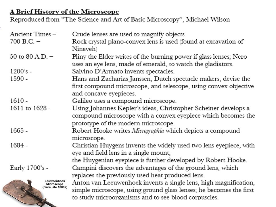
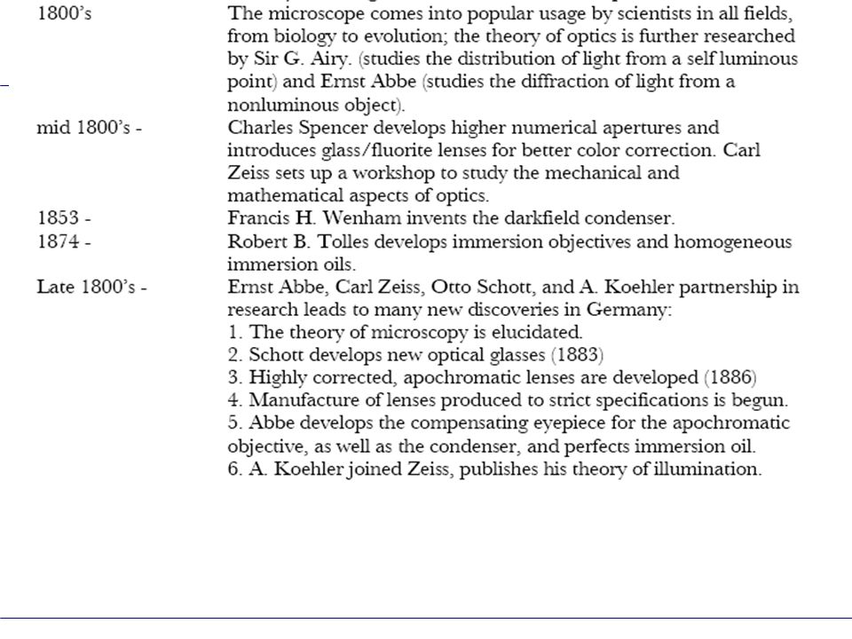
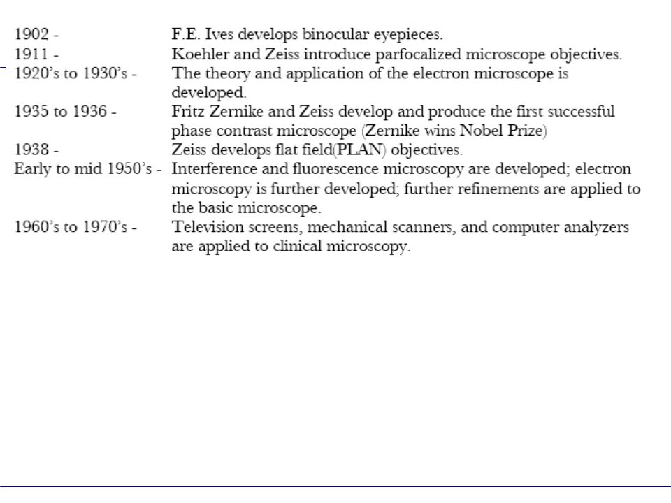
Brief History of the Microscope What‘s a microscope? Definition of Magnification Conventional Viewing Distance Leeuwenhoek > Compound > Stereo Microscope The “Telescope”, a simple detour How to make the specimen visible – Contrast! Definition of Contrast Techniques: Brightfield Phase Darkfield Pol DIC (Differential Interference Contrast) Fluorescence Optical Sectioning – an expansion of Fluorescence Setting up the Microscope (Lab) Koehler Illumination Resolution & Empty Magnification Agenda
 Fluorescence Optical Sectioning – an expansion of Fluorescence Setting up the Microscope (Lab) Koehler Illumination Resolution & Empty Magnification Agenda.")
6
Objects appear to the eye at different magnifications, depending on their distance from the eye. Accommodation (lens) will make it possible. M B ~ 2x M A A B What is “Magnification”?
 will make it possible. M B ~ 2x M A A B What is Magnification .")
7
Conventional Viewing Distance 250 mm 1x ?

8
“Magnification” 1x f = 250 mm 1x

9
Magnification via Single Lens f = 250 mm 1x Example: f=50mm 5x Magnifying Glass (Loupe)
")
10
The “simple” microscope Leeuwenhoek Microscope

11
The current -corrected Compound Microscope Tube lens (Zeiss: f=164.5mm) Objective Eyepiece
 Objective Eyepiece")
12
Q: What happens if we take the objective away? Tube lens (Zeiss: f=164.5mm) Objective Eyepiece Tube f 250mm f M Answer: We have created a “Telescope” ∞ ∞
 Objective Eyepiece Tube f 250mm f M Answer: We have created a Telescope ∞ ∞.")
13
AxioImager Upright Research Microscope

14
Axiovert 200 Inverted Research Microscope

15
The basic light microscope types Upright microscope. Inverted microscope

16
Illumination via Transmitted Light The specimen must be transparent !

17
Upright microscope. Inverted microscope

18
Illumination via “Reflected” (Incident) Light Eg. Fluorescence, Opaque Samples
 Light Eg. Fluorescence, Opaque Samples")
19
Upright microscope. Inverted microscope

20
Upright microscope. Inverted microscope Mixed Illumination

21
Other Ways to Illuminate Reflectors Ring Lights Fiber Optics LED’s Etc.

22
“Couldn’t one build a microscope for both eyes, and thereby generate spatial images?” Question addressed to Ernst Abbe in 1896 by Horatio S. Greenough

23
1896: Drawing by Horatio S. Greenough 1897 – the first Stereo Microscope in the world, built by Zeiss, according to the “Greenough” principle

24
Greenough TypeTelescope Type Introduced first by Zeiss - 1946 Introduced first by Zeiss - 1897

25
Greenough Stereo Microscopes SteMi DV4

26
Greenough Stereo Microscopes SteMi 2000 (2000-C, 2000-CS)
")
27
Research Stereo Microscopes SteREO Discovery V12SteREO Lumar V12

28
How to make the specimen visible – CONTRAST! Definition of Contrast Techniques: Brightfield Phase Darkfield Pol DIC (Differential Interference Contrast) Fluorescence Optical Sectioning – an expansion of Fluorescence Agenda
 Fluorescence Optical Sectioning – an expansion of Fluorescence Agenda.")
29
C ONTRAST 50 – 0 / 50 + 0 = 1 50 – 100 / 50 + 100 = -0.33 50 – 50 / 50 + 50 = 0 50 Units0 Units100 Units 50 Units 50

30
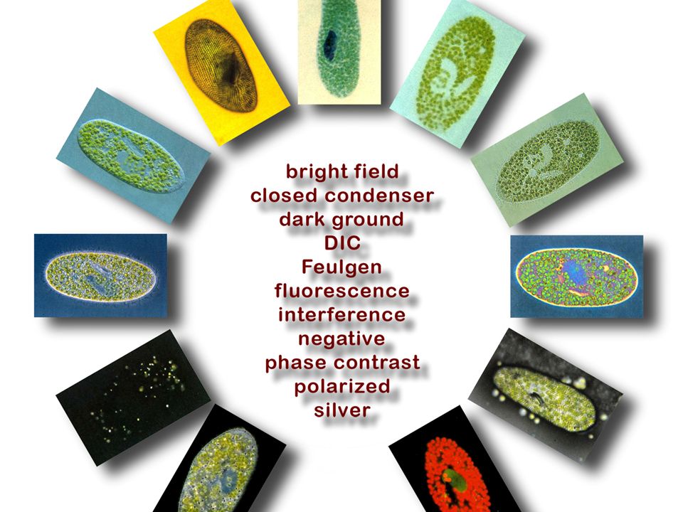
Brightfield Darkfield Phase Contrast Polarized Light DIC (Differential Interference Contrast) Fluorescence (and related techniques) Common Illumination Techniques
 Fluorescence (and related techniques) Common Illumination Techniques")
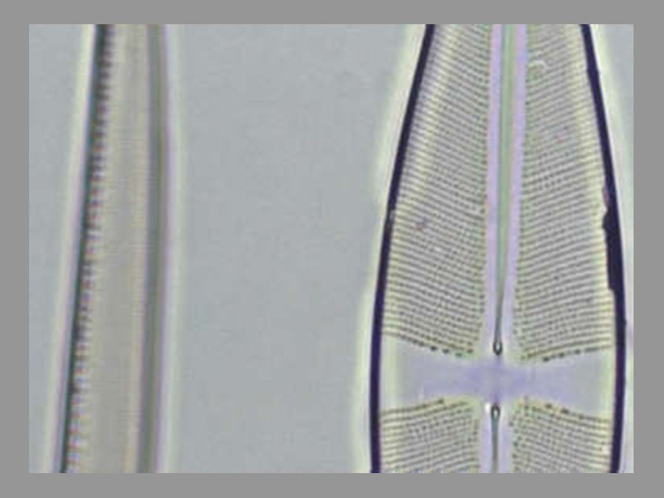
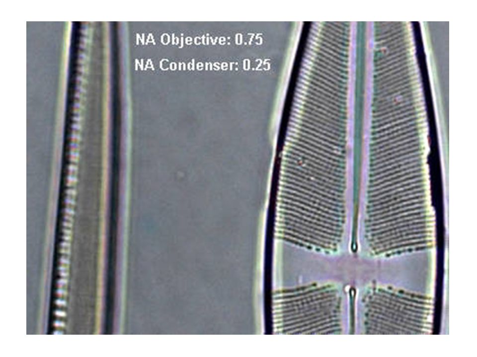
32
Brightfield For naturally absorbing or stained samples True Color Representation Proper Technique for Measurements Spectral Dimensional

35
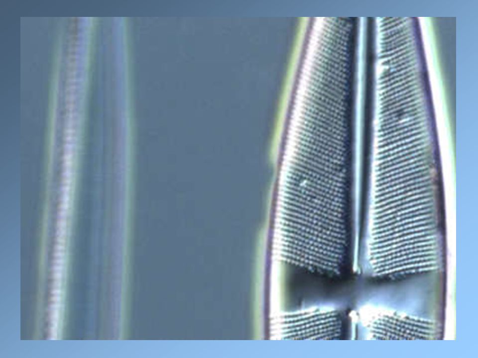
Bacillaria

36
Paramecium bursaria Condenser diaphragm openCondenser Diaphragm almost closed

37
Paramecium bursaria Indian Ink StainingFeulgen StainingSilver Staining Different Staining Techniques

39
Phase Contrast (Frits Zernike 1934) - “Halo” effect > Reduced resolution + No staining necessary + Good Depth of Field + Easy alignment + Orientation independent + Repeatable setup + Works with plastic dishes
 - Halo effect > Reduced resolution + No staining necessary + Good Depth of Field + Easy alignment + Orientation independent + Repeatable setup + Works with plastic dishes")
40
Required Adjustment: Superimpose Phase Ring of condenser over (dark) phase plate of objective (after Koehler Illumination) Required Components for Phase Contrast: 1.Objective with built-in Phase Annulus 2.Condenser or Slider with Centerable Phase Ring for illumination (Ph0, 1, 2 or 3)
 phase plate of objective (after Koehler Illumination) Required Components for Phase Contrast: 1.Objective with built-in Phase Annulus 2.Condenser or Slider with Centerable Phase Ring for illumination (Ph0, 1, 2 or 3)")
41
Phase Shifts: Cells have higher n than water. Light moves slower in higher n, consequently resulting in a phase retardation Phase shift depends on n and on thickness of specimen detail Illumination bypasses Specimen > no phase shift Illumination passes through thin part of Specimen > small phase retardation Illumination passes through thick part of Specimen > larger phase retardation

42
1.Illumination from Condenser Phase Ring (“0” Order) > meets phase ring of objective 2.Objective Phase Ring a) attenuates the non-diffracted 0th Order b) shifts it ¼ wave forward 3.Affected rays from specimen, expressed by the higher diffraction orders, do not pass through phase ring of objective >¼ wave retarded 4.Non-diffracted and diffracted light are focused via tube lens into intermediate image and interfere with each other; ¼+¼= ½ wave shift causes destructive interference i.e. Specimen detail appears dark Condenser Objective Specimen Tube Lens

43
Paramecium bursaria Phase Contrast

44
Rhipidodendron Phase Contrast

45
Cochliopodium Phase Contrast

46
Lyngbya Bacteria Phase Contrast

48
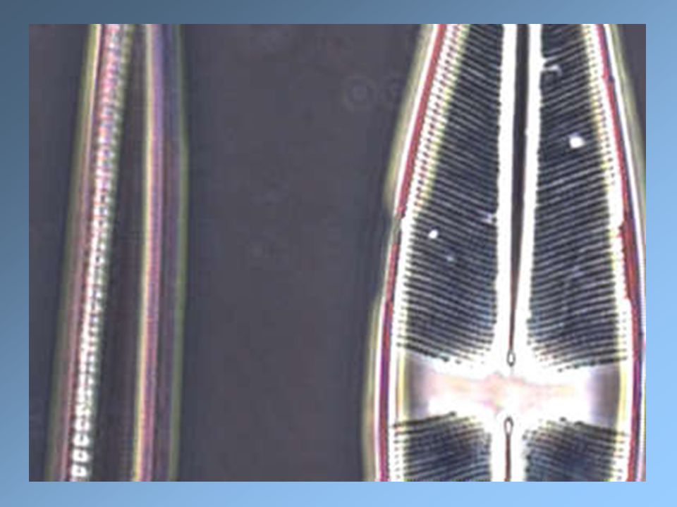
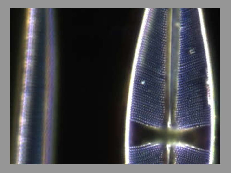
Darkfield No staining necessary Detection of sub-resolution details possible Excellent, reversed contrast Central Darkfield via “hollow cone” Oblique Darkfield via Illumination from the side Not useful for Measurements (sizes exaggerated)
")
49
Required conditions for Darkfield: Illumination Aperture must be larger than objective aperture I.e. direct light must bypass observer Iris Diaphragm Low NA Objective High NA Objective

50
Paramecium bursaria Polarized Light Darkfield

51
Polarized Light Specimen is placed between 2 crossed polarizers. Only light produced by birefringent particles (e.g. crystals) or coming from the edges of particles (“edge birefringence”) is visible. Looks sometimes like Darkfield Orientation-specific (linear Pol) Linear / circular Polarized Light
 or coming from the edges of particles ( edge birefringence ) is visible. Looks sometimes like Darkfield Orientation-specific (linear Pol) Linear / circular Polarized Light.")
52
Brightfield Background Birefringent Material Polarized Light Pol + Red I Color of sample and background modified by wave plate

53
When Polarizers are crossed, only items that rotate the plane of polarization reach the detector. Wave plate adds color Polarized Light Polarizer 1 Polarizer 2 (Analyzer) Specimen
 Specimen.")
54
Required / Recommended Components: Polarizer (fixed or rotatable) Analyzer (fixed or rotatable) Strain-free Condenser and Objective Rotating, centerable Stage Wave plate and/or Compensator Crossline Eyepiece
 Analyzer (fixed or rotatable) Strain-free Condenser and Objective Rotating, centerable Stage Wave plate and/or Compensator Crossline Eyepiece")
56
DIC (Differential Interference Contrast after Nomarski) High Contrast and high resolution High Contrast and high resolution Control of condenser aperture for optimum contrast Control of condenser aperture for optimum contrast Changes GRADIENTS into brightness differences Changes GRADIENTS into brightness differences 3-D Image appearance 3-D Image appearance Color DIC by adding a wave plate Color DIC by adding a wave plate Best contrast / resolution via different DIC sliders Best contrast / resolution via different DIC sliders Orientation-specific > orient fine details perpendicular to DIC prism Orientation-specific > orient fine details perpendicular to DIC prism
 High Contrast and high resolution High Contrast and high resolution Control of condenser aperture for optimum contrast Control of condenser aperture for optimum contrast Changes GRADIENTS into brightness differences Changes GRADIENTS into brightness differences 3-D Image appearance 3-D Image appearance Color DIC by adding a wave plate Color DIC by adding a wave plate Best contrast / resolution via different DIC sliders Best contrast / resolution via different DIC sliders Orientation-specific > orient fine details perpendicular to DIC prism Orientation-specific > orient fine details perpendicular to DIC prism")
57
DIC Observing local differences in phase retardation

58
9 Image 8 Tube lens 7 Analyzer (7a with Wave Plate) 6 Wollaston Prism behind objective 5 Objective 4 Specimen 3 Condenser with receptacle for prisms 2 Wollaston Prism before condenser 1 Polarizer
 6 Wollaston Prism behind objective 5 Objective 4 Specimen 3 Condenser with receptacle for prisms 2 Wollaston Prism before condenser 1 Polarizer")
59
Wollaston Prism Birefringence (Different refractive index for differentpolarization orientations)Polarized beam, under 45˚ to prism,gets split into “ordinary” and“extraordinary” beam
Polarized beam, under 45˚ to prism,gets split into ordinary and extraordinary beam")
60
Required Components for DIC: Nosepiece with DIC receptacles Polarizer (or Sénarmont Polarizer) Low Strain Condenser and Objective* DIC Prisms for Condenser (# I or II or III) Appropriate DIC Slider for each objective Analyzer (or Sénarmont Analyzer) *Not needed for New Plas-DIC (up to 40x)
 Low Strain Condenser and Objective* DIC Prisms for Condenser (# I or II or III) Appropriate DIC Slider for each objective Analyzer (or Sénarmont Analyzer) *Not needed for New Plas-DIC (up to 40x)")
61
Paramecium bursaria DIC Interference

62
Fluorescence Easy to set up > Objective = Condenser Highly specific technique, wide selection of markers Detection and Identification of Proteins, Bacteria, Viruses Basics for –Special Techniques eg. TIRF, FRET, FRAP etc. –3-D imaging –Deconvolution –Structured Illumination –Confocal Techniques

63
Epi - Fluorescence Example: Specimen containing green fluorescing Fluorochrome Dichromatic Mirror Emission Filter Excitation Filter Observation port FL Light Source

64
Epi - Fluorescence Filter Sets Curve for a typical GFP filter set Example

65
Epi - Fluorescence (Specimen containing green fluorescing Fluorochrome) Dichromatic Mirror Emission Filter Excitation Filter Observation port FL Light Source Specimen containing green fluorescing Fluorochrome
 Dichromatic Mirror Emission Filter Excitation Filter Observation port FL Light Source Specimen containing green fluorescing Fluorochrome")
66
Paramecium bursaria Fluorescence

67
How to improve Fluorescence Imaging in a major way: Optical Sectioning

68
Optical sectioning – increased contrast and sharpness

69
Overview of Optical sectioning Methods 1.Confocal and Multi-photon Laser Scanning Microscopy –Pinhole prevents out-of-focus light getting to the sensor(s) (PMT - Photomultiplier) (30 – 70 µm) –Multi Photon does not require pinhole (90 – 500 µm) 2.Spinning disk systems –A large number of pinholes (used for excitation and emission) is used to prevent out-of-focus light getting to the camera –E.g. Perkin Elmer, Solamere ( up to 30 µm) 3.Structured Illumination –Moving grid represents the reference for in-focus information –Zeiss Apotome (10-50 µm)
 3.Structured Illumination –Moving grid represents the reference for in-focus information –Zeiss Apotome (10-50 µm).")
70
Overview of Optical sectioning Methods - cont‘d - 4.Total Internal Reflection Fluorescence (TIRF) –High NA Objective projects beam at angle which exceeds critical angle. –Area touching cover slip (evanescent field) is typically smaller than 200 nm 5.Deconvolution –Point-Spread function (PSF) information is used to calculate light back to its origin –Post processing of an image stack
 is typically smaller than 200 nm 5.Deconvolution –Point-Spread function (PSF) information is used to calculate light back to its origin –Post processing of an image stack.")
71
Limited Depth of Field With Standard Microcopy Amber fossil (Chironomide) Thickness app. 300 µm Conventional incident light

72
Amber fossil (Chironomide) Thickness app. 300 µm Conventional incident light 3D reconstruction Optical Sectioning + Extended Focus Software

Similar presentations
 iris diaphragm.>")


, Phase Contrast, DIC 3.Newer.>")


 Supplemental instruction Designed by Pyeongsug Kim ©2010 Picture from>")

