Download presentation
Presentation is loading. Please wait.

1
Ch. 10 DNA, RNA, PROTEIN SYNTHESIS

2
Discovery of DNA In 1928, Fredrick Griffith (a British medical officer) was studying a bacterium called Streptococcus pneumoniae. He was trying to develop a vaccine for the virulent strain of the bacterium. The virulent strain is protected by a polysaccaharide capsule which will protect it from the body’s defense system. He grew 2 strains in petri dishes the virulent strain was called the S strain; the non-virulent was called the R strain and lacked a capsule.

3
© 2008 by Sinauer Associates, Inc. ; http://www. nature

4
Griffith concluded that some hereditary information could be transferred from the virulent bacteria to the non-virulent which would transform the non-virulent into the virulent strain. Transformation- is the transfer of genetic material from one cell/organism to another cell/organism.

5
In 1940’s Oswald Avery, an American researcher, and his colleagues wanted to determine what the transformation agent in Griffith’s experiment was: Protein, DNA, or RNA. The scientists used separate enzymes in heat-killed S cells to destroy the three molecules separately to determine agent. Used protease enzyme to destroy the protein Used RNase enzyme to destroy the RNA Used DNase enzyme to destroy the DNA They then mixed the individual heat-killed S cells batches separately with live R cells then injected the mixture into mice. The researchers found the cells missing protein and RNA were able to transform the R cells into virulent S cells. The cells absent of DNA did not transform R cells. Conclusion: DNA was responsible for the transformation in bacteria.

6
In 1952, Martha Chase and Alfred Hershey, American researchers, researched whether DNA or protein was the hereditary material a virus transfers when the virus enters a bacterium. Viruses which infect bacteria are known as bacteriophages. They utilized E. coli (Escherischia coli) and determined DNA was the hereditary material utilized.
 and determined DNA was the hereditary material utilized.")
7
DNA Structure In the 1950’s, James Watson and Francis Crick teamed together to determine the structure of DNA. By 1953 they had concluded DNA was made of 2 strands and was shaped like a spiral staircase and they illustrated their findings in a model which was correct and explained how DNA replicated. Watson and Crick utilized other scientists work and findings, to direct their modeling research, such as X-Ray diffraction by Maurice Wilkins and Rosalind Franklin. In 1962, Watson, Crick, Wilkins received the Nobel Peace Prize in Medicine for their work Franklin was not included as she had passed away in 1958 no one is honored with a Nobel Award post-humously.

8
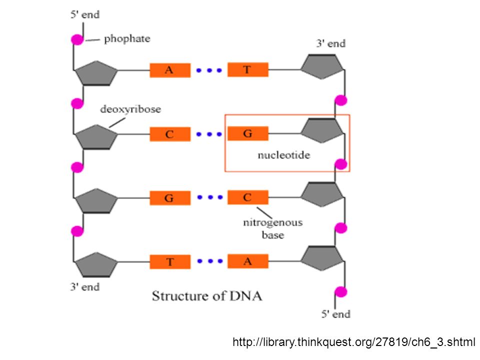
DNA- Deoxyribonucleic Acid
Nucleic Acid made of 2 strands in the form of a double helix (spiral staircase) Consists of nucleotides which has 3 parts: A phosphate group A 5-carbon sugar (Deoxyribose) 1 of 4 different Nitrogenous bases Adenine- Purine (double ring) always binds w/ Thymine Thymine- Pyrimidine (single ring) always binds w/ Adenine Guanine- Purine; always binds w/ Cytosine Cytosine- Pyrimidine; always binds w/ Guanine The double helix is held together by hydrogen bonds between the nitrogen bases attached to the 2 different strands. The nitrogenous bases make the steps of the spiral staircase structure. The rails (backbone) consists of phosphate groups bound to the 5-carbon sugars; the 5-carbon sugars bond to the nitrogenous bases.
 Consists of nucleotides which has 3 parts: A phosphate group. A 5-carbon sugar (Deoxyribose) 1 of 4 different Nitrogenous bases. Adenine- Purine (double ring) always binds w/ Thymine. Thymine- Pyrimidine (single ring) always binds w/ Adenine. Guanine- Purine; always binds w/ Cytosine. Cytosine- Pyrimidine; always binds w/ Guanine. The double helix is held together by hydrogen bonds between the nitrogen bases attached to the 2 different strands. The nitrogenous bases make the steps of the spiral staircase structure. The rails (backbone) consists of phosphate groups bound to the 5-carbon sugars; the 5-carbon sugars bond to the nitrogenous bases.")
10
X-ray diffraction image from:
Watson and Crick and their tin-and-wire model of DNA, image from:

11
In 1949, the nitrogenous bases were found to be in complimentary percentages by Erwin Chargaff.
He discovered that adenine was always present in the same percentages as thymine; he also found the remaining percentage of bases consisted equally of guanine and cytosine. These pairs of bases are known as complimentary base pairs. The observations led to the development to base-pairing rules in DNA. Adenine – Thymine (A – T) Cytosine – Guanine (C – G) The order of nitrogenous bases on a DNA chain is known as base sequence. Ex: A C C T G T G A G A C T G G A C A C T C T G
 Cytosine – Guanine (C – G) The order of nitrogenous bases on a DNA chain is known as base sequence. Ex: A C C T G T G A G A C. T G G A C A C T C T G.")
12
DNA Replication Is the process by which DNA is copied within a cell before a cell undergoes division. Step 1: Helicases (an enzyme) separate the DNA strands by breaking the Hydrogen bonds which connect the nitrogenous bases. Creating the replication fork (the Y-shaped region created when the DNA strands separate. Step 2: DNA Polymerases (an enzyme) bring free floating nitrogenous bases to the now exposed DNA strands creating new hydrogen bonds between the original strands and the new bases creating 2 new DNA molecules. Step 3: DNA Polymerase finish creating replicating the 2 exposed strands and then falls off, resulting in 2 new DNA molecules. Replication occurs at the replication fork in two separate directions at the same time; always in the 3’ to 5’ direction. Replication errors are rare about 1 in 1 billion paired nucleotides added (similar to typing our Biology textbook 1000 times!)
 separate the DNA strands by breaking the Hydrogen bonds which connect the nitrogenous bases. Creating the replication fork (the Y-shaped region created when the DNA strands separate. Step 2: DNA Polymerases (an enzyme) bring free floating nitrogenous bases to the now exposed DNA strands creating new hydrogen bonds between the original strands and the new bases creating 2 new DNA molecules. Step 3: DNA Polymerase finish creating replicating the 2 exposed strands and then falls off, resulting in 2 new DNA molecules. Replication occurs at the replication fork in two separate directions at the same time; always in the 3’ to 5’ direction. Replication errors are rare about 1 in 1 billion paired nucleotides added (similar to typing our Biology textbook 1000 times!)")
13
Protein Synthesis 1st begins with the synthesis of RNA (Ribonucleic Acid) called Transcription. Transcription is the process of RNA polymerase (an enzyme) copying certain sections of DNA. These sections are known as genes which control particular enzymes or proteins. The polymerase will begin transcription at a promoter and end at a termination signal. (this would be the length of a gene) RNA is a single strand of nucleotide bases. RNA nitrogenous bases: Cytosine – Guanine Adenine – Uracil There are 3 types of RNA: mRNA: MessengerRNA - carries the message to be translated. tRNA: TransferRNA - translates the message found w/in mRNA. rRNA: RibosomalRNA - structural component of ribosomes.
 copying certain sections of DNA. These sections are known as genes which control particular enzymes or proteins. The polymerase will begin transcription at a promoter and end at a termination signal. (this would be the length of a gene) RNA is a single strand of nucleotide bases. RNA nitrogenous bases: Cytosine – Guanine. Adenine – Uracil. There are 3 types of RNA: mRNA: MessengerRNA - carries the message to be translated. tRNA: TransferRNA - translates the message found w/in mRNA. rRNA: RibosomalRNA - structural component of ribosomes.")
14
The newly formed RNA molecule (mRNA molecule) is encoded with valuable instructions called the genetic code. The genetic code is the base sequence of an RNA molecule as it relates to the 3 base sequences called codons which represent amino acids. Each codon represents a start or stop signal, or an amino acid. AUG (start codon) – Methionine UAA, UAG, UGA (stop codons) There are 64 mRNA codons which code for 20 amino acids. Amino acids are the subunits of polypeptides (proteins). The # and sequence of amino acids will determine what protein is created.
 – Methionine. UAA, UAG, UGA (stop codons) There are 64 mRNA codons which code for 20 amino acids. Amino acids are the subunits of polypeptides (proteins). The # and sequence of amino acids will determine what protein is created.")
15
Translation The synthesis of a protein from the message carried within the mRNA molecule. mRNA molecule attaches to ribosome where tRNA molecules w/ an anticodon subunit on one end which is complementary to a codon on the mRNA strand carries the amino acid coded for by the mRNA codon

16
tRNA molecules bind to the mRNA molecule one at a time, when the 2nd attaches the first transfers its amino acid to the 2nd’s amino acid by forming a peptide bond. After the 1st tRNA releases its amino acid it will release from the mRNA molecule. Each subsequent tRNA molecule connection will restart the events above. The synthesis of the protein will end when the ribosome reaches a stop codon at which point the newly formed protein is released.

17
DNA RNA = Transcription RNA Protein = Translation
DNA DNA = Replication DNA RNA = Transcription RNA Protein = Translation DNA RNA 5-Carbon Sugar: Deoxyribose Ribose Nitrogenous Base: Adenine – Thymine Adenine – Uracil Guanine – Cytosine Guanine – Cytosine Structure: Double Helix Single Strand Function: Genetic Information Protein Synthesis (Heredity)
")
Similar presentations


 Harmless bacteria (rough colonies) Heat-killed, disease- causing bacteria (smooth colonies) Control (no growth)>")


