Download presentation
Presentation is loading. Please wait.

1
Sensory and Motor Mechanisms AP Chapter 50

2
Notice You do not need to know the specific neuroanatomy of the sensory organs, rather the mechanisms of how they work, ie: type of receptors, generally how signal is carried (by vibrations, photopigments, etc), signaling mechanisms or opening of ions channels. Even though more information is included in the power point, just read for information purposes.

3
The brain’s processing of sensory input and motor output is cyclical rather than linear The way it ISN’T: sensing brain analysis action. The way it is: sensing, analysis, and action are ongoing and overlapping processes. Sensations begin as different forms of energy that are detected by sensory receptors. –This energy is converted to action potentials that travel to appropriate regions of the brain. The limbic region plays a major role in determining the importance of a particular sensory input.

4
Sensory receptors transduce stimulus energy and transmit signals to the nervous system

5
4 Functions common to all sensory pathways 1.Sensory Reception 2.Sensory transduction 3.Transmission 4.Perception

6
Sensory receptors are categorized by the type of energy they transduce.

7
Categories of sensory receptors 1.Mechanoreceptors – pressure, touch, motion, sound, hair cells 2.Chemoreceptors general – solute conc specific – molecules; gustatory (taste), olfactory (smell) 3.Electromagnetic 4.Thermoreceptors 5.Photoreceptors 6.Pain receptors – in humans, nociceptors in epidermis, located in skin and other areas, aspirin/ibuprofen blocks prostaglandins
, olfactory (smell) 3.Electromagnetic 4.Thermoreceptors 5.Photoreceptors 6.Pain receptors – in humans, nociceptors in epidermis, located in skin and other areas, aspirin/ibuprofen blocks prostaglandins")
8
Mechanoreceptors for hearing and equilibrium Utilize moving fluid and settling particles Mammals – pressure waves picked up by ears and converted into nerve impulses Fish – lateral line systems Invertebrates – statocysts with ciliated receptor cells with sand granules Insects – body hairs that vibrate, some have ears

9
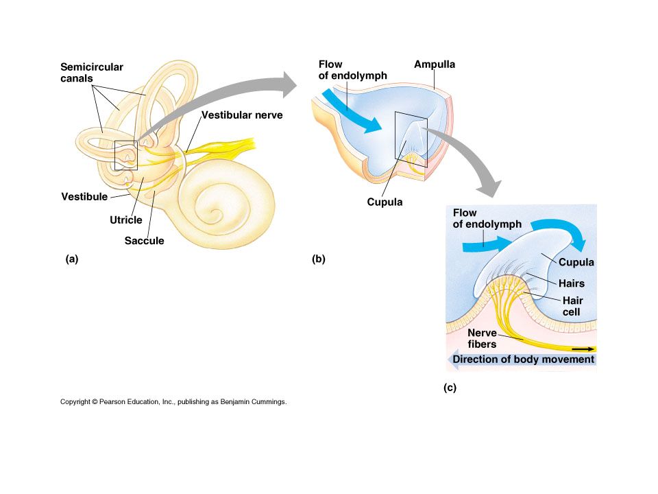
Our Hearing and Balance Energy of fluid into energy of action potentials Uses sensitive hair cells True organ of hearing – the organ of Corti located in the cochlea Balance – semicircular canals Hearing animation

11
Hearing http://msjensen.cehd.umn.e du/1135/Links/Animations/F lash/0019- swf_effect_of_soun.swf The three small bones transmit vibrations To the inner ear which contains fluid-filled canals.

12
Air pressure vibrates fluid in canals which vibrate the basilar membrane, bending the hairs of its receptor cells against the tectorial membrane which opens ion channels and allows K+ to enter the cells and cause a depolarization and releases neurotransmitters to continue to the auditory nerve to the brain.

13
Fig. 50-9 “Hairs” of hair cell Neuro- trans- mitter at synapse Sensory neuron More neuro- trans- mitter (a) No bending of hairs(b) Bending of hairs in one direction(c) Bending of hairs in other direction Less neuro- trans- mitter Action potentials Membrane potential (mV) 0 –70 0 1 2 3 4 5 6 7 Time (sec) Signal –70 –50 Receptor potential Membrane potential (mV) 0 –70 0 1 2 3 4 5 6 7 Time (sec) –70 –50 Membrane potential (mV) 0 –70 0 1 2 3 4 5 6 7 Time (sec) –70 –50 Signal
 No bending of hairs(b) Bending of hairs in one direction(c) Bending of hairs in other direction Less neuro- trans- mitter Action potentials Membrane potential (mV) 0 – Time (sec) Signal –70 –50 Receptor potential Membrane potential (mV) 0 – Time (sec) –70 –50 Membrane potential (mV) 0 – Time (sec) –70 –50 Signal.")
14
Frequency (pitch) determined by areas of basilar membrane that vibrate at different frequencies; areas are thick and thin
 determined by areas of basilar membrane that vibrate at different frequencies; areas are thick and thin")
15
Volume is controlled by amplitude of wave – stronger bends hair cells more and more action potentials

16
Balance in the semicircular canals is also a response to hair cells; different head angles stimulate different. Hair cells; lateral line systems in fish and some amphibians work like this too. The inner ear also contains the organs of equilibrium Copyright © 2002 Pearson Education, Inc., publishing as Benjamin Cummings

19
Statocysts are mechanoreceptors that function in an invertebrates sense of equilibrium. –Statocyst function is similar to that of human semi- circular canals –Use ciliated (hair- like cells) Many invertebrates have gravity sensors and are sound-sensitive Copyright © 2002 Pearson Education, Inc., publishing as Benjamin Cummings Fig. 49.21
 Many invertebrates have gravity sensors and are sound-sensitive Copyright © 2002 Pearson Education, Inc., publishing as Benjamin Cummings Fig")
20
A diversity of photoreceptors has evolved among invertebrates. Planaria – eyecup for light and direction Insects/crustaceans- compound eyes (ommatidia) Jellyfish, spider, mollusks – single lens eye
 Jellyfish, spider, mollusks – single lens eye.")
21
Taste and Smell Odor/taste molecules bind to ciliated receptor cells and trigger a signal- transduction pathway that involves a G- protein and, often, adenylyl cyclase and cyclic AMP’s. cAMP to open Na + channels, depolarizing the membrane and sending action potentials to the brain.

22
Copyright © 2002 Pearson Education, Inc., publishing as Benjamin Cummings Fig. 49.24

23
Fig. 50-13 G protein Sugar molecule Phospholipase C Tongue Sodium channel PIP 2 Na + IP 3 (second messenger) Sweet receptor ER Nucleus Taste pore SENSORY RECEPTOR CELL Ca 2+ (second messenger) IP 3 -gated calcium channel Sensory receptor cells Taste bud Sugar molecule Sensory neuron
 Sweet receptor ER Nucleus Taste pore SENSORY RECEPTOR CELL Ca 2+ (second messenger) IP 3 -gated calcium channel Sensory receptor cells Taste bud Sugar molecule Sensory neuron.")
24
Vertebrate eyes Rods and cones are photoreceptors located in the retina of the eye. Rods are more light sensitive and are concentrated toward the edge of the retina. Cones are more color sensitive and are concentrated in the center of the visual field called the

25
Vertebrates have single-lens eyes Is structurally analogous to the invertebrate single-lens eye.

26
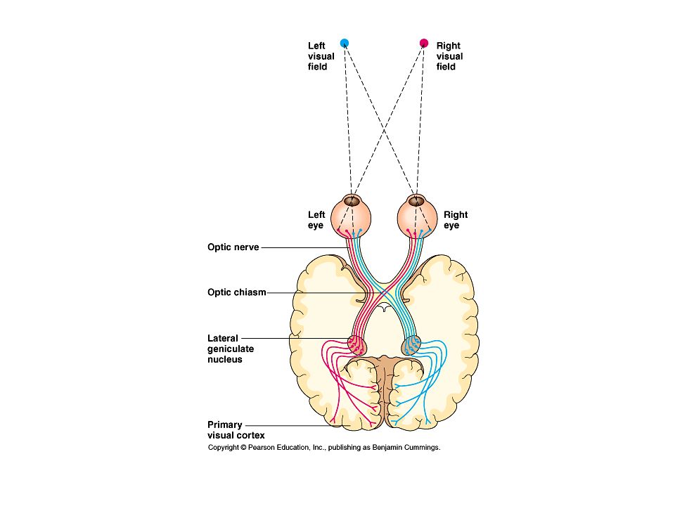
How does this work? Rods and cones synapse with bipolar cells in the retina, which synapse with ganglion cells, whose axons form the optic nerve. R/C BP Ganglion Cells

27
Light hits the retina and then comes back through the cells to the optic nerve.

28
Photoreceptors

29
Rods and cones have visual pigments embedded in a stack of folded membranes or disks in each cell. Retinal is the light-absorbing pigment and is bonded to a membrane protein – opsin. Combo – rhodopsin.

30
When retinal absorbs light, it changes shape and separates from opsin. In the dark, the retinal is converted back to its original shape.

31
Copyright © 2002 Pearson Education, Inc., publishing as Benjamin Cummings Opsin activates a G protein and opens/closes Na channels to continue/discontinue the nerve impulse. Fig. 49.13

32
Notice in the light, the Na + channels are closed.

34
remember… The ultimate perception of the stimulus depends on the area of the brain that is stimulated!

35
In summary: Touch – mechanoreceptors, dendrites of neurons pick up ions Smell, taste – chemoreceptors, gen (solute conc), specific (individual molecules), G protein activates a second messenger that controls a Na or K ion channel Sight (light) – electroreceptors, trans retinal activates a G protein cascade that opens/closes Na channels
, specific (individual molecules), G protein activates a second messenger that controls a Na or K ion channel Sight (light) – electroreceptors, trans retinal activates a G protein cascade that opens/closes Na channels")
36
Hearing, balance – mechanoreceptors, moving fluid, settling particles, bending of hair cells open ion channels

37
Locomotion and muscle action

38
The muscle cell’s structure is conducive to its purpose which is to contract upon receiving a stimulus.

39
How the muscle cell is organized Animations This is ONE muscle cell, called a muscle fiber. for energy The muscle fiber (cell) is made up of many myofibrils which in turn are made up of sarcomeres, the units of contraction.
 is made up of many myofibrils which in turn are made up of sarcomeres, the units of contraction..")
40
The sarcoplasmic reticulum (SR) is a special type of smooth ER found in smooth and striated muscle. The SR contains large stores of calcium ions, which it releases when the cell is depolarized. Action potential Is spread in the T tubules

41
Fig. 50-25b TEM Thick filaments (myosin) M line Z line Thin filaments (actin) Sarcomere 0.5 µm The contracting unit is the sarcomere. This is what gives skeletal muscles and heart muscles their striated appearance.
 M line Z line Thin filaments (actin) Sarcomere 0.5 µm The contracting unit is the sarcomere. This is what gives skeletal muscles and heart muscles their striated appearance..")
42
When the sarcomere contracts, the filaments slide over each other. The sliding filament model Animation: Sarcomere Contraction

43
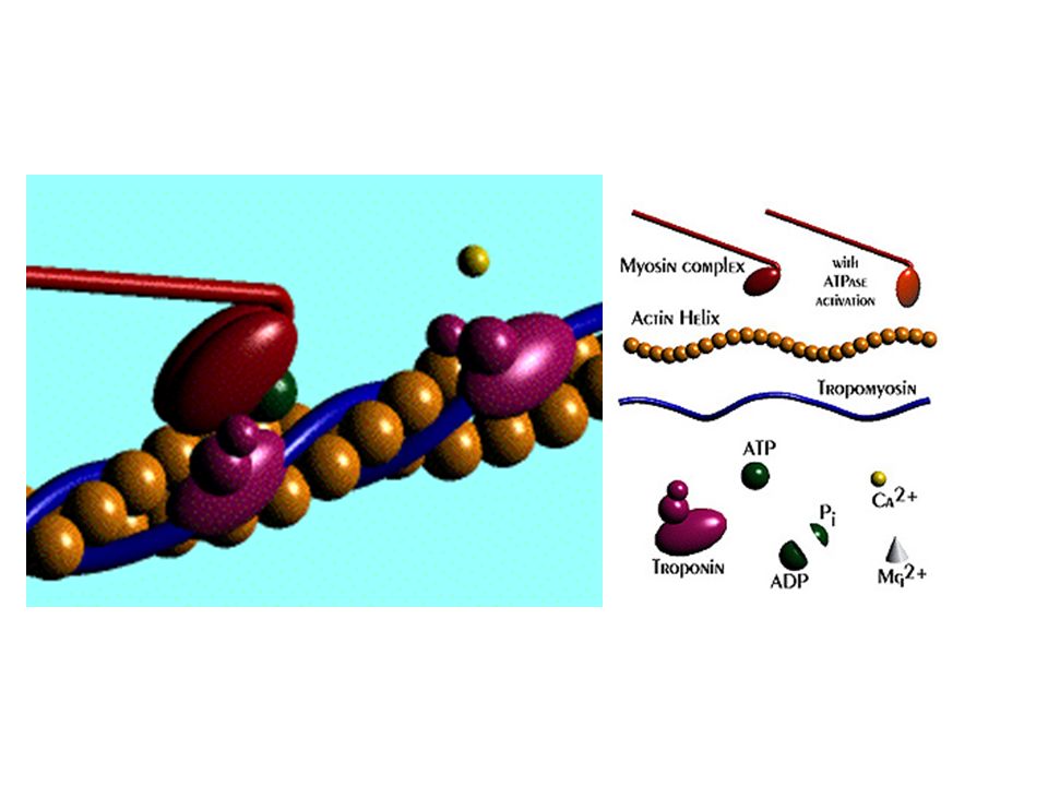
How does this happen? a closer look: Myosin Myosin is made of polypeptides twisted to form a fiber helix with a globular end, which has ATPase activity & an affinity to bind to actin.ATPase

44
a closer look: Actin Actin is a globular protein; each globular actin unit contains a myosin binding site. Remember – Actin – Ac”thin”

45
Mechanism of action 1. The Neuromuscular Junction – neuron to muscle Signal travels from motor neuron by acetycholine (excitatory) to the skeletal muscle cell and depolarizes it. An action potential is spread in the T tubules and changes the permeability of the sarcoplasmic reticulum which releases Ca +.
 to the skeletal muscle cell and depolarizes it. An action potential is spread in the T tubules and changes the permeability of the sarcoplasmic reticulum which releases Ca +..")
46
2.Actin involvement Myosin-binding sites are blocked by a strand of tropomyosin whose position is controlled by Troponin complex molecules. Ca + ions bind to the complex and move the tropomyosin and expose the binding sites for myosin.

47
3. Myosin Involvement -The globular heads of the myosin are energized by ATP and bind to actin forming a cross-bridge -When relaxing to its low-energy state, the myosin head bends and pulls the attached actin toward the center of the sarcomere

48
4. Completion When its’s over, Ca + returns to the sarcoplasmic reticulum and tropomyosin recovers binding sites on actin. Acetylcholine is degraded at the synapse.

49
Mechanism of Filament Sliding at the Neuromuscular Junction Animation: Action Potentials and Muscle Contraction

51
The sliding-filament model of muscle contraction. Interactions between myosin and actin generate force during uscle contractions Copyright © 2002 Pearson Education, Inc., publishing as Benjamin Cummings Fig. 49.33

53
Protein models of muscle action

54
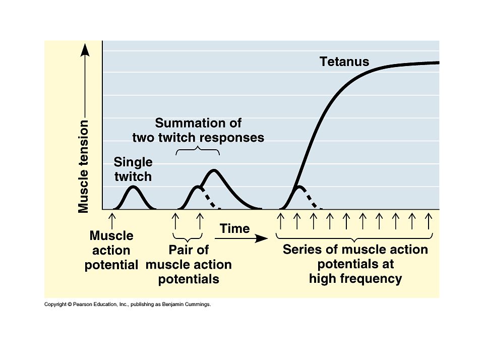
Contraction Of single muscle fiber – all or none Twitch: slow – less SR, Ca in longer, fibers must have many mitochondria, a good blood supply (myoglobin better which picks up O 2 better and stores it) Fast – rapid and powerful contraction Tetanus – smooth, sustained contraction; action potentials arrive rapidly
 Fast – rapid and powerful contraction Tetanus – smooth, sustained contraction; action potentials arrive rapidly")
56
Muscle Fatigue Depletion of ATP, loss of ion gradient, accumulation of lactic acid

57
Hydrostatic skeleton: consists of fluid held under pressure in a closed body compartment. –Form and movement is controlled by changing the shape of this compartment. –Advantageous in aquatic environments and can support crawling and burrowing. –Does not allow for running or walking. Skeletons support and protect the animal body and are essential to movement

58
Exoskeletons – supportive, protective but do not grow (molted) Endoskeletons – supportive, grow with the organism, less protective
 Endoskeletons – supportive, grow with the organism, less protective")
Similar presentations


, which.>")


http://www.youtube.com/watch?v=SCasruJT->")


