Download presentation
Presentation is loading. Please wait.

1
Anatomy of the Diencephalon,
NEUROANATOMY Lecture : 6 Anatomy of the Diencephalon, Limbic System Pituitary Gland & Prepared and presented by: Dr. Iyad Mousa Hussein, MD, Ph.D in Neurology Head of Neurology Department Nasser Hospital

2
LECTURE OBJECTIVES: 1. Definition, Site, Surfaces, and Subdivisions of the Diencephalon. 2. Definition, site, Parts, nuclei, and functions of the Thalamus. 3. Definition, Site, Parts, and Nuclei and functions of the Hypothalamus. 4. The Hypothalamic Releasing and Inhibitory Hormones. 5. Definition, Site, Structures, and functions of the Epithalamus. 6. Definition, Site, and Structures of the Metathalamus. 7. Definition, Site, and Structures of the Subthalamus. 8. Anatomy and Functions of the Pituitary Gland. 9.Definition, Site structures, Connections and functions of the Limbic System.

3
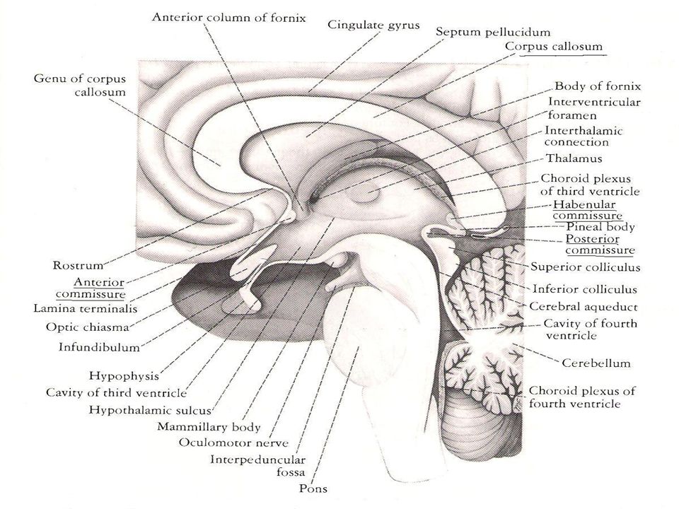
The Diencephalon Definition and Site:
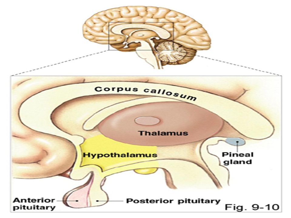
It is the part of the brain which lies above the midbrain and between the lower parts of the two cerebral hemispheres. Cavity of the diencephalon: is the third ventricle.

6
Surfaces of the Diencephalon
Two lateral surfaces: each lateral surface related to the internal capsule. The Lower surface: a. The middle (largest) part: is the subthalamus, which lies upper the midbrain. b. The anterior part: is formed by the hypothalamus and is connected with pituitary gland. C. The posterior part: is formed by the metathalamus. 3. Upper surface: it is formed by the two thalami and the third ventricle in the middle.
 part: is the subthalamus, which. lies upper the midbrain. b. The anterior part: is formed by the hypothalamus. and is connected with pituitary gland. C. The posterior part: is formed by the metathalamus. 3. Upper surface: it is formed by the two thalami and the third ventricle in the middle.")
8
Subdivisions of the Diencephalon
It is subdivided into five parts: 1. Thalamus (bilateral): the largest part. 2. Subthalamus: it lies directly above midbrain. 3. Hypothalamus: it lies infront of the subthalamus. 4. Metathalamus (bilateral). 5. Epithalamus.
: the largest part. 2. Subthalamus: it lies directly above midbrain. 3. Hypothalamus: it lies infront of the subthalamus. 4. Metathalamus (bilateral). 5. Epithalamus.")
9
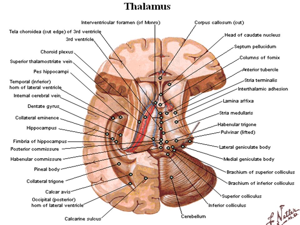
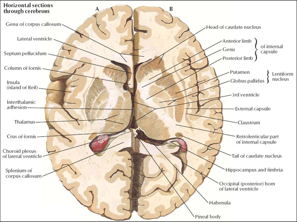
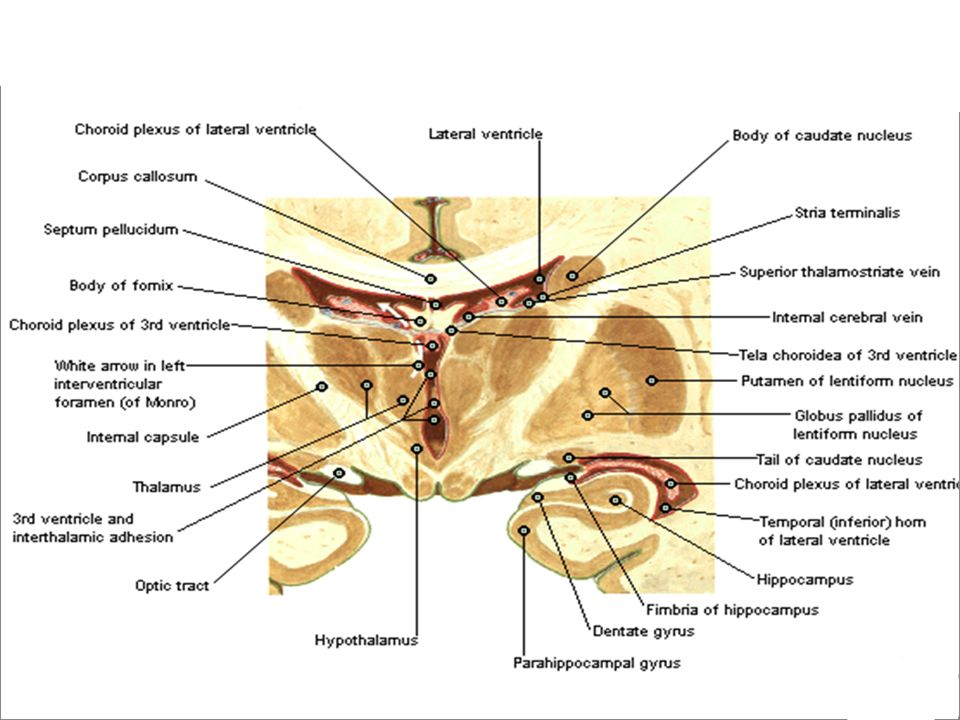
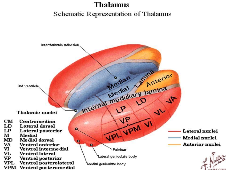
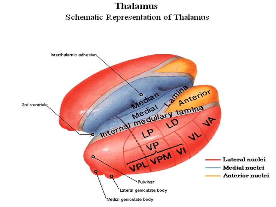
The Thalamus Definition: it is a large, egg-shaped mass of gray
matter lying in the middle of the cerebrum. Site: it lies on each side of the third ventricle, immediately above the subthalamus. Parts of the Thalamus: 1. Anterior part. 2. Medial part. 3. Lateral part.

14
Functions of the Thalamus
Sensory Function: the thalamus is the great sensory relay station on the pathway of all sensations to cerebral cortex (except smell). 2. Consciousness level. 3. Emotional function.
. 2. Consciousness level. 3. Emotional function.")
15
Nuclei of the Thalamus 1. Medial group. 3. Lateral group.
4. Anterior group.

16
Nuclei of the Thalamus Anterior nucleus. Dorsomedial nucleus.
Lateral posterior nucleus. Lateral dorsal nucleus. Lateral pulvinar nucleus. Ventral anterior nucleus. Ventral posterior nucleus (Ventral Posterolateral & Posteromedial nuclei). Ventral lateral nucleus. Inttralaminar nucleus. Midline reticular nucleus.
. Ventral lateral nucleus. Inttralaminar nucleus. Midline reticular nucleus.")
18
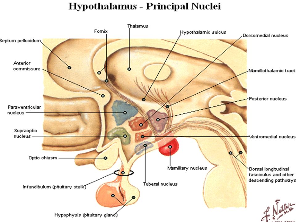
The Hypothalamus Definition and Site:
It is the part of the diencephalon witch lies infront of subthalamus and anterioinferior to the thalamus. The hypothalamus, although small (0,3 of the total brain), is very important part of the central nervous System (Neuroendocrine organ).
, is very important part of the central nervous. System (Neuroendocrine organ).")
21
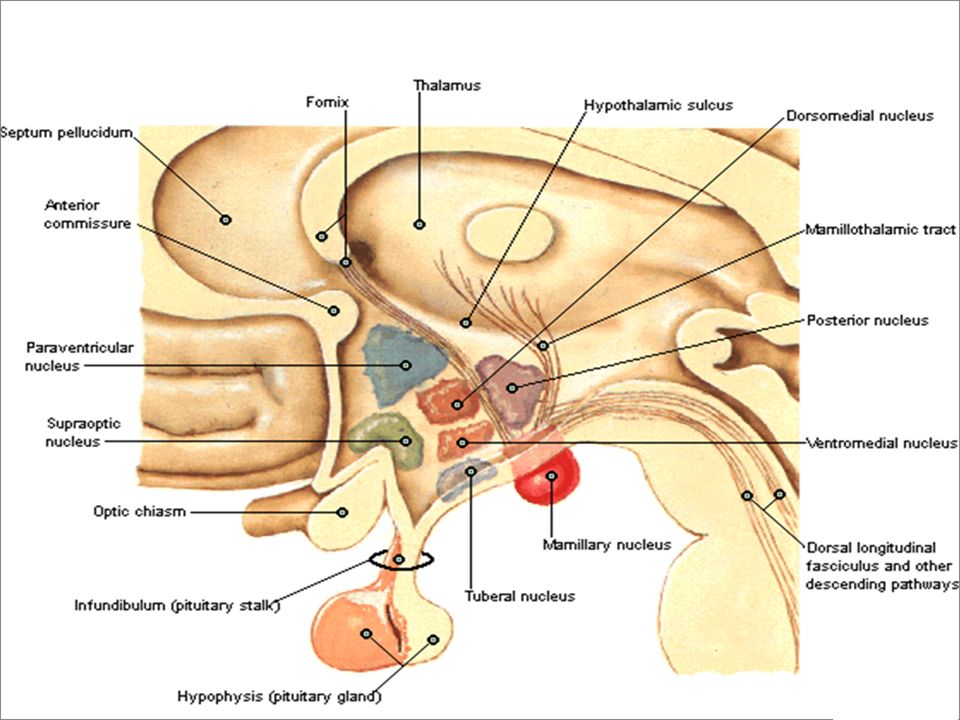
Parts and Nuclei of the Hypothalamus
Optic part: immediately related to optic chiasma. It consists of two nuclei: a. Supraoptic nucleus. b. Paraventricular nucleus. 2. Tuberal part: it consists of three nuclei: a. Ventromedial nucleus. b. Dorsomedial nucleus. c. Tuberal nucleus. 3. Mamillary part: it consists of two nuclei: a. Posterior nucleus. b. Lateral nucleus. 4. Posterior perforated substance.

23
Connections of the Hypothalamus
A. Afferent fibers: 1. Somato and Visceral afferents. 2. Visual afferents. 3. Olfaction. 4. Auditory afferents. 5. Thalamohypothalamic fibers. 6. Amygdaloidhypothalamic fibers. 7. Tegmental fibers: from midbrain. B. Efferent fibers: 1. Descending fibers to the brain stem and spinal cord. 2. To the limbic system. 3. To the pituitary gland.

24
Functions of the Hypothalamus
Hypothalamus has important regulatory functions: 1. Temperature. 2. Emotional regulation. 3. Growth (via thyroid stimulating hormone). 4. Hunger and thirst. 5. Sexual behaviour. 6. Control of various endocrine and activity rhythms (via hormones). 7. Memory (visual and verbal memory).
. 4. Hunger and thirst. 5. Sexual behaviour. 6. Control of various endocrine and activity. rhythms (via hormones). 7. Memory (visual and verbal memory).")
25
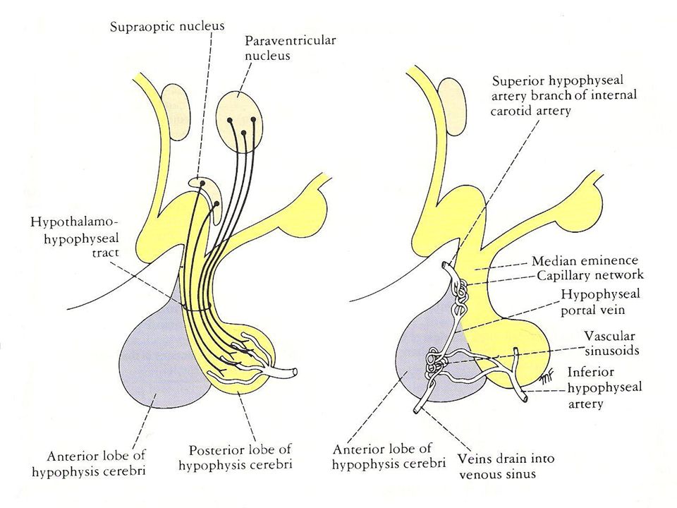
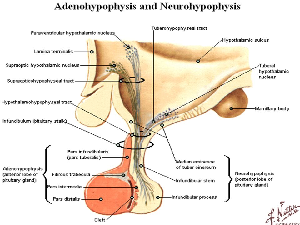
Connections of the Hypothalamus with the Hypophysis (Pituitary Gland)
1. The hormones vasopressin (antidiuretic hormone) and oxytocin are synthesized in the hypothalamus → the posterior lobe of the hypophysis. 2. The hypothalamus is play important role in the production of the releasing hormones and release-inhibitory hormones.
 and oxytocin are synthesized in the hypothalamus → the posterior lobe of the hypophysis. 2. The hypothalamus is play important role in the production of the releasing hormones and release-inhibitory hormones.")
27
The Hypothalamic Releasing and Inhibitory Hormones
Anterior Pituitary Hormones Hypothalamic Regulatory Hormones Growth hormone (GH) Growth hormone-releasing hormone (GHRH) Growth hormone (reduced production) Growth hormone-inhibiting hormone (GHIH) or Somatostatin Prolactin hormone Prolactin-releasing hormone (PRH) Prolactin hormone (reduced production) Prolactin-inhibiting hormone (PIH) Adrenocorticotropic hormone (ACTH) Corticotropin-relasing hormone (CRH) Thyroid-stimulating hormone (TSH) Thyrotropin-relasing hormone (TRH) Luteinizing hormone (LH) and follicle-stimulating hormone Lutenizing hormone-relasing hormone (LHRH)
 Growth hormone-releasing hormone (GHRH) Growth hormone (reduced production) Growth hormone-inhibiting hormone (GHIH) or Somatostatin. Prolactin hormone. Prolactin-releasing hormone (PRH) Prolactin hormone (reduced production) Prolactin-inhibiting hormone (PIH) Adrenocorticotropic hormone (ACTH) Corticotropin-relasing hormone (CRH) Thyroid-stimulating hormone (TSH) Thyrotropin-relasing hormone (TRH) Luteinizing hormone (LH) and follicle-stimulating hormone. Lutenizing hormone-relasing hormone (LHRH)")
28
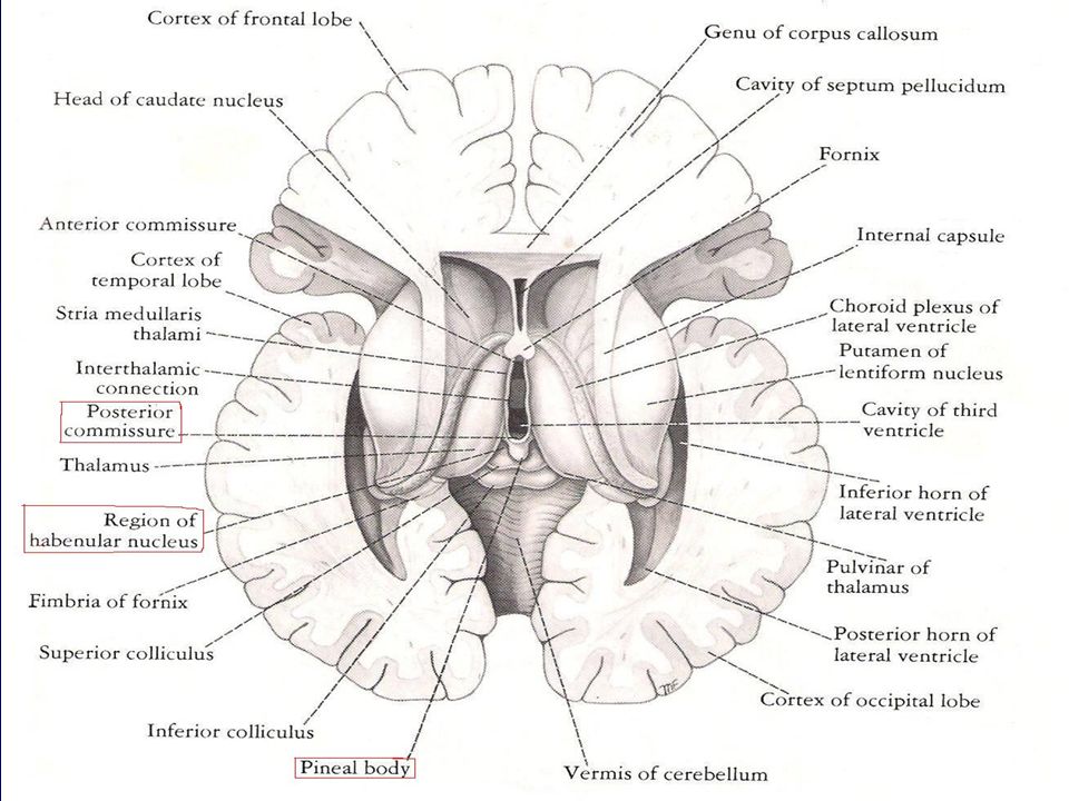
The Epithalamus Definition and Site: it is a part of the diencephalon which is attached to posterior end of the upper surface of the diencephalon. Parts of the Epithalamus: 1. Right and left Habenular nucleus. 2. Habenular commissure. 3. Posterior commissure. 4. Pineal body.

31
The Metathalamus Definition and Site:
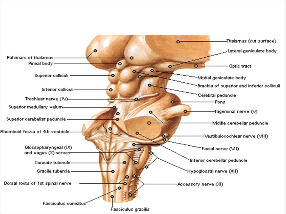
It is the part of the diencephalon which is attached to posterior part of the inferior surface of the thalamus. Parts of the Metathalamus: 1. Lateral geniculate body (LGB): visual function. 2. Medial geniculate body (MGB): auditory function.
: visual function. 2. Medial geniculate body (MGB): auditory function.")
36
The Subthalamus Definition and Site: it is the part of the diencephalon which located between the thalamus and midbrain. Parts of the Subthalamus: A. Posterior smaller part: containing of five bundles: 1. Medial lemniscus. 2. Spinal lemniscus. 3. Trigeminal lemniscus. 4. Reticuo-thalamic tract. 5. Superior cerebellar peduncle. B. Anterior larger part: containing of five bundles. 1. The upper end of the red nucleus. 2. The upper end of the substantia nigra. 3. The subthalamic nucleus. 4. The ansa lenticularis bundle. 5. The fasciculus lenticularis.

39
Pituitary Gland (Hypophysis or Master Gland)
Site: it lies below the hypophyseal fossa below the diaphragm sella. Shape: it is an avoid body, its transverse diameter is 12mm and its anteroposterior diameter is 8mm. Relation: 1. Above: diaphragm sella which separates the gland from optic chiasma. 2. Below: body of sphenoid and sphenoid sinus separating the gland from nasopharynx. 3. On each side: cavernous sinus and its contains. Connection: with hypothalamus. Components: 1. Anterior lobe (Adenohypophysis). 2. Posterior lobe (Neurohypophysis).
. 2. Posterior lobe (Neurohypophysis).")
42
Functions of the Pituitary Gland
Production of the following hormones: A. Anterior pituitary hormones (from anterior lobe): 1. Growth hormone (GH). 2. Prolactin hormone. 3. Adrenocorticotropic hormone (ACTH). 4. Thyroid-stimulating hormone (TSH). 5. Luteinizing hormone (LH). 6. Follicle-stimulating hormone. B. Posterior pituitary hormones (from posterior lobe): 1. Vasopressin (Antidiuretic hormone). 2. Oxytocin.
: 1. Growth hormone (GH). 2. Prolactin hormone. 3. Adrenocorticotropic hormone (ACTH). 4. Thyroid-stimulating hormone (TSH). 5. Luteinizing hormone (LH). 6. Follicle-stimulating hormone. B. Posterior pituitary hormones (from posterior lobe): 1. Vasopressin (Antidiuretic hormone). 2. Oxytocin.")
44
The Limbic System (Emotional Brain)
Definition and Site: It is the number of cortical and subcortical structures lying between the cerebral cortex and the hypothalamus.

45
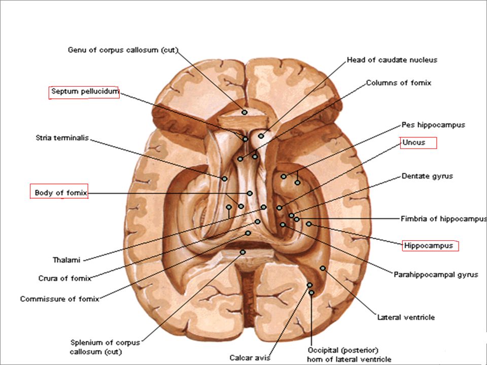
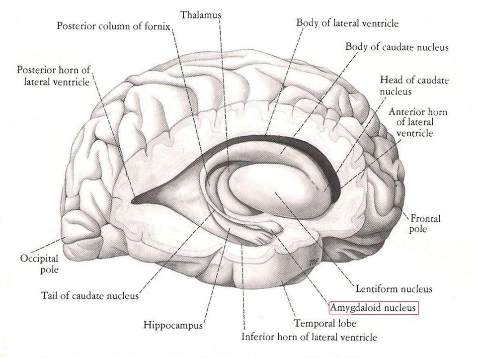
Structures of the Limbic System
Cingulate gyrus. Parahippocampal gyrus. Uncus. Hipocampal formation. Mammilary bodies. Septum pollucidum. Amygdaloid nucleus. The fornix.

50
Connections of the Limbic System
Cerebral cortex. Thalamus. Hypothalamus. Epithalamus.

51
Functions of the Limbic System
1. Control the endocrine system. 2. Control the emotional behavior. 3. Recent memory (hippocampus). 4. There is no evidence that the limbic system has an olfactory function.
. 4. There is no evidence that the limbic system has. an olfactory function.")
52
Thank You

Similar presentations


 from pituitary disturbances? 1)Soldier # 1 2)Soldier # 2 3)Soldier # 3 4)Soldiers.>")


 Pia mater -inner membrane, contains.>")


>")
