Download presentation
Presentation is loading. Please wait.

2
Lecturer name: DR ALBADR Chairman of radiology department Lecture Date: 2011 Introduction to 365 rad

3
Lecture Objectives.. 1- to learned different type of radiology modalities. 2- to have the principle of the indication and contra- indication for different radiology investigation. 3- Usage of different type of contrast.

4
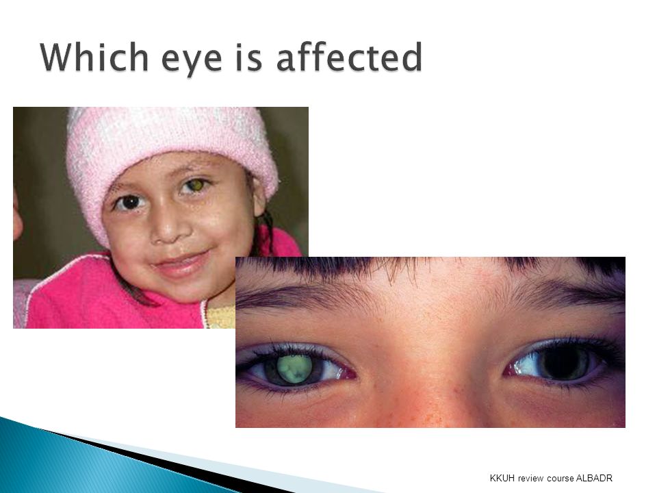
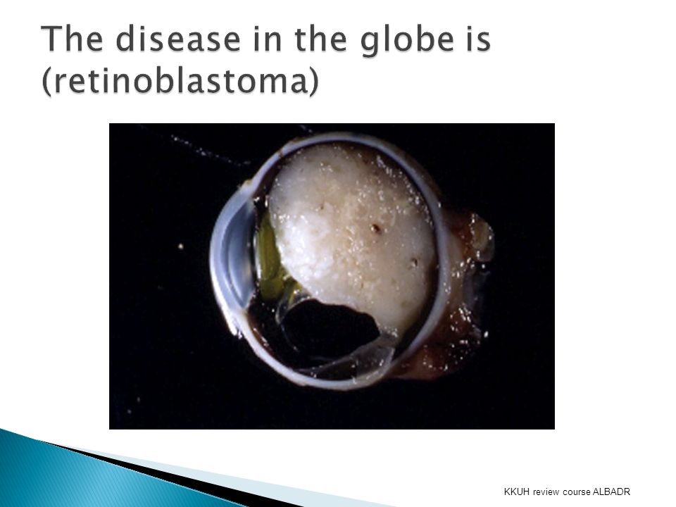
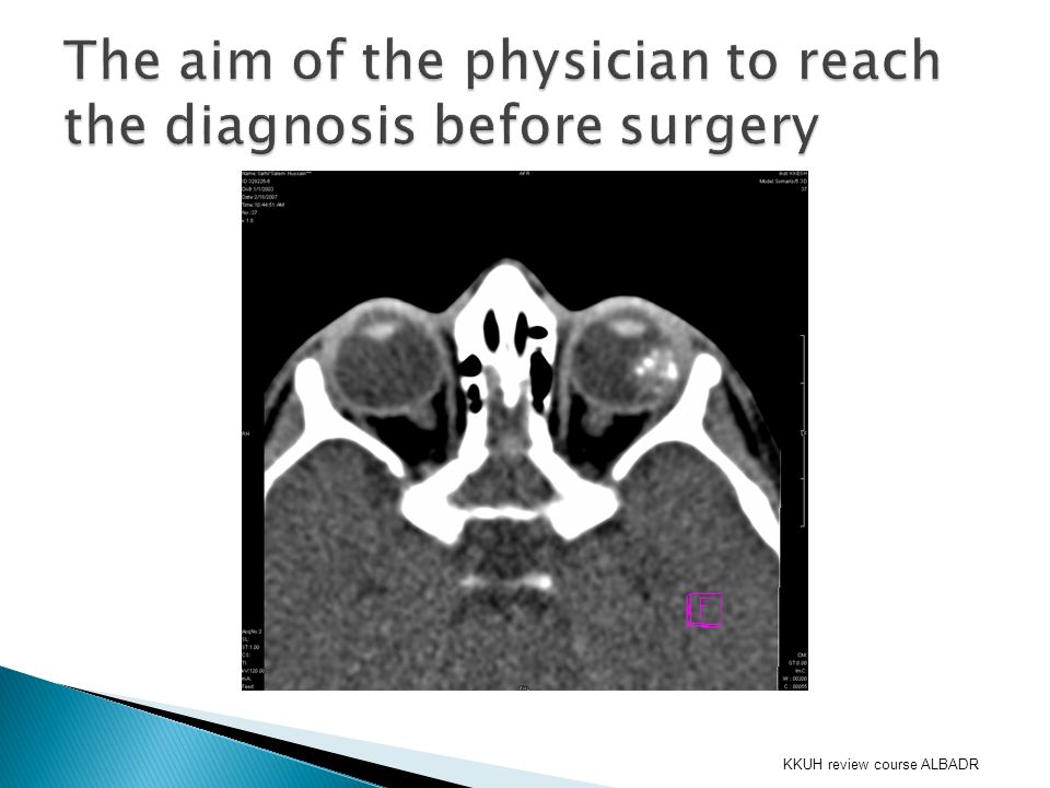
KKUH review course ALBADR

10
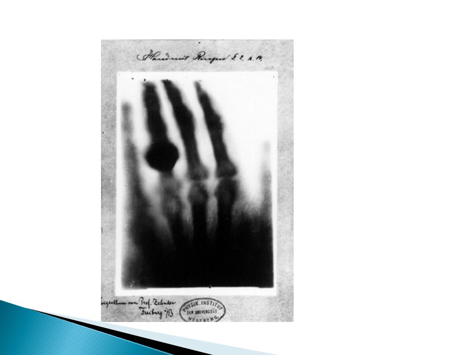
WHAT is X RAY ? ELECTROMAGNATIC RADIATION CAUSING IONIZATION IN THE BODY. X RAY IS COMING FROM ??? WHERE ? You need : 1- machine. 2-Patient. 3-detector.

14
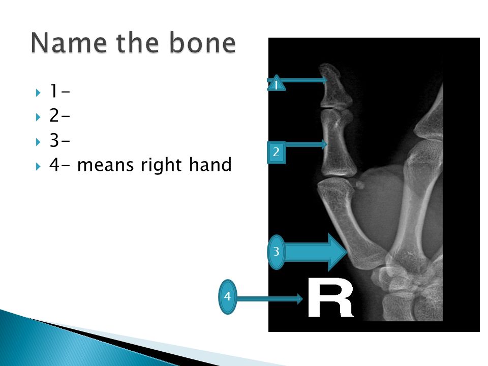
1- 2- 3- 4- means right hand 1 2 3 4

15
1 9 7 8 6 5 3 2 10 4 Name the bone

18
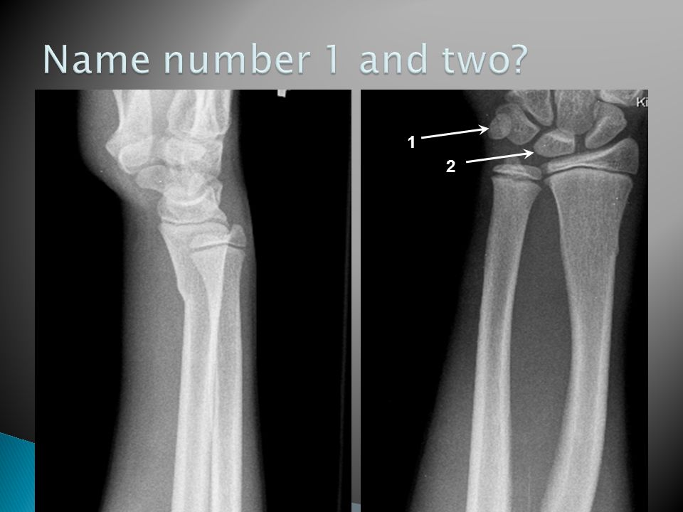
1 2

20
1 2

21
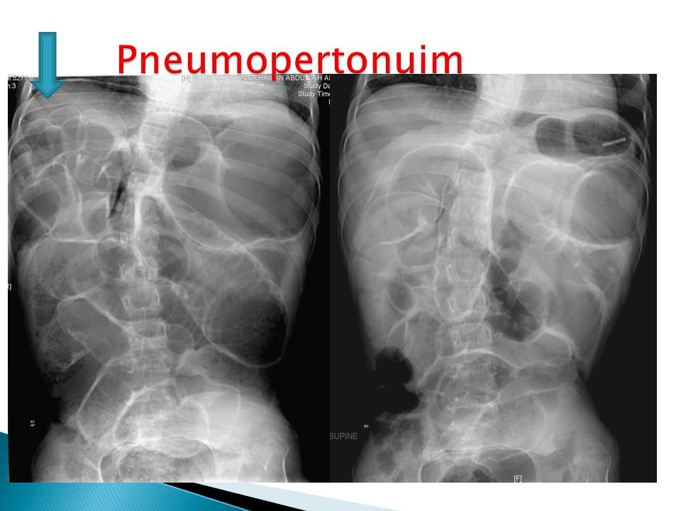
2- Is the air inside the bowel or outside the bowel

24
Pneumothorax : A pneumothorax is an abnormal collection of air or gas in the pleural space that separates the lung from the chest wall Pnuemoperitoneum : Pneumoperitoneum is the presence of air or gas in the abdominal (peritoneal) cavity. It is often seen on X-ray, but small amounts are often missed, and CT is needed...

25
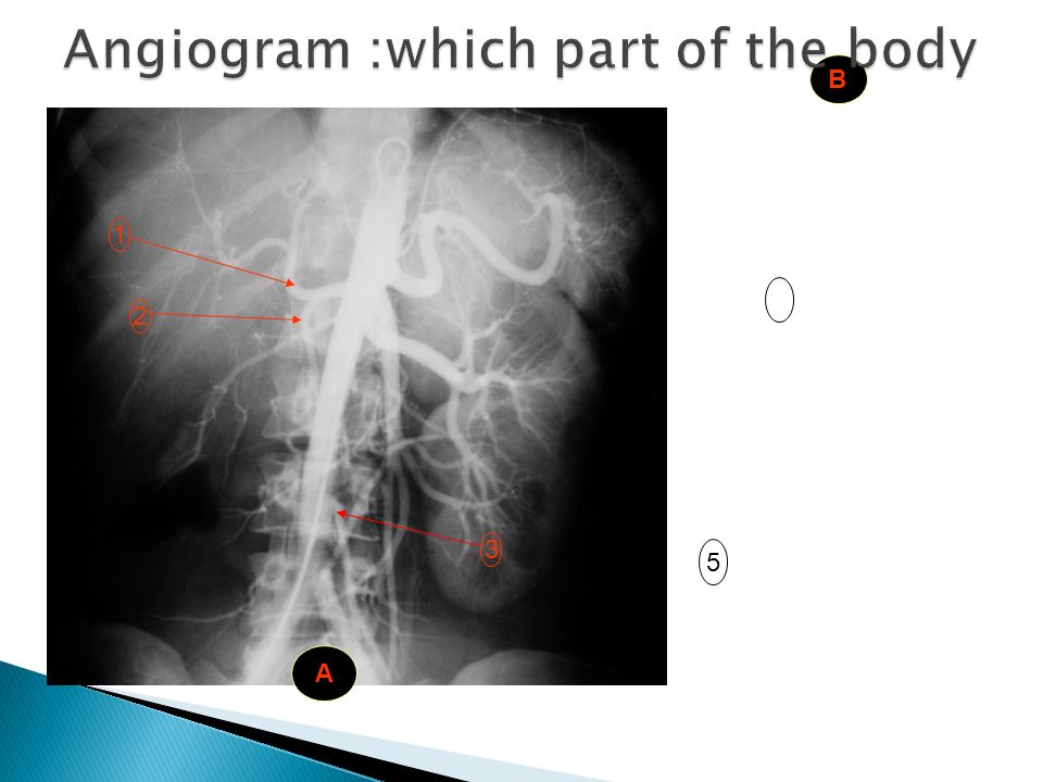
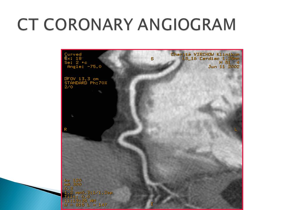
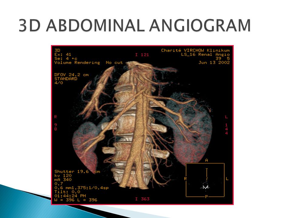
Oral contrast: Baruim swallow :A barium swallow (or esophagography) is a medical imaging procedure used to examine the upper GI (gastrointestinal) tract Baruim meal : A barium meal, also known as an upper gastrointestinal series is a procedure in which radiographs of the esophagus, stomach and duodenum Baruim enema :A barium enema, or lower gastrointestinal (GI) examination. Contrast is radio-opaque I V Contrast : Angiogram Angiography or arteriography is a medical imaging technique used to visualize the inside, or lumen, of blood vessels.

26
1-Myelogram 2-sialogram 3-mamogram 4-sinogram 5- MRCP Magnetic resonance cholangiopancreato graphy 6-ERCP Endoscopic Retrograde Cholangio- Pancreatography

28
5 1 2 3 A B

30
Ultrasound An ultrasound machine creates images that allow various organs in the body to be examined. The machine sends out high-frequency sound waves, which reflect off body structures. A computer receives these reflected waves and uses them to create a picture. Unlike with an x-ray or CT scan, there is no ionizing radiation exposure with this test.

31
Ultrasound Aim Tech Advantage vs disadvantage Organ limitation Uses

35
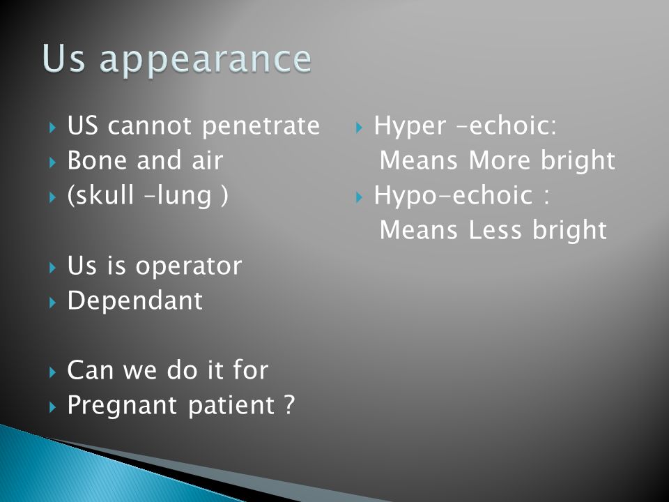
US cannot penetrate Bone and air (skull –lung ) Us is operator Dependant Can we do it for Pregnant patient ? Hyper –echoic: Means More bright Hypo-echoic : Means Less bright

39
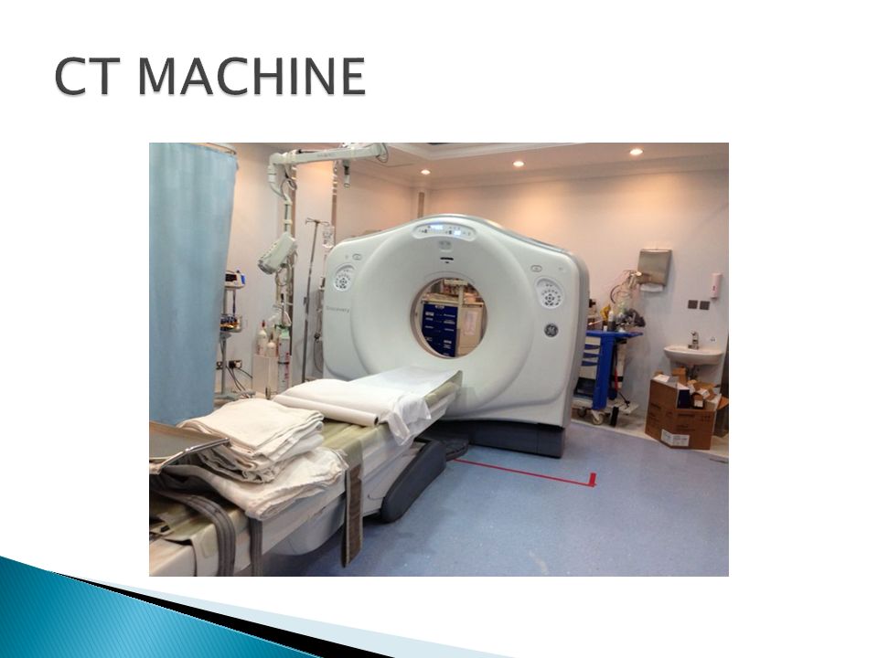
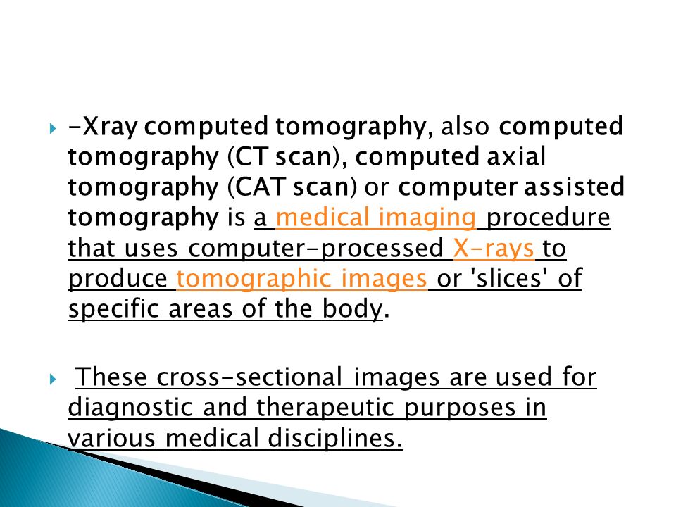
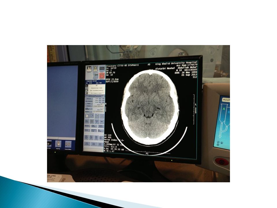
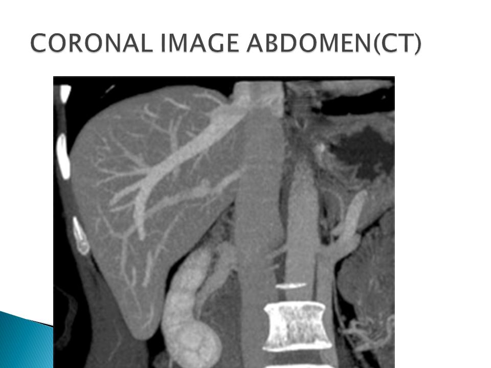
-Xray computed tomography, also computed tomography (CT scan), computed axial tomography (CAT scan) or computer assisted tomography is a medical imaging procedure that uses computer-processed X-rays to produce tomographic images or 'slices' of specific areas of the body.medical imagingX-raystomographic images These cross-sectional images are used for diagnostic and therapeutic purposes in various medical disciplines.
, computed axial tomography (CAT scan) or computer assisted tomography is a medical imaging procedure that uses computer-processed X-rays to produce tomographic images or slices of specific areas of the body.medical imagingX-raystomographic images These cross-sectional images are used for diagnostic and therapeutic purposes in various medical disciplines.")
40
RADITION RISK- pregnancy ? CONTRAST RISK THERE IS A LARGE AMOUNT OF RADITION IN CT EXAMINATION DURING THE STUDY : iv CONTRAST IS USEDE WITH PRECAUTION ORAL CONTRAST IS SAFE

49
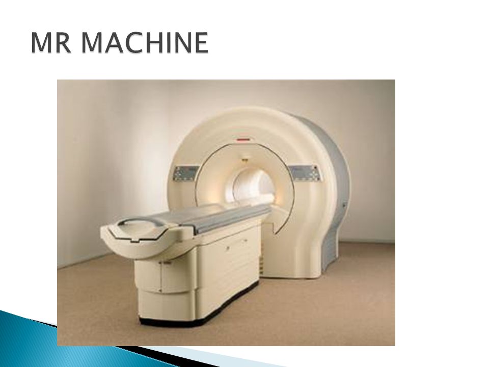
is a Medical imaging used in radiology to visualize internal structures of the body in detail. MRI can create more detailed images of the human body than are possible with X- rays.

50
An MRI scanner is a device in which the patient lies within a large, powerful magnet where the magnetic field is used to align the magnetization of some atomic nuclei in the body, magnet magnetizationatomic nuclei Radiofrequency magnetic fields are applied to systematically alter the alignment of this magnetization.This causes the nuclei to produce a rotating magnetic field detectable by the scanner—and this information is recorded to construct an image.

52
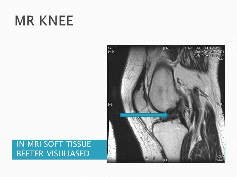
IN MRI SOFT TISSUE BEETER VISULIASED

53
ONLY BONE CAN BE SEEN

54
IN MRI SOFT TISSUE BEETER VISUALISED IN X-RAY YOU CAN NOT SEE THE LIGMENT

58
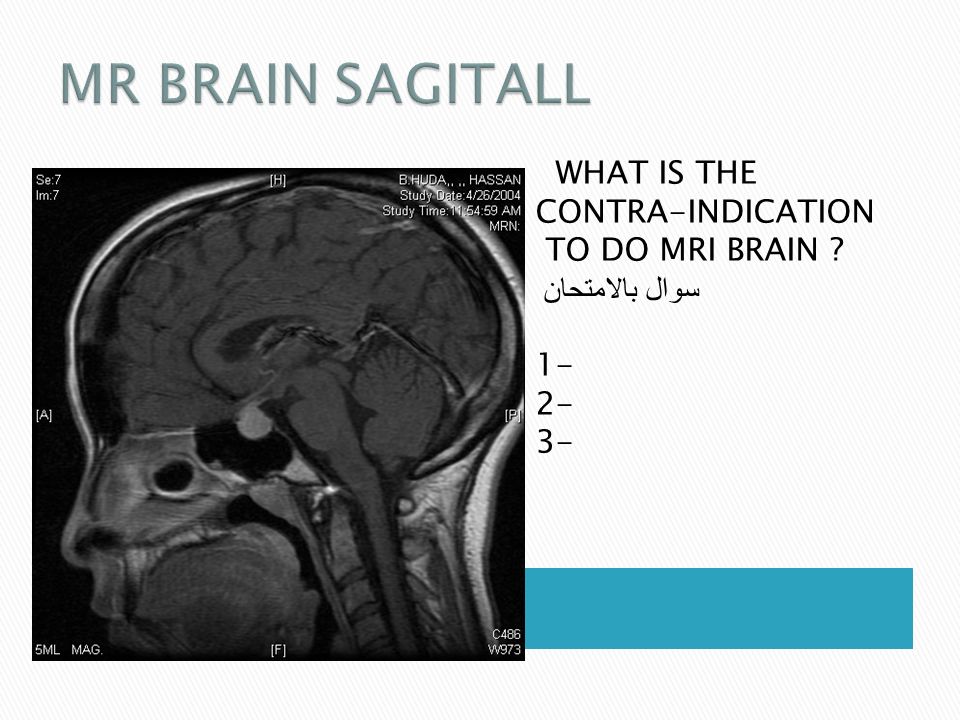
WHAT IS THE CONTRA-INDICATION TO DO MRI BRAIN ? سوال بالامتحان 1- 2- 3-

59
Radiology secret page 6-30 Diagnostic imaging Peter armstrong page 1-13

Similar presentations


 CT scanning or (CAT scanning) is using X-rays to create a 3D image of the inside of an object. CT stands for computed tomography.>")


