Download presentation
Presentation is loading. Please wait.

1
The "pH-Activated Trigger" Mechanism of Colicin E1 Channel Domain Abdi Musse MSc. Final Examination Supervisor Dr. A. R. Merrill Advisory committee Dr. G. Harauz Dr. F. J. Sharom

2
Outline 1.Introduction 2.Research Objectives 3.Results and Discussion 4.Summary and Conclusions

3
Overview The Biology of Pore-forming Colicins Antimicrobial proteins that are secreted by Escherichia coli Targets the cytoplasmic membrane Forms lethaly depolarizing ion channels Dissipations of the cationic gradients (H +, K +, Na + ) Colicin E1
 Colicin E1")
4
R C T Colicin Ia Wiener et al. (1997) Structure and Function H2NH2N COOH RCT BtuB Receptor Tol Network (TolC, A and Q) Channel-forming
 Structure and Function H2NH2N COOH RCT BtuB Receptor Tol Network (TolC, A and Q) Channel-forming.")
5
The Channel Domain Elkins et al. (1997) H1 H2 H3 H10 H4 H7 H6 H5 H8 H9 2.5 Å Structure of P190 Three- layered sandwich structure
 H1 H2 H3 H10 H4 H7 H6 H5 H8 H9 2.5 Å Structure of P190 Three- layered sandwich structure.")
6
Interactions with Membranes Activated-intermediate Membrane-anchored Precursor

7
Formations of the Open Channel The precursor The open channel Monomer 4 – 9 Å diameter Voltage-gated

8
Mechanism of Activation Acid-induced activation is common to most toxins Onset of Protein unfolding Increased structural flexibility Potentiates the massive unfloding events requisite for membrane insertion and channel formation

9
The pH-Activated Trigger Hypothesis The trigger motif: helices 4 and 5a Activating helix-to-coil transition of the trigger motif Disruption of the critical H-bonds formed by D-408, D-410 and D-423 Merrill et al. (1997) H4 H5a
 H4 H5a.")
10
The Research Objectives Purpose To test the proposed pH-activated trigger mechanism Approaches 1.Replacements of the critical acidic residues with serine 2.Incorporation of a disulphide bond within the trigger motif Tools Membrane binding Insertion kinetics Channel activity Structural elucidations

11
Mutant Proteins of Colicin E1 Asp Ser D410S D408S D408S/D410S D408S/D423S D410S/D423S D408S/D410S/D423S Ala Cys A407C/A411C Single Trp F413W F413W/D408S/D423S

12
Cytotoxicity Cytotoxicity

13
Structural Integrity WT (folded) WT (7 M GnHCl)
 WT (7 M GnHCl)")
14
Probing Free Sulfahydral Side-chains in A407C/A411C with MIANS Probing Free Sulfahydral Side-chains in A407C/A411C with MIANS Non-fluorescent

15
Presence of a Disulfide Bond in A407C/A411C Channel Peptide MIANS fluorescence WT (GnHCl) A407C/A411C (GnHCl) WT (folded) A407C/A411C (folded) Stoichiometry of MIANS Conjugation
 A407C/A411C (GnHCl) WT (folded) A407C/A411C (folded) Stoichiometry of MIANS Conjugation")
16
Membrane Binding Membrane Binding TNP Fluorescence Quenching

17
Typical Binding Profile for the WT Channel Peptide pK a 4

18
The Expected profile for the Asp Ser Mutants

19
b

20
The Expected Profile for the Disulphide Bonded Mutant b c

21
The Binding Profile for the WT protein Expected pH-binding profile The effective pK a 4.1 (0.1)
")
22
The Binding Profiles of the Double Asp Ser Mutants Alkaline-directed shift in binding profile Consistent with the predicted profile of an altered trigger mechanism

23
The Binding Profiles of the Disulphide Bonded Mutant A407C/A411C Un-expected binding profile At pH 4.5: K a = 1.4 (0.2) M -1 (reduced) 1.7 (0.3) M -1 (oxidized)
 M -1 (reduced) 1.7 (0.3) M -1 (oxidized)")
24
Membrane Insertion Fluorescence Quenching

25
Time Course of the Fluorescence Quenching

26
Apparent Rates of Membrane Insertion D408 H-bond D410 Salt bridge

27
In vitro Channel Activity Cl - Efflux Cl - Fluorescence Dequenching

28
Time Course of the Fluorescence Dequenching

29
The Initial Rate of Cl - Efflux

30
W-424 W-413

31
The Time-resolved and Steady-state Fluorescence of the Single Trp Mutants

32
Time-resolved and Steady-state Fluorescence Parameters

33
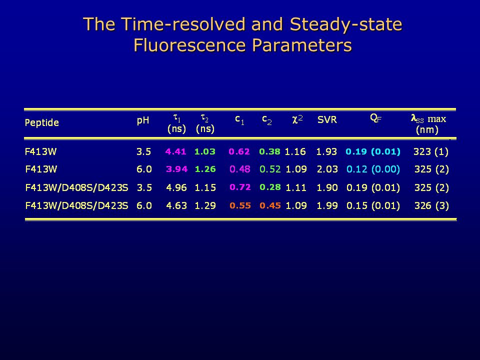
The Time-resolved and Steady-state Fluorescence Parameters

35
Time-resolved and Steady-state Fluorescence Parameters

36
The Trigger Residues

37
The Topology of the Trigger Motif

38
Possible Implications for the in vivo Mechanism of Activation H1 Docking site Trigger

39
Summary and Conclusions These observations confirm the proposed pH-activated trigger mechanism of colicin E1 Asp Ser mutations disrupted criticall H-bonds within the tirgger motif Elevated binding, insertion, and channel activities at near-neutral pH Shift in the helix-to-coil transition of the trigger motif toward random Coil-like conformational state for helix 4

40
Acknowledgements Advisor Dr. A. R. Merrill Examining Committee Dr. G. Harauz Dr. P. D. Josephy Dr. M. Palmer Colleagues in the Merrill Laboratory Tanya Brodeur Susan Yates Tania Roberts Gerry Prentice* Dave Teal Zahir Hussein *Special thanks Advisory Committee Dr. G. Harauz Dr. F. J. Sharom

Similar presentations


- Application to Voltage-Gated K + Channels Lauriane Angué 1,2 Stephen J. Tucker 1 / Mark I. Wallace.>")

>")
