Download presentation
Presentation is loading. Please wait.

2
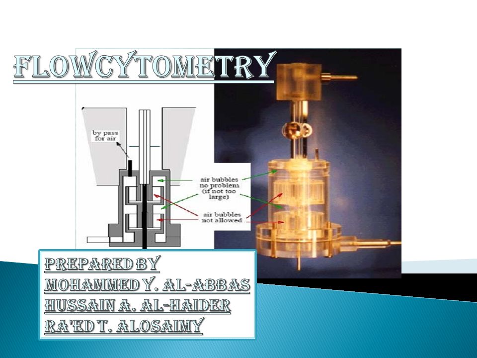
Flow cytometry is a technique for counting, examining, and sorting microscopic particles suspended in a stream of fluid.

3
When the laser beam strikes the stream, the majority of the photons will pass through unobstructed. Some of these photons will diverge slightly, primarily via light diffraction, from their path as they contact the membranes of passing cells.

4
The result will be: Forward scatter(FSC): is proportional to cell size; the bigger the cell, the more light is scattered. Side scatter(SSC): is proportional to cell complexity; the more organelles inside the cytoplasm, the more light scatter.
: is proportional to cell complexity; the more organelles inside the cytoplasm, the more light scatter..")
5
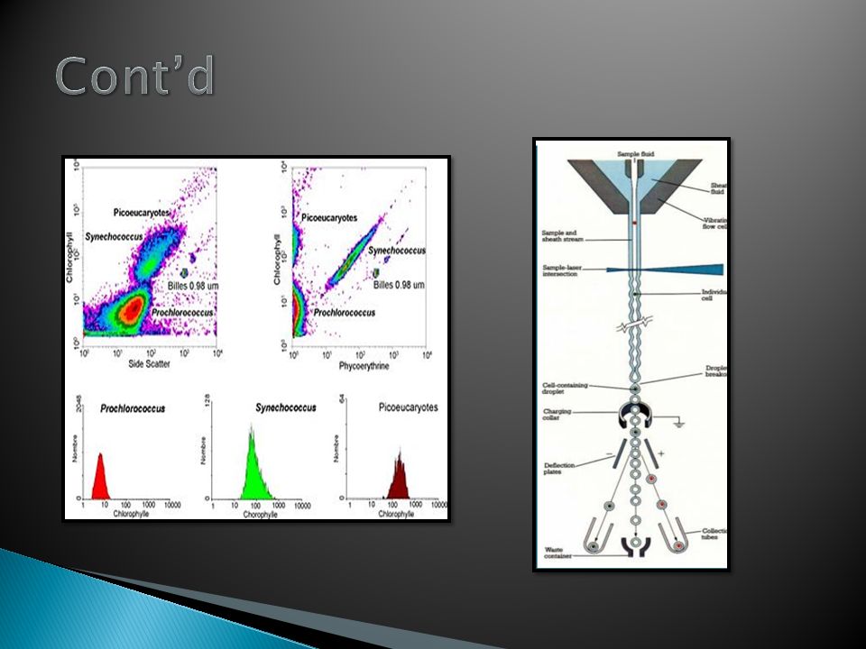
1. liquid stream (sheath fluid) 2. a light source 3. filter or prism 4. detector and Analogue to Digital Conversion (ADC) system 5. a computer for analysis of the signals. Fl ow Ce ll La se r Be a m FS Sen sor Fluore scenc e Picku p Lens SS Se ns or FL 1 Se ns or 52 5B P FL 2 Se ns or 57 5B P FL 3 Se ns or 62 0B P FL 4 Se ns or 67 5B P 48 8D L 48 8B K 55 0D L 60 0D L 64 5D L
 system 5. a computer for analysis of the signals. Fl ow Ce ll La se r Be a m FS Sen sor Fluore scenc e Picku p Lens SS Se ns or FL 1 Se ns or 52 5B P FL 2 Se ns or 57 5B P FL 3 Se ns or 62 0B P FL 4 Se ns or 67 5B P 48 8D L 48 8B K 55 0D L 60 0D L 64 5D L.")
6
No reagents or probes required (Structural) ◦ Cell size(Forward Light Scatter) ◦ Cytoplasmic granularity(90 degree Light Scatter) ◦ Photsynthetic pigments Reagents are required. ◦ Structural DNA content DNA base ratios RNA content ◦ Functional Surface and intracellular receptors. DNA synthesis DNA degradation (apoptosis) Cytoplasmic Ca++ Gene expression IntrinsicExtrinsic
 Cytoplasmic Ca++ Gene expression IntrinsicExtrinsic.")
7
First, let’s talk about how the sample is delivered to the laser. It is important that particles or cells are passed through the laser beam one at a time. Most flow cytometers accomplish this by injecting the sample stream containing the cells into a flowing stream of sheath fluid or saline solution. The sample stream becomes compressed to roughly one cell in diameter. This is called hydrodynamic focusing. As a cell passes through the laser, it will refract or scatter light at all angles. Forward scatter, or low- angle light scatter, is the amount of light that’s scattered in the forward direction as laser light strikes the cell. forward scatter is roughly proportional to the size of the cell.

8
Light is quantified by a detector that converts intensity into voltage. In most cytometers, a blocking bar (called an obscuration bar) is placed in front of the forward scatter detector. As a cell crosses the laser, light is scattered around the obscuration bar and is collected by the detector. Small cells produce a small amount of forward scatter and large cells produce a large amount of forward scatter, the magnitude of the voltage pulse recorded for each cell is proportional to the cell size. If we plot a histogram of these data, smaller cells appear toward the left and larger cells appear toward the right.
 is placed in front of the forward scatter detector. As a cell crosses the laser, light is scattered around the obscuration bar and is collected by the detector. Small cells produce a small amount of forward scatter and large cells produce a large amount of forward scatter, the magnitude of the voltage pulse recorded for each cell is proportional to the cell size. If we plot a histogram of these data, smaller cells appear toward the left and larger cells appear toward the right..")
9
Light scattering at larger angles, for example to the side, is caused by granularity and structural complexity inside the cell. This side-scattered light is focused through a lens system and is collected by a separate detector, usually located 90 degrees from the laser’s path. The signals collected by the side- scatter detector can be plotted on one dimensional histograms like we saw for forward scatter. A scatter plot using forward and side scatter data from a typical peripheral blood cell run. include lymphocytes which are small cells possessing low internal complexity; monocytes which are medium- sized cells with slightly more internal complexity, and neutrophils and other granulocytes which are large cells that have a lot of internal complexity.

10
One of the most common ways to study cellular characteristics using flow cytometry involves the use of fluorescent molecules such as fluorophore-labeled antibodies the labeled antibody is added to the cell sample. The antibody then binds to a specific molecule on the cell surface when laser light of the right wavelength strikes the fluorophore, a fluorescent signal is emitted and detected by the flow cytometers The fluorescent light coming from labeled cells as they pass through the laser then it is directed through a series of filters and mirrors delivered to the appropriate detectors. Fluorescence data is collected in generally the same way as forward and side scatter data. In a population of labeled cells, some will be brighter than others. As each cell crosses the path of the laser, a fluorescence signal is generated. The fluorescent light is then directed to the appropriate detector where it is translated into a voltage pulse proportional to the amount of fluorescence emitted. All of the voltage pulses are recorded and can be presented graphically.

12
1. molecular biology 2. pathology 3. immunology 4. medicine (especially in transplantation, hematology, tumor immunology and chemotherapy, genetics and sperm, sorting in IVF). 5. plant biology
. 5. plant biology.")
13
Flow Cytometry and sorting

Similar presentations


>")

 of blood...>")

 Optical Light Scatter and Flow Cytometry.>")


 Dr. Hayder Kh. Q. Ali 1.>")
