Download presentation
Presentation is loading. Please wait.

1
Immunologic Laboratory Tests Kristine Krafts, M.D.

2
Agglutination reactions DAT IAT Immunofluorescence ELISA Western blot Flow cytometry Immunologic Lab Tests Outline

3
What does it measure? Where does the Ag-Ab interaction occur? How is the Ag-Ab complex detected? Things to Remember About Each Test

4
Agglutination reactions Immunologic Lab Tests Outline

5
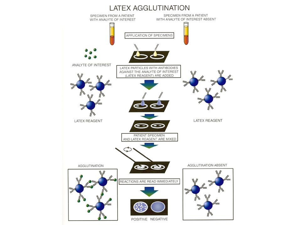
Detection of Ag or Ab in patient specimen Examples: testing for antibodies to infectious agents testing for Hemophilus influenzae type B capsular antigen in CSF Agglutination Reactions: Purpose

6
Use particles coated with Ag or Ab Add patient’s serum (containing Ab or Ag) See if particles clump Agglutination Reactions: Method
 See if particles clump Agglutination Reactions: Method")
9
Clumping = patient has the antibody (or antigen) Agglutination Reactions: Interpretation
 Agglutination Reactions: Interpretation")
10
negativepositive

11
Agglutination reactions DAT Immunologic Lab Tests Outline

12
Detection of Ab (or complement) on patient’s red cells Also called the direct Coombs Test Performed in patients with hemolytic anemia DAT: Purpose
 on patient’s red cells Also called the direct Coombs Test Performed in patients with hemolytic anemia DAT: Purpose")
13
Use patient’s red cells (coated with Ab) Add anti-human globulin (AHG) (Coombs reagent) Look for agglutination DAT: Method
 Add anti-human globulin (AHG) (Coombs reagent) Look for agglutination DAT: Method")
14
patient red cells+AHG=agglutination

15
Clumping = patient red cells are coated with antibody and/or complement DAT: Interpretation

16
Agglutination reactions DAT IAT Immunologic Lab Tests Outline

17
Detection of antibodies to red cell antigens Also called the indirect Coombs Test Performed as part of pre-transfusion testing antibody screen cross-match IAT: Purpose

18
Use patient serum (containing Ab) Add donor RBCs (coated with Ag) Add anti-human globulin (Coombs reagent) Look for agglutination IAT: Method
 Add donor RBCs (coated with Ag) Add anti-human globulin (Coombs reagent) Look for agglutination IAT: Method")
19
patient Ab +AHG = agglutination donor RBC = Ab-coated donor RBC +

20
patient serum (without red cell Ab) AHG reagent RBC (with red cell Ag) patient serum (with red cell Ab) AHG reagent RBC (with red cell Ag) ANTIBODY SCREENING no agglutination (negative test) agglutination (positive test)
 AHG reagent RBC (with red cell Ag) patient serum (with red cell Ab) AHG reagent RBC (with red cell Ag) ANTIBODY SCREENING no agglutination (negative test) agglutination (positive test)")
21
Clumping = patient has an antibody to the donor (or reagent) red cells IAT: Interpretation
 red cells IAT: Interpretation")
22
Agglutination reactions DAT IAT Immunofluorescence Immunologic Lab Tests Outline

23
Detection of a specific antigen or antibody in a histologic specimen Examples: detection of bacterial organisms detection of antigen-antibody complexes Immunofluorescence: Purpose

24
Fix specimen on slide Add antibody specific for the desired antigen Look for fluorescence Fix specimen on slide Add antibody specific for the desired antigen Add second antibody Look for fluorescence DirectIndirect Immunofluorescence: Methods

30
Fluorescence = patient has the antigen Immunofluorescence: Interpretation

31
Agglutination reactions DAT IAT Immunofluorescence ELISA Immunologic Lab Tests Outline

32
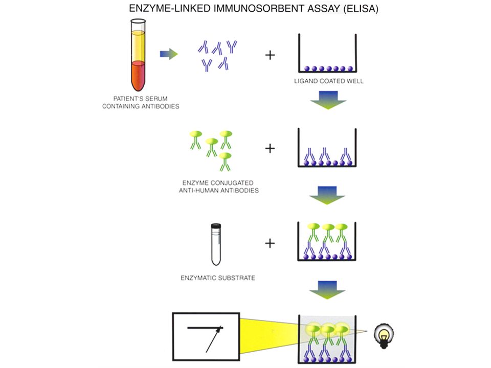
Detection of antibodies in patient specimen Examples: home pregnancy tests HIV tests tests for some coagulation factors, cytokines, and autoantibodies ELISA: Purpose

33
Add patient specimen to well coated with ligand Add AHG with enzyme attached Add substrate Measure color change ELISA: Method

35
Color change = patient has the antibody ELISA: Interpretation

36
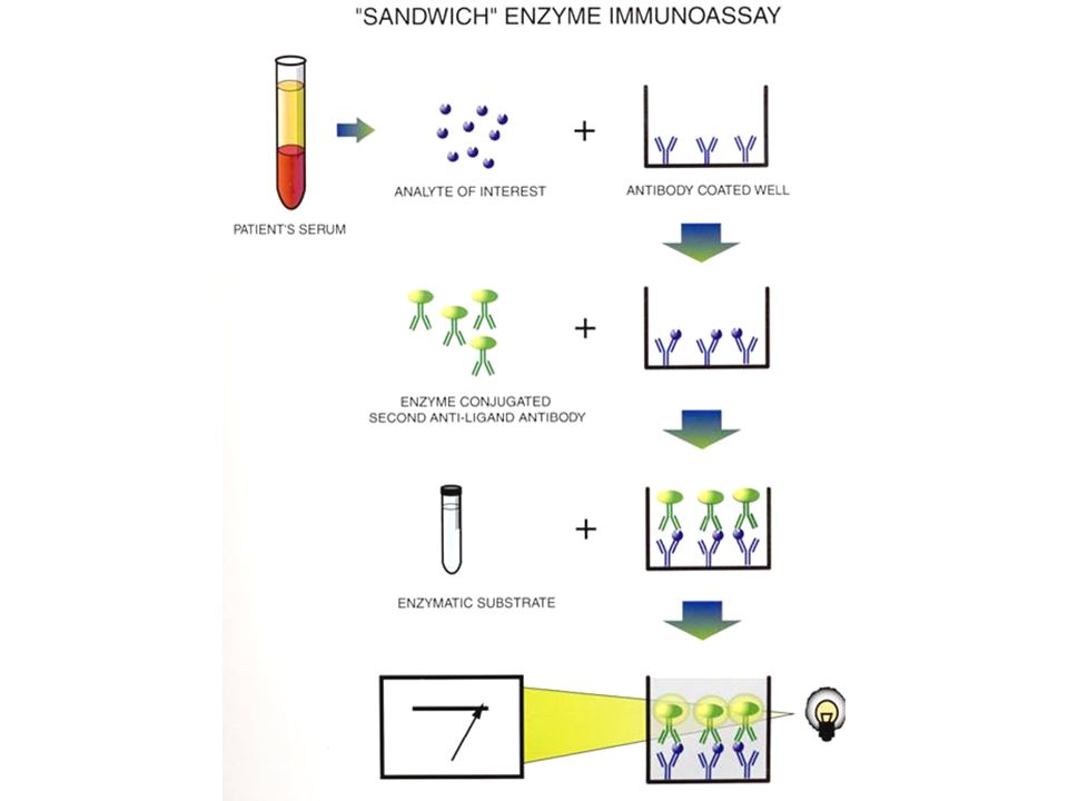
Sandwich immunoassay detects antigen (not antibody) coat well with antibody rest is like ELISA Radioimmunoassay detects antibody or antigen detector is a radioactive substance otherwise like ELISA or sandwich immunoassay ELISA: Variations
 coat well with antibody rest is like ELISA Radioimmunoassay detects antibody or antigen detector is a radioactive substance otherwise like ELISA or sandwich immunoassay ELISA: Variations")
38
Agglutination reactions DAT IAT Immunofluorescence ELISA Western blot Immunologic Lab Tests Outline

39
Detection of antibodies in patient specimen Most common example: HIV test Western Blot: Purpose

40
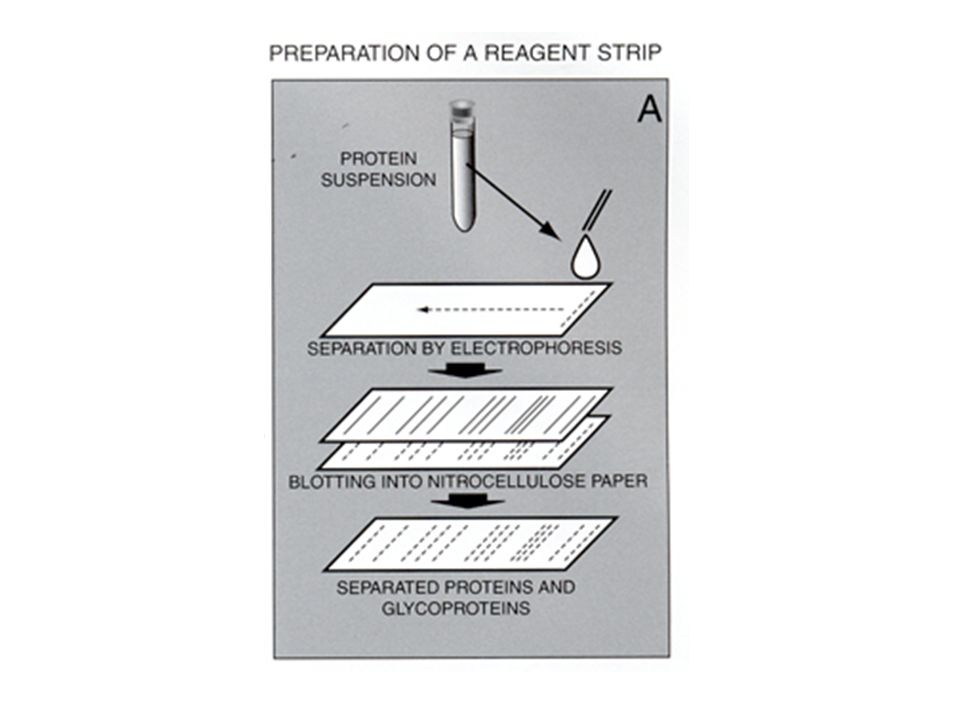
Make a protein suspension of the target of the antibody you’re looking for (e.g., HIV) Electrophorese the suspension onto a little gel strip Apply the patient’s specimen (containing antibodies) to the strip Add AHG that has an enzyme attached Add substrate and look for bands Western Blot: Method
 Electrophorese the suspension onto a little gel strip Apply the patient’s specimen (containing antibodies) to the strip Add AHG that has an enzyme attached Add substrate and look for bands Western Blot: Method")
43
Bands on strip = patient has antibodies to corresponding proteins Western Blot: Interpretation

44
Enough bands = patient is “positive”

45
Agglutination reactions DAT IAT Immunofluorescence ELISA Western blot Flow cytometry Immunologic Lab Tests Outline

46
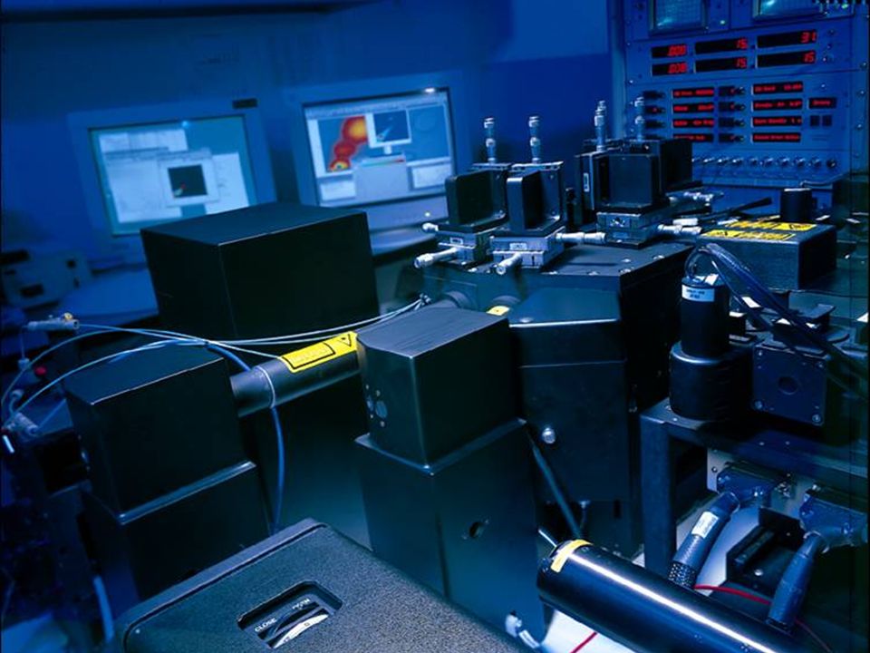
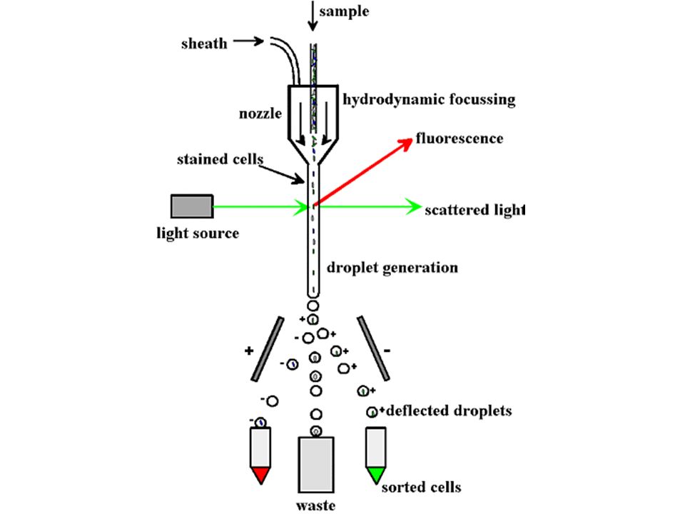
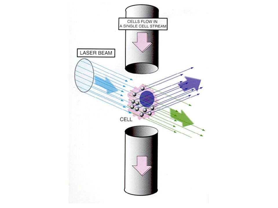
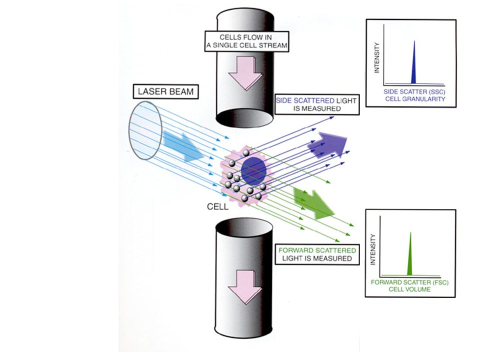
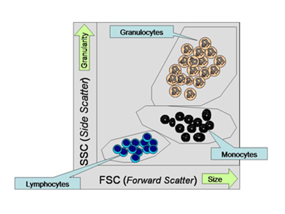
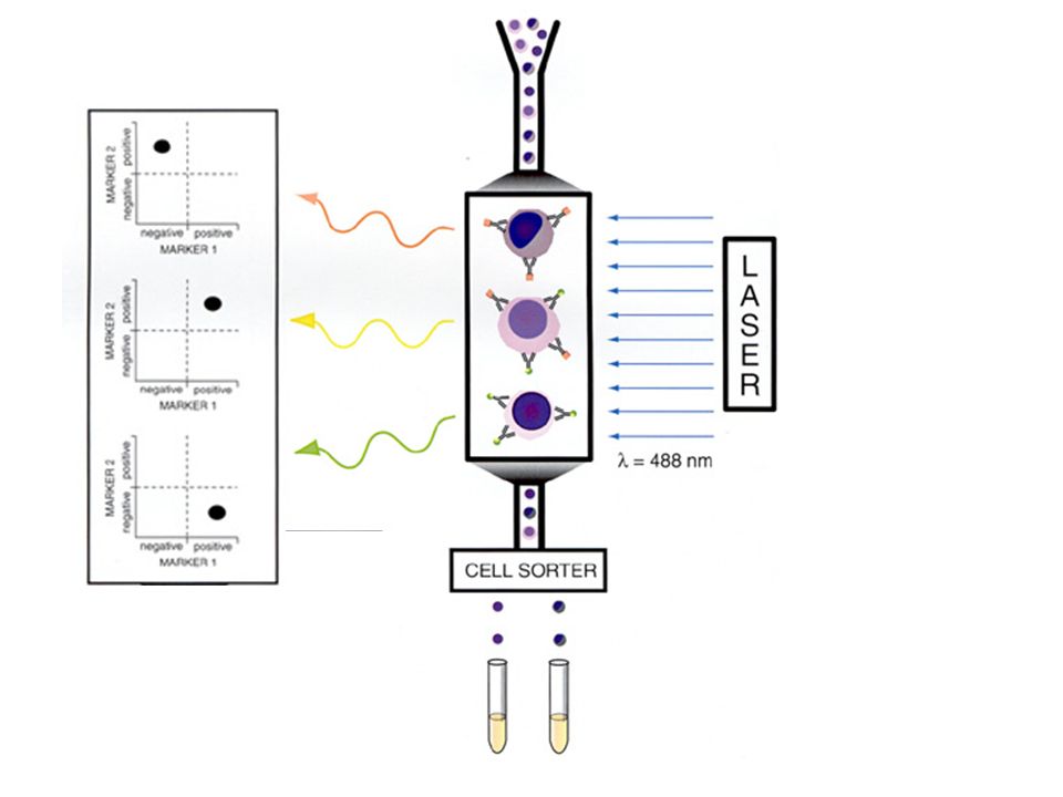
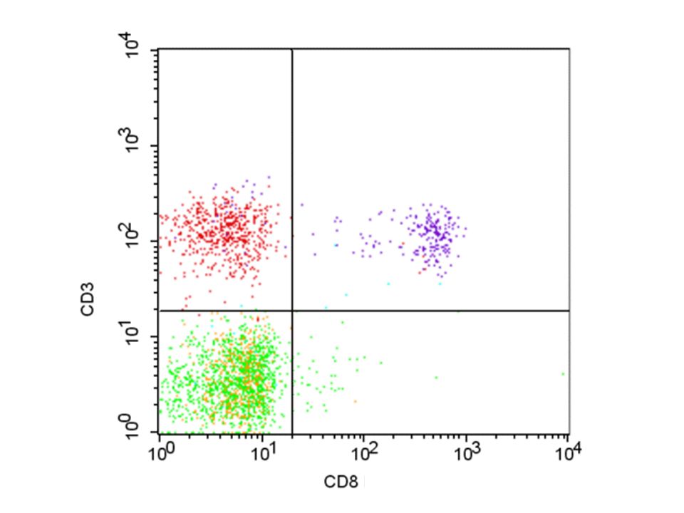
Characterization of cell size, complexity, antigens Examples: diagnosis of leukemia and lymphoma determination of CD4/CD8 counts in patients with HIV Flow Cytometry: Purpose

58
Complicated! Combine size, complexity and antigen expression data to come up with meaningful description of cells. Flow Cytometry: Interpretation

Similar presentations


>")


>")


 Or to precipitate.>")


