Download presentation
Presentation is loading. Please wait.

1
The Eye

3
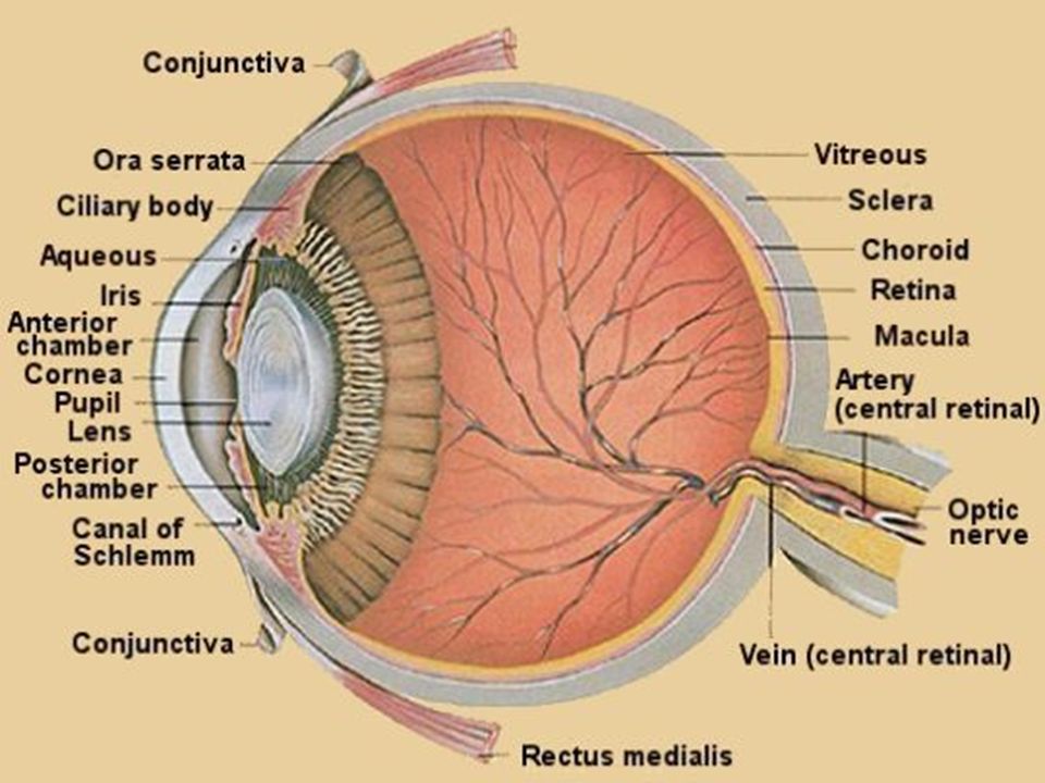
Major Parts of the Eye Cornea - Iris -
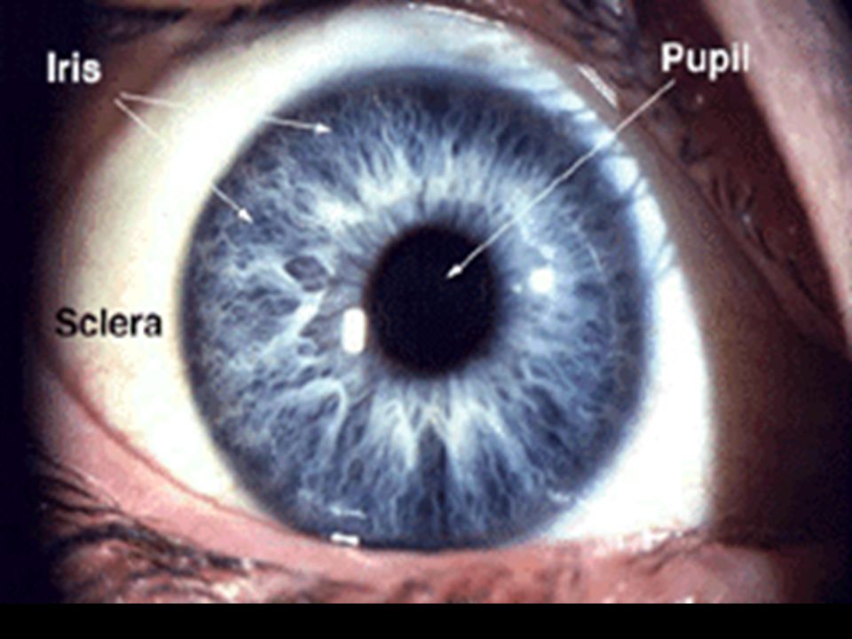
clear, transparent portion of the outer coat of the eyeball through which light passes to the lens. Iris - gives our eyes color smooth muscles that open or close an apeture called the PUPIL.

4
Pupil - Opening in middle of iris. Lens
Eye's primary light-focusing structure located behind pupil. Muscles attach to it that contract causing lens to contract enabling lens to focus light.

6
Draw this

7
Aqueous vs Vitreous Humour
aqueous is a clear, watery anterior and posterior chamber (anterior compartment)
")
9
vitreous is a transparent, colorless mass of soft, gelatinous material
behind the lens - posterior “compartment” Note: your book only mentions anterior compartment (aqueous) and posterior compartment (vitreous)
 and posterior compartment (vitreous)")
11
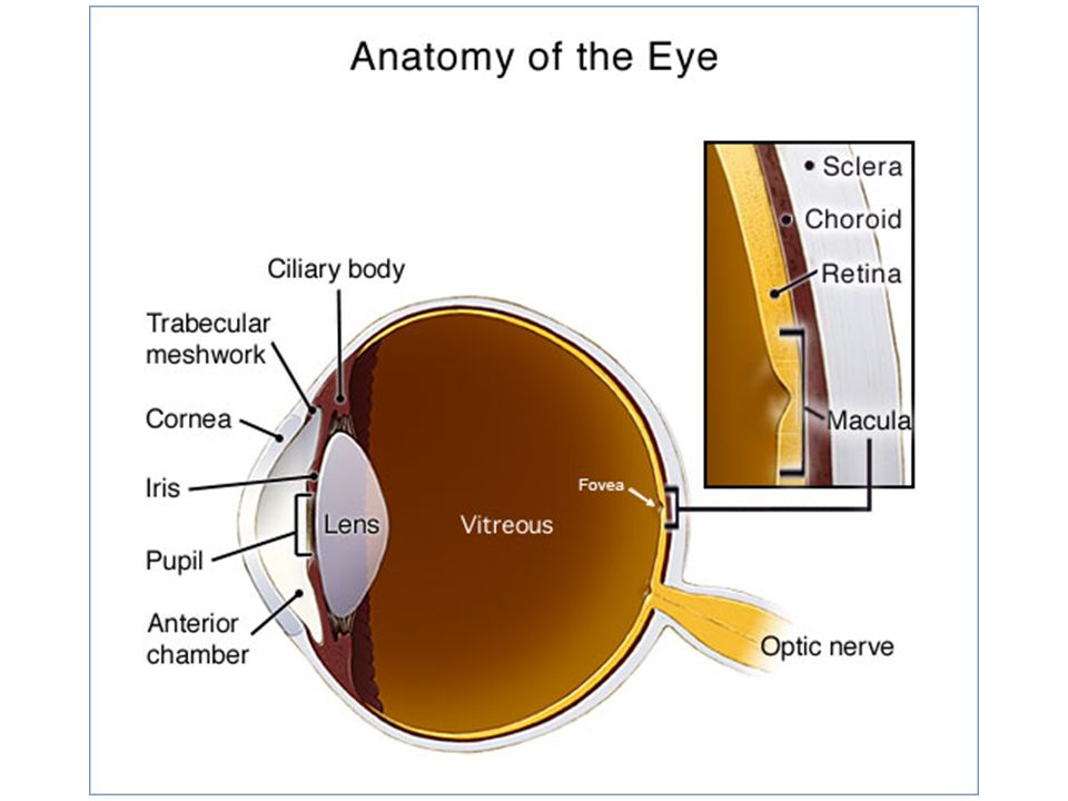
Eye layers Conjunctiva Sclera Choroid
is a clear membrane covering the white of the eye (sclera) Sclera is the white of the eye Choroid carries blood vessels, is the inner coat between the sclera and the retina
 Sclera. is the white of the eye. Choroid. carries blood vessels, is the inner coat between the sclera and the retina.")
12
Retina A thin membrane on back of eye that contains light-sensitive receptors (rods & cones).
.")
14
Cones Rods Light receptors that are sensitive only in bright light.
They can distinguish form & colour very well. Rods Light receptors that are sensitive in dim light. They cannot distinguish colour & therefore, you only see shades of gray in dim light.

16
Fovea is the region of keenest vision.
Macula is a small area in the retina that provides our most central, acute vision. Small depressed area in centre of macula directly in line with centre of cornea & lens is called the FOVEA. Fovea is the region of keenest vision. Light-sensitive cones are concentrated in fovea.

18
Optic Nerve Carries nerve impulses to the brain created when light strikes rods & cones.

19
Blind spot the point on the retina where the approximately 1 million axons converge on the optic nerve, there are no rods or cones. This spot, called the blind spot, is thus insensitive to light.

20
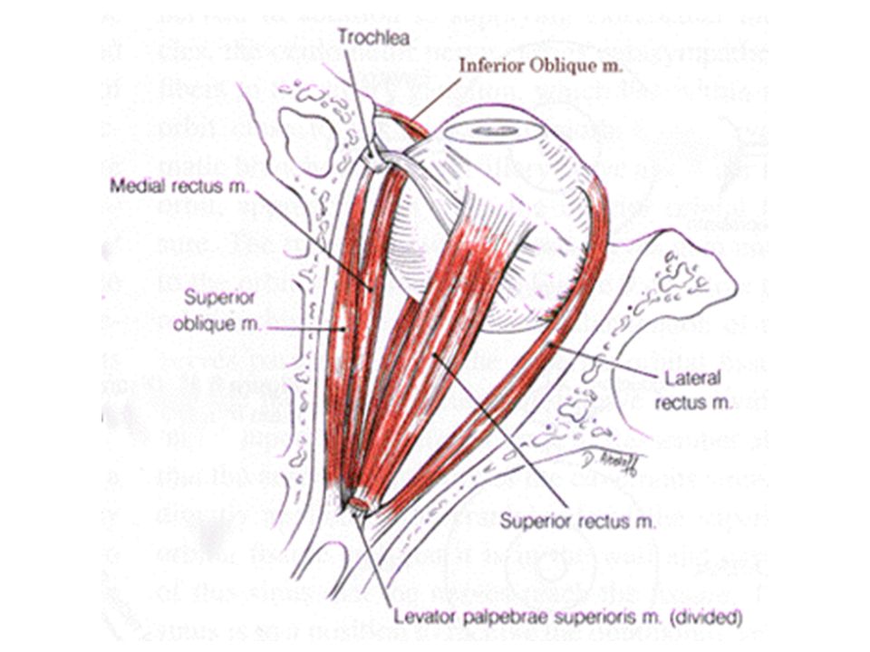
Eye Muscles Movement of the eye is controlled by six muscles:
Medial rectus- lies on the inner side Lateral rectus- lies on the outer side Superior rectus- lies above the eye Inferior rectus- lies below the eye Superior oblique- lies above and runs obliquely inferior oblique- lies below and runs obliquely

23
Why do we see? Light reflected from object enters your eye.
Cornea & lens bend light rays together. Rays cross & focus on retina which appears upside down & backward. Light strikes rods & cones creates nerve impulses that are carried by optic nerve to brain to be interpreted. Retinal image reversed in brain so we see object right side up.

24
Retinal Layers Composed of three layers: 1. Ganglion cell layer
2. Bipolar cell layer 3. Rod and cone cell layer

25
rods and cones - actual photoreceptors
ganglion cells - transmit to the brain; the axons of these ganglion cells make up the optic nerve. bipolar cells - process input from photoreceptors and transmit the signal to ganglion cells.

Similar presentations


 This tough layer creates the white of the eye except in the front where it forms the transparent cornea. The.>")

 17 April 2017 Biology Matters textbook page 281 Concept Map.>")


. 2. *The sclera is the.>")
 than.>")


