Download presentation
Presentation is loading. Please wait.

1
Chest Pain

3
What must we learn? 1. Epidemiology 2. Pathophysiology 3. Diagnostic Approach 4. Diagnostic Table 5. Management and Disposition

4
Case آقاي 42 ساله با شكايت درد قفسه سينه از شب گذشته به بخش اورژانس مراجعه نموده است. بيمار ذكر مينمايد از شب گذشته درد هايي در سمت چپ قفسه سينه دارد كه در حدود 1 دقيقه طول ميكشد ماهيت تيزدارد به محل قفسه سينه دارد كه در حدود 1 دقيقه طول ميكشد ماهيت تيزدارد به محل خاصي تير نميكشد و در حالت استراحت و فعاليت تفاوتي نمينمايد. درد ها از شب گذشته مكررا تكرار شده همراه با انها تهوع استفراغ و تعريق را ذكر شب گذشته مكررا تكرار شده همراه با انها تهوع استفراغ و تعريق را ذكر نمينمايد.درد با تنفس عميق تشديد ميشود. بيمار محل درد را نقطه اي در زير نمينمايد.درد با تنفس عميق تشديد ميشود. بيمار محل درد را نقطه اي در زير نيپل چپ نشان ميدهد. بيمار سابقه بيماري خاصي را ذكر نميكند بجز سرماخوردگي در چند روز اخير همچنين داروي خاصي نيز مصرف نمينمايد. سابقه مصرف سيگار الكل و مواد مخدر را هم نميدهد. در معاينه ديسترس خمصي ندارد BP= 110/85 mmHg PR=92/ Min RR= 16/Min T= 37.1 c ( oral ) در معاينه بجز تندرنس موضعي بر روي ناحيه Apical نكته حاصي ندارد. تشخيس شما چيست؟
 در معاينه بجز تندرنس موضعي بر روي ناحيه Apical نكته حاصي ندارد. تشخيس شما چيست؟.")
5
Epidemiology > 5 million/year patients of Emergency rooms > 5 million/year patients of Emergency rooms A symptom caused by several life threatening disease A symptom caused by several life threatening disease Accurately discerning the correct diagnosis and treatment of a chest pain is one of the most difficult tasks

6
Epidemiology Catastrophic causes are: Acute coronary syndromes (ACS) Aortic dissection Pulmonary embolus Pneumothorax Pericarditis with tamponade Esophageal rupture ACS is the most significant potential diagnosis Emergency physicians reportedly have missed 3% to 5% of MI accounting for 25% of malpractice Most of the chest pains presenting to the ED have a benign origin
 Aortic dissection Pulmonary embolus Pneumothorax Pericarditis with tamponade Esophageal rupture ACS is the most significant potential diagnosis Emergency physicians reportedly have missed 3% to 5% of MI accounting for 25% of malpractice Most of the chest pains presenting to the ED have a benign origin")
7
Pathophysiology Afferent fibers from the heart, lungs, great vessels, and esophagus enter the same thoracic dorsal ganglia. Dorsal segments overlap three segments above and below a level Disease of a thoracic origin can produce pain anywhere from the jaw to the epigastrium Radiation of pain is explained by somatic afferent fibers synapsing in the same dorsal root ganglia

8
Pathophysiology The quality of visceral chest pain has been described as: Burning Aching Stabbing Pressure

9
Typical ischemic chest pain 1. Retrosternal chest pressure 1. Burning or heaviness 2. Radiating occasionally to neck, jaw, epigastrium, shoulders, or left arm. 2. Precipitated by exercise, cold weather, or emotional stress 3. Duration 2-10 minutes

10
Atypical Chest Pain 1. Pleuritic chest pain 2. In the middle or lower abdominal region. 3. Localized at the tip of one finger, particularly over the left ventricular ( LV ) apex. 4. Reproduced with movement or palpitation of chest wall or arms. 5. Constant for many hours 6. Very brief episodes 7. Radiates into the lower extremities
 apex. 4. Reproduced with movement or palpitation of chest wall or arms. 5. Constant for many hours 6. Very brief episodes 7. Radiates into the lower extremities.")
11
Diagnostic Approach Rapid Assessment and Stabilization The first questions: 1. 1. What are the life-threatening possibilities in this patient 2. 2. Must I intervene immediately? Assessing the patient's appearance and vital signs Tension Pneumothorax

12
Diagnostic Approach 80% to 90% of information pertinent to the differential diagnosis is obtained by the history, physical examination and ECG. 1. 1. History 2. 2. Physical Examination 3. 3. Ancillary Studies

13
History A useful initial approach is to classify patients into three categories: Chest wall pain Pleuritic or respiratory chest pain Visceral chest pain Pain, Associated syncope/near-syncope, Associated dyspnea, Associated hemoptysis, nausea and vomiting

14
Differential Diagnosis 1. Cardiac : Angina unstable angina unstable angina Acute MI Acute MI Pericarditis Pericarditis 2. vascular: Aortic Dissection Pulmonary Embolism Pulmonary Embolism pulmonary hypertension pulmonary hypertension 3. pulmonary: Pleuritis and/or pneumonia Tracheobronchitis Tracheobronchitis Spontaneous pneuomothorax Spontaneous pneuomothorax 4. GI: Esophageal reflux Peptic ulcer Peptic ulcer Gallbladder disease Gallbladder disease Pancreatitis Pancreatitis 5. Musculoskeletal: Costochondritis Cervical disc disease Cervical disc disease 6. Infectious: Herpes Zoster 7. Psychological: Panic Disorder

15
Ancillary Studies Chest radiograph and 12-lead ECG ECG should be performed in all patients 30 years old and older within 10 min of arrival

16
Ancillary Studies; Serum Markers CK-MB values in healthy controls may be up to L and up to 5% of total CK.

17
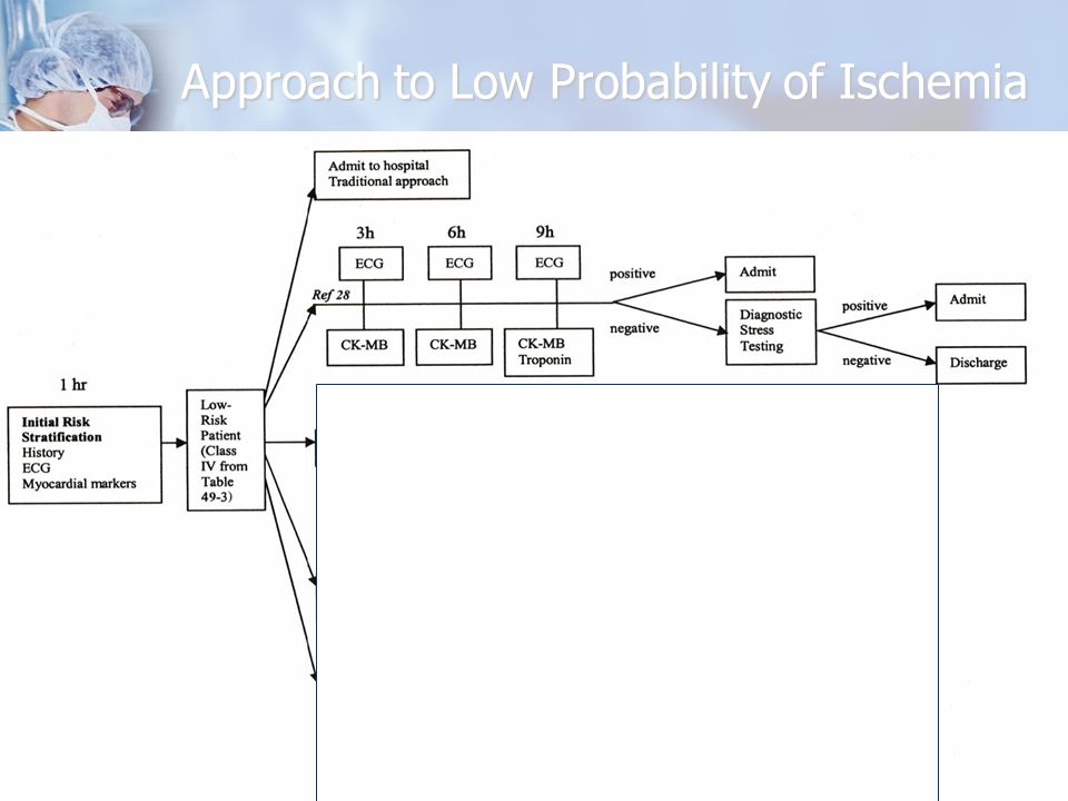
Approach to Low Probability of Ischemia 1. 1. History alone is not adequate to exclude the presence of acute ischemia 2. 2. The goal should always be "zero tolerance" for missed AMI.

18
Approach to Low Probability of Ischemia

20
Initial assessment of critical diagnoses

Similar presentations


 Wheezing Cough Blood-streaked sputum (hemoptysis)>")

 Risk factors (associated diseases) Physical signs Investigations Complications and treatment.>")


 pericarditis (irritation of pericardium) thoracic aortic dissection.>")
 Definition of ACS Signs and symptoms of ACS Gender and age related difference in ACS Pathophysiology.>")