Download presentation
Presentation is loading. Please wait.

1
Efficient Generation of Human iPSCs by a Synthetic Self-Replicative RNA
Naohisa Yoshioka,1 Edwige Gros, Hai-Ri Li, Shantanu Kumar, Dekker C. Deacon, Cornelia Maron, Alysson R. Muotri, Neil C. Chi, Xiang-Dong Fu, Benjamin D. Yu and Steven F. Dowdy1, cell stem cell. Presented by S. Andleeb Fatima Ph.D (1st sem.) Deptt. Of Zoology
 Deptt. Of Zoology.")
2
Outline Introduction VEE-RNA Generation of iPSCs: procedure
Generation of iPSCs by Retroviruses, Sendai virus, RF-mRNA VEE-RNA Structure Engineering GFP ORF Reprogramming factors ORFs Generation of iPSCs: procedure Characterization of iPSC clones Differentiation of clones In-vitro In-vivi

3
eliminates integrative DNA-based approaches for use in RMT
INTRODUCTION The generation of human induced pluripotent stem cells (hiPSCs) has great potential for the development of personalized stem cell therapies. 1 Generation of hiPSCs by retroviral expression of four reprogramming factors opened the potential for regenerative medicine therapies based on patient-specific, personalized stem cells. eliminates integrative DNA-based approaches for use in RMT insertional mutagenic potential of retroviruses combined with the potential for latent reprogramming factor gene activation, especially c-MYC,
 has great potential for the development of personalized stem cell therapies. 1. Generation of hiPSCs by retroviral expression of four reprogramming factors opened the potential for regenerative medicine therapies based on patient-specific, personalized stem cells. eliminates integrative DNA-based approaches for use in RMT. insertional mutagenic potential of retroviruses combined with the potential for latent reprogramming factor gene activation, especially c-MYC,")
4
appear inherently safer methods for future clinical applications
Several methods based on DNA, RNA, miRNAs, and proteins have been developed to generate integration-free iPSCs 2 Of all these methods, RNA-based iPSC approaches using Sendai virus, miRNAs, and mRNA transfection avoid potential integration problems associated with DNA-based approaches appear inherently safer methods for future clinical applications 3

5
Using Sendai virus (SV)
Expression of pluripotent factors by infection with Sendai virus, a negative-sense, ssRNA virus (does n’t go through a DNA intermediate) offers an efficient iPS approach in the absence of concerns for integration into the genome. Due to persistent SV replication in iPSC clones, this approach requires a negative selection step followed by one or more recloning steps from the single-cell level to isolate virus-free iPSCs. A temperature-sensitive mutant of Sendai virus is a successful alternative method to remove the virus though it requires a higher biosafety due to production of infectious virus particles.
 offers an efficient iPS approach in the absence of concerns for integration into the genome. Due to persistent SV replication in iPSC clones, this approach requires a negative selection step followed by one or more recloning steps from the single-cell level to isolate virus-free iPSCs. A temperature-sensitive mutant of Sendai virus is a successful alternative method to remove the virus though it requires a higher biosafety due to production of infectious virus particles.")
6
RF RF(Reprogramming factor) mRNA
One of more-promising non-DNA-based iPS approaches transfection of four individual RF mRNAs generated by in vitro transcription . due to the rapid degradation of RF-mRNAs it requires repetitive daily transfection of four individual mRNAs into the same target cells over the 14-day reprogramming period.

7
Although both Sendai virus and mRNA transfection approaches generate iPSCs, there remains a need for a simple, highly reproducible, non-DNA-based approach to generate hiPSCs

8
(4) can be selectively retained and degraded in a controlled fashion.
To develop an RNA-based iPSC generation strategy such an approach was focused that (1) utilizes a single RNA species capable of self-replicating for a limited number of cell divisions, thereby reducing the number of transfections; (2) is capable of encoding at least four reprogramming factor open reading frames (ORFs); (3) consistently expresses all reprogramming factor genes at high threshold levels over multiple cellular divisions (4) can be selectively retained and degraded in a controlled fashion.
 utilizes a single RNA species capable of self-replicating for a limited number of cell divisions, thereby reducing the number of transfections; (2) is capable of encoding at least four reprogramming factor open reading frames (ORFs); (3) consistently expresses all reprogramming factor genes at high threshold levels over multiple cellular divisions. (4) can be selectively retained and degraded in a controlled fashion.")
9
VEE RNA To express all four reprogramming factors, a noninfectious (nonpackaging), self-replicating Venezuelan equine encephalitis (VEE) virus RNA replicon was modified. (also being investigated as an expression platform for vaccine development ) The VEE replicon: a positive-sense, ssRNA mimics cellular mRNA with a 5’ cap and poly(A) tail does not utilize a DNA intermediate, so there is no potential for genomic integration
, self-replicating Venezuelan equine encephalitis (VEE) virus RNA replicon was modified. (also being investigated as an expression platform for vaccine development ) The VEE replicon: a positive-sense, ssRNA. mimics cellular mRNA with a 5’ cap and poly(A) tail. does not utilize a DNA intermediate, so there is no potential for genomic integration.")
10
VEE RNA; structure To develop a single RNA iPSC generation approach, we modified a polycistronic, self-replicative RNA system that would consistently express the reprogramming factors over multiple cellular divisions. VEE RNA encodes four nonstructural replication complex proteins (nsPs) as a single ORF in the 5’ end of the RNA that is separated from the viral structural protein ORFs in the 3’ end.
 as a single ORF in the 5’ end of the RNA that is separated from the viral structural protein ORFs in the 3’ end.")
11
Analysis of exogenous protein expression by VEE RNA
It has the ability to express exogenous proteins by replacing the 3’ structural protein ORFs with GFP To evaluate the VEE RNA replicon in primary human fibroblasts, we replaced the 3’ ORF with GFP, followed by an internal ribosomal entry site (IRES) and a Puromycin-resistance gene (Puror).
 and a Puromycin-resistance gene (Puror).")
12
VEE-GFP RNA was produced using either SP6 or T7 RNA pol
VEE-GFP RNA was produced using either SP6 or T7 RNA pol. from a standard in vitro transcription kit followed by 5’ capping, and poly(A) tail addition resulting in a high-yield, full-length, RNA transcript.
 tail addition resulting in a high-yield, full-length, RNA transcript.")
13
Exposure of cells to single-stranded VEE RNA induces a strong interferon (IFN)-α/β innate immune response. To mitigate the innate immune response to VEE-GFP RNA, B18R protein from Western vaccinia virus was utilized that binds to and neutralizes type I IFNs. We compared GFP expression in primary human foreskin fibroblasts (HFFs) transfected with VEEGFP RNA alone or cotransfected with B18R mRNA.
 transfected with VEEGFP RNA alone or cotransfected with B18R mRNA.")
14
Consistent with induction of a strong innate immune response to cells exposed to single-stranded RNA, in the absence of B18R, we observed little-to-no GFP expression 1 day after transfectionn. In contrast, cotransfection of VEE-GFP RNA replicon with B18R mRNA resulted in high levels of GFP expression in HFFs, B18R is required for efficient expression of proteins from the VEE RNA replicon.

15
The generation of iPSCs requires consistent, high-level expression of reprogramming factors for >7 days; To continuously suppress the innate immune response while avoiding daily transfection of B18R mRNA, we prepared conditioned media harvested from human fibroblasts expressing B18R protein (B18R-CM). Therefore, the persistence of the VEE-GFP RNA replicon in human primary fibroblasts over 7 days was examined
. Therefore, the persistence of the VEE-GFP RNA replicon in human primary fibroblasts over 7 days was examined.")
16
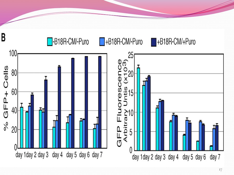
HFFs were cotransfected with VEE-GFP RNA replicon and B18R mRNA (3:1 ratio) on day 0,
then cultured in the presence or absence of 20% B18R-CM (conditioned media) plus/minus puromycin on day 1. Puromycin selection in the presence of B18R-CM resulted in a >90% GFP-positive population, whereas puromycin selection in the absence of B18R-CM resulted in <1% viable GFP cells . The level of GFP expression in the presence of B18R-CM gradually decreased from day 1 to day 4 but then remained steady out to day 7 In contrast, the level of GFP expression in the absence of B18RCM continuously dropped to <10% intensity .
 plus/minus puromycin on day 1. Puromycin selection in the presence of B18R-CM resulted in a >90% GFP-positive population, whereas puromycin selection in the absence of B18R-CM resulted in <1% viable GFP cells . The level of GFP expression in the presence of B18R-CM gradually decreased from day 1 to day 4 but then remained steady out to day 7. In contrast, the level of GFP expression in the absence of B18RCM continuously dropped to <10% intensity .")
18
B18R-CM and puromycin are required for retention of VEE-GFP RNA
B18R-CM and puromycin are required for retention of VEE-GFP RNA. Photographs of GFP expression on day 7

19
VEE GFP replicon persistence was dose dependent on B18R-CM .
Persistence of high levels of GFP expression from VEE-GFP RNA-treated fibroblasts for over a month was noted when continuously cultured in the presence of B18R-CM and puromycin . Taken together, these results showed both the necessity of B18R protein to overcome the VEE RNA-induced innate immune response the ability to selectively retain or degrade the VEE RNA replicon from cells by exposure to or withdrawal from B18R-CM.

20
Development of Self-Replicative VEE RNA to Express Reprogramming Factors
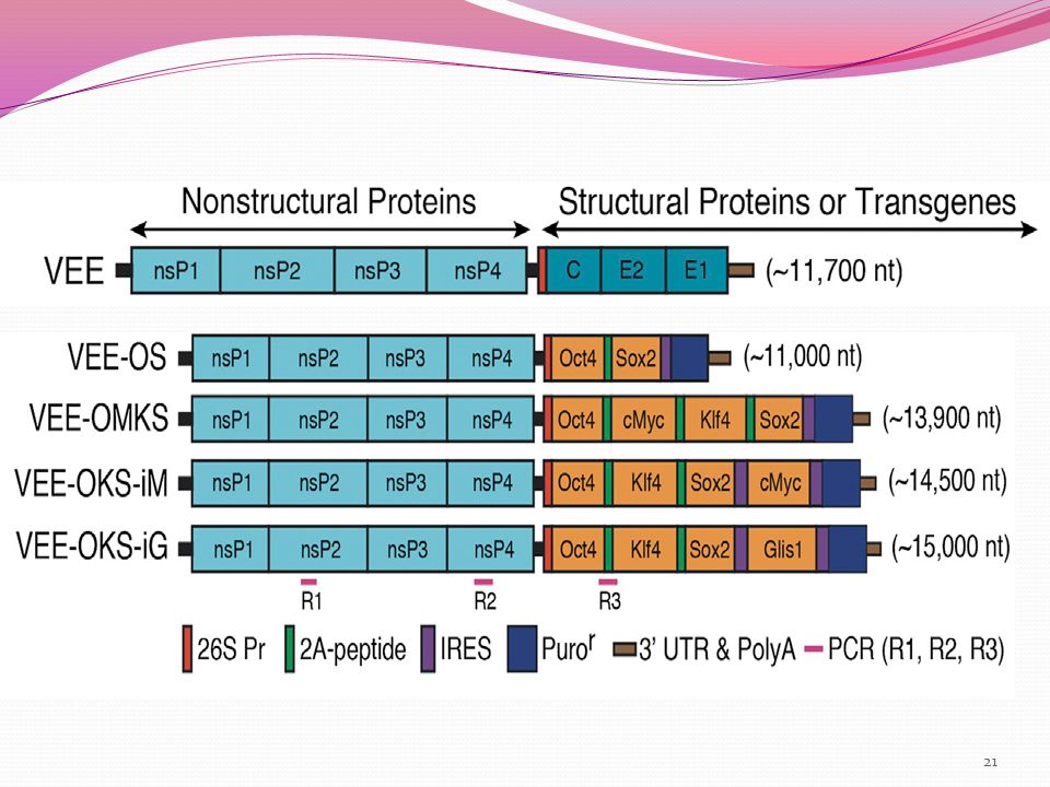
VEE RNA replicon 3’ ORF was engineered to encode four reprogramming factors OCT4, KLF4, SOX2, with c-MYC or GLIS1, (which avoids the potential genomic instability induced by c-MYC.) We generated and compared several VEE-RNA construct configurations using the following nomenclature: VEE-OMKS = OCT4, c-MYC, KLF4, SOX2 separated by internal ribosomal skipping 2A peptides followed by an IRES and Puror ORF; VEE-OKS-iM = OCT4, KLF4, SOX2 separated by 2A peptides followed by an IRES then c-MYC and a second IRES and Puror ORF; VEE-OKS-iG = OCT4, KLF4, SOX2 separated by 2A peptides followed by IRES then GLIS1 and a second IRES and Puror ORF.
 We generated and compared several VEE-RNA construct configurations using the following nomenclature: VEE-OMKS = OCT4, c-MYC, KLF4, SOX2 separated by internal ribosomal skipping 2A peptides followed by an IRES and Puror ORF; VEE-OKS-iM = OCT4, KLF4, SOX2 separated by 2A peptides followed by an IRES then c-MYC and a second IRES and Puror ORF; VEE-OKS-iG = OCT4, KLF4, SOX2 separated by 2A peptides followed by IRES then GLIS1 and a second IRES and Puror ORF.")
22
VEE-RF RNAs were produced by SP6 or T7 in vitro transcription, 5’ capping, and poly(A) tail addition resulting in full-length VEE-OKS-iM RNA, VEE-OMKS RNA, and VEE-OKS-iG RNA . Transfection of various VEE-RF RNA replicons with B18R mRNA into human fibroblasts resulted in high levels of expression of all four reprogramming factors that exceeded reprogramming factor expression levels from retroviruses on day 1 and day 10

23
Together, these observations demonstrated the ability to express four reprogramming factors and a Puro gene from a single, synthetic VEE-RF RNA replicon in primary human cells, while utilizing B18R to block the innate immune response.

24
To develop a VEE-RF RNA replicon-based iPSC protocol, several parameters wereevaluated, including number and timing of VEE-RF RNA transfections, selection for VEE-RF RNA replicon retention by puromycin, and the genetic organization of the VEE-RF RNA replicon. Human HFFs or BJ fibroblasts were cotransfected with VEE-RF RNA replicons and B18R mRNA on day 1, then cultured in the presence of 20% B18R-CM plus puromycin. Based on expression results of VEERF RNAs , we initially compared iPSC generation from either twice (days 1 and 3) or five-times (days 1, 3, 5, 7, and 9) transfections of VEE-OMKS RNA or VEE-OSK-iM RNA.
 or five-times (days 1, 3, 5, 7, and 9) transfections of VEE-OMKS RNA or VEE-OSK-iM RNA.")
25
Strikingly, alkaline phosphatase (AP)-positive staining iPS colonies were only generated from VEE-OKS-iM RNA in both BJ and HFFs, and no iPSC colonies were observed from VEEOMKS RNA
-positive staining iPS colonies were only generated from VEE-OKS-iM RNA in both BJ and HFFs, and no iPSC colonies were observed from VEEOMKS RNA")
26
Reason 1 1. One significant difference between the two RNA vectors is the relative level of c-MYC expression to the other reprogramming factors lower c-MYC levels expressed from the VEE-OKS-iM RNA, where c-MYC is the last ORF with a 5’ IRES, higher levels of c-MYC expressed from the VEE-OMKS RNA non-iPS-generating replicon, where c-MYC is the second ORF and utilizes a 2A ribosome-skipping peptide.

27
An inverse c-MYC expression sensitivity to generating iPSCs using retroviral vectors is previously reported, where high c-MYC levels were correlated with a decreased number of iPSC colonies Using VEE-OKS-iM RNA,conditions were further optimized for iPSC generation and found that B18R-CM was required until the appearance of iPSC colonies on feeding culture, whereas puromycin could be removed at the point of plating onto feeder cultures 2. 2

28
To avoid the potential for genomic instability induced by c-MYC, we also generated a related VEE-OKS-iG RNA construct that substituted GLIS1 for c-MYC . several transfections of either VEE-OKS-iM RNA or VEEOKS- iG RNA in the presence of B18R-CM and puromycin selection over the first 7 days resulted in the highest generation of AP-positive colonies.

29
Starting with one well of a 6-well format ( cells/well), we generally observed >100 iPS colonies per starting well from both HFFs and BJ fibroblasts when using three (or more) VEE-RF RNA transfections . Although iPSCs were generated in the absence of puromycin selection, we observed a substantially reduced efficiency. To further refine the approach, the type of media used during the first 7 days after transfection was changed to either Advanced-DME or Pluriton, both of which resulted in a large number of AP-positive colonies from a single transfection of either VEE-OKS-iM RNA or VEE-OKS-iG RNA in BJ or HFFs (Table 1).
.")
30
Moreover, a single transfection of VEE-OKS-iM RNA or VEE-OKS-iG RNA produced from either SP6 or T7 RNA polymerases into human adult cells of normal human dermal fibroblasts (NHDF-c) (aged 50) and human dermal fibroblasts (HDFs) (aged 58) generated AP-positive colonies with a characteristic iPSC morphology. Thus, methodology was refined to generate iPSCs from a single transfection of the VEE-RF RNA replicon into both newborn and adult human fibroblasts.

31
Characterization of iPSC Clones
1. Retention of ESC morphology through multiple cell divisions >100 iPSC colonies were mechanically isolated from multiple independent VEE-OKS-iM RNA and VEE-OKS-iG RNA protocols and had a >95% success rate for the ability of isolated iPS clones to continuously divide and retain a human embryonic stem cell (hESC) morphology.
 morphology.")
32
2. Expression of stem cell markers
Of the >100 iPS morphologylike clones isolated 30 clones were analysed for expression of stem cell markers by immunofluorescence. All 30 VEE-RF RNA iPS clones analyzed showed strong nuclear staining of endogenous OCT4, SOX2, and NANOG and strong cell surface staining of SSEA4, TRA-1-60, and TRA-1-81, with negative staining of SSEA1 (Figure 2E). In addition, six clones from HDF human adult fibroblasts and three clones from NHDF-c human adult fibroblasts were examined by immunofluorescence and found that all clones expressed TRA-1-60, TRA-1-81, SSEA4, and NANOG but did not express SSEA1.
. In addition, six clones from HDF human adult fibroblasts and three clones from NHDF-c human adult fibroblasts were examined by immunofluorescence and found that all clones expressed TRA-1-60, TRA-1-81, SSEA4, and NANOG but did not express SSEA1.")
33
3. Loss of VEE-RNA Continuous exposure to B18R-CM was essential for both retention of the VEE-RF RNA replicon and iPSC generation. Withdrawal of B18R-CM from iPS culture medium resulted in the elimination of the VEE-RF RNA replicon. To confirm the complete loss of VEE-RF-RNA replicons, we developed a highly sensitive and specific PCR protocol capable of detecting <10 fg of the VEE-RF-RNA replicon.

34
RT-PCR analysis of isolated RNAs showed that all iPSC clones had lost the VEE-RF-RNA replicon by passage 8, whereas most clones lost the RNA replicon in passage 5 or 6 (Figure 3; Table S2).
.")
35
4. Examination of hES marker gene
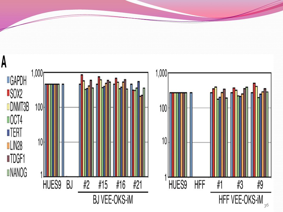
To further characterize the established iPSC clones, expression of hES marker genes were examined by qRT-PCR. Consistent with expression levels in human HUES9 ESCs, iPSC clones generated from both parental BJ and HFFs with either the VEE-OKS-iM RNA or VEE-OKS-iG RNA protocol expressed robust levels of endogenous OCT4, SOX2, NANOG, LIN28, TDGF1, DNMT3B, and TERT, in contrast to low or no expression levels in starting parental BJ and HFFs. Likewise, VEE-OKS-iM RNA or VEE-OKS-iG RNA-generated iPS clones from human adult HDFs and NHDF-c fibroblasts also expressed OCT4, SOX2, NANOG, LIN28, TDGF1, DNMT3B, and TERT by qRT-PCR.

37
5. Demethylation of OCT4 & NANOG promoter
A hallmark of induced pluripotency is reduced DNA methylation of CpG dinucleotides in the OCT4 and NANOG promoter regions Bisulfite genomic sequencing of both the OCT4 and NANOG promoter regions showed extensive demethylation in iPSC clones compared to parental fibroblasts

38
6. mRNA expression profile
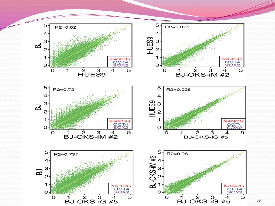
To investigate mRNA expression profiles in iPSC clones, we performed whole-genome RNA sequencing (RNA-seq). All four iPSC clones analyzed by RNA-seq showed unsupervised hierarchical clustering and expression signatures characteristic of human HUES9 ESCs that were highly divergent from parental human fibroblasts . Together, these results demonstrate that VEE-RF RNA replicon-generated iPSCs have all of the expression profile hallmarks of hESCs.
. All four iPSC clones analyzed by RNA-seq showed unsupervised hierarchical clustering and expression signatures characteristic of human HUES9 ESCs that were highly divergent from parental human fibroblasts . Together, these results demonstrate that VEE-RF RNA replicon-generated iPSCs have all of the expression profile hallmarks of hESCs.")
40
Concerns 1 Geuking et al. (2009) reported that nonretroviral RNA viruses under extreme conditions can recombine with endogenous retrotransposon genetic elements and result in a reverse transcription into DNA followed by genomic integration. Therefore, we examined VEE-RF iPSC clones for the presence of DNA copies. However, consistent with an RNA-only vector that does not go through a DNA intermediate, we did not detect any genomic integrations of VEE-RF by genomic PCR analysis or by Southern blot analysis (Figures S5B–S5E).
 reported that nonretroviral RNA viruses under extreme conditions can recombine with endogenous retrotransposon genetic elements and result in a reverse transcription into DNA followed by genomic integration. Therefore, we examined VEE-RF iPSC clones for the presence of DNA copies. However, consistent with an RNA-only vector that does not go through a DNA intermediate, we did not detect any genomic integrations of VEE-RF by genomic PCR analysis or by Southern blot analysis (Figures S5B–S5E).")
41
2 A consistent concern for iPS generation protocols is the generation of aneuploid or tetraploid iPSC clones. By flow cytometry DNA analysis, we observed several tetraploid iPSC clones generated from VEE-RF OKS-iM RNA, but no tetraploid colonies were detected from VEE-RF OKS-iG RNA. However, karyotype analysis of four independent iPSC clones generated from both OKS-iM and OKS-iG VEE-RF RNA replicons that showed normal DNA content by flow cytometry contained normal diploid karyotypes .

42
Differentiation of VEE-RNA Replicon-Generated iPSC Clones
Finally, we tested the pluripotency of VEE-RF RNA replicon generated hiPSC clones to differentiate in vitro and in vivo. First, iPSCs were differentiated in vitro into cardiomyocytes Four independent iPSC clones derived from either the OKS-iM or OKS-iG VEE-RF replicons were treated with activin A, bone morphogenetic protein 4 (BMP4), basic fibroblast growth factor (bFGF), vascular endothelial growth factor (VEGF), and dickkopf homolog 1 (DKK1) in serum-free media.
, basic fibroblast growth factor (bFGF), vascular endothelial growth factor (VEGF), and. dickkopf homolog 1 (DKK1) in serum-free media.")
43
Differentiated embryoid bodies (EBs) from all four iPS clones began spontaneously contracting at day 7 and were stably contracting on day 15. Using antibodies against cTNT and a-actinin (Figure 5A), immunohistochemistry revealed that all four differentiated iPS EBs were positive for cardiomyocyte markers. These data combined with the spontaneously contracting EBs confirm the ability of VEE-RNA replicon- generated iPSC clones to differentiate into cardiomyocytes
, immunohistochemistry revealed that all four differentiated iPS EBs were positive for cardiomyocyte markers. These data combined with the spontaneously contracting EBs confirm the ability of VEE-RNA replicon- generated iPSC clones to differentiate into cardiomyocytes.")
44
STAINING FOR CARDIOMYOCYTE MARKERS

45
DIFFERENTIATION IN VIVO
To test for in vivo pluripotency to differentiate into cells of all three germ layers, human VEE-RF RNA iPSC clones were injected into immunodeficient mice to generate differentiated teratomas. H&E of sections from two independent iPSC clones contained representative cell types of all three germ layers (ectoderm, endoderm, and mesoderm) that are spread throughout the sections. Immunohistochemistry staining of four additional, independent iPSC-derived teratomas was positive for ectoderm markers AE1/AE3 (cytokeratin), NF-1 (neuronal cells), and GFAP (neuronal cells), mesoderm marker Desmin (muscle cells), and endoderm marker AFP (primitive and definitive endoderm) (Figure 5C).
 that are spread throughout the sections. Immunohistochemistry staining of four additional, independent iPSC-derived teratomas was positive for. ectoderm markers AE1/AE3 (cytokeratin), NF-1 (neuronal cells), and GFAP (neuronal cells), mesoderm marker Desmin (muscle cells), and. endoderm marker AFP (primitive and definitive endoderm) (Figure 5C).")
46
Collectively, these observations confirm the ability of both VEE-OKS-iM and VEE-OKS-iG RNA replicons to efficiently generate pluripotent hiPSCs

47
conclusion This study reported the generation of hiPSCs by a single transfection of a self-replicating VEERNA species that expresses four reprogramming factor ORFs (OCT4, KLF4, SOX2, with either c-MYC or GLIS1) VEE-reprogramming factor (VEE-RF) RNA generated hiPSCs that were free of VEE RNA and had all the hallmarks of human stem cells expression of embryonic stem cell [ESC] markers, global gene expression, and differentiation in vivo into all three germ lineages. The VEE-RF RNA can also be selectively retained or removed from cells
 VEE-reprogramming factor (VEE-RF) RNA generated hiPSCs that were free of VEE RNA and had all the hallmarks of human stem cells. expression of embryonic stem cell [ESC] markers, global gene expression, and differentiation in vivo into all three germ lineages. The VEE-RF RNA can also be selectively retained or removed from cells.")
Similar presentations


>")
 –Use.>")


 is a member of the herpesvirus family. About 70-90% of the population is infected with HCMV. In healthy.>")
 from somatic cells also requires transformation.>")
>")


>")
>")


>")