Download presentation
Presentation is loading. Please wait.

1
2.4 Membranes 2.4.1 Draw a diagram to show the fluid mosaic model of a biological membrane. (1) The diagram should show the phospholipid bilayer, cholesterol, glycoproteins and integral and peripheral proteins. Use the term plasma membrane not cell surface membrane for the membrane surrounding the cytoplasm. Integral proteins are embedded in the phospholipid of the membrane whereas peripheral proteins are attached to its surface.

2
2.4 Membranes 2.4.2 Explain how the hydrophobic and hydrophilic properties of phospholipids help to maintain the structure of cell membranes. (3) Hydrophobic – ‘afraid of water’ Hydrophilic – ‘loves water’

3
2.4 Membranes

4
2.4 Membranes

5
2.4 Membranes

6
2.4 Membranes Fibers of the extracellular matrix
Carbohydrate (of glycoprotein) Glycoprotein Microfilaments of cytoskeleton Phospholipid Cholesterol Proteins Plasma membrane Glycolipid Cytoplasm
 Glycoprotein. Microfilaments of cytoskeleton. Phospholipid. Cholesterol. Proteins. Plasma membrane. Glycolipid. Cytoplasm.")
7
2.4 Membranes

8
2.4 Membranes 2.4.3 List the functions of membrane proteins including
hormone binding sites – Insulin on liver Immobilized enzymes – epithelial villi cells Cell adhesion – Tight junctions channels for passive transport – facilitated diffusion channels pumps for active transport.

10
Concept Check Membranes organize cell activities. The proteins imbedded in the membranes are essential to their function. These membrane proteins have properties that allow them to “float” in the membrane. Which of the following describe those properties? The surface region of the protein in the interior of the membrane is mostly hydrophobic. The surface region of the protein in the interior of the membrane is mostly hydrophillic. The surface region exposed to the outer environment is hydrophobic. The surface region exposed to the interior environment is hydrophobic.

11
Answer Membranes organize cell activities. The proteins imbedded in the membranes are essential to their function. These membrane proteins have properties that allow them to “float” in the membrane. Which of the following describe those properties? The surface region of the protein in the interior of the membrane is mostly hydrophobic.

12
2.4 Membranes 2.4.4 Define diffusion (1)
Diffusion is the passive movement of particles from a region of high concentration to a region of low concentration (down a concentration gradient), until there is an equal distribution. Define osmosis Osmosis is the passive movement of water molecules, across a partially permeable membrane, from a region of lower solute concentration (high water concentration) to a region of higher solute concentration (low water concentration).
, until there is an equal distribution. Define osmosis. Osmosis is the passive movement of water molecules, across a partially permeable membrane, from a region of lower solute concentration (high water concentration) to a region of higher solute concentration (low water concentration).")
13
2.4 Membranes 2.4.5 Explain passive transport across membranes in terms of diffusion. (3) Requires no energy Moves from down the concentration gradient Some molecules pass through the membrane Some molecules use channels for facilitated diffusion

14
Passive transport is diffusion across a membrane
In passive transport, substances diffuse through membranes without work by the cell Spreading from areas of high concentration to areas of low concentration Equilibrium Membrane Molecules of dye Figure 5.14A Figure 5.14B

15
Passive Transport

16
Small nonpolar molecules such as O2 and CO2
Diffuse easily across the phospholipid bilayer of a membrane

17
Transport proteins may facilitate diffusion across membranes
Many kinds of molecules Do not diffuse freely across membranes For these molecules, transport proteins Provide passage across membranes through a process called facilitated diffusion Solute molecule Transport protein Figure 5.15

18
Osmosis is the diffusion of water across a membrane
In osmosis Water travels from a solution of lower solute concentration to one of higher solute concentration Lower concentration of solute Higher concentration of solute Equal concentration of solute H2O Solute molecule Selectively permeable membrane Water molecule Solute molecule with cluster of water molecules Net flow of water Figure 5.16

19
Osmosis

20
Concept Check This diagram represents osmosis of water across a semipermeable membrane. The U-tube on the right shows the results of the osmosis. What could you do to level the solutions in the two sides of the right hand U-tube? Add more water to the left hand side. Add more water to the right hand side. Add more solute to the left hand side. Add more solute to the right hand side.

21
Answer This diagram represents osmosis of water across a semipermeable membrane. The U-tube on the right shows the results of the osmosis. What could you do to level the solutions in the two sides of the right hand U-tube? Add more solute to the left hand side.

22
2.4 Membranes

23
2.4 Membranes 2.4.6 Explain the role of protein pumps and ATP in active transport across membranes. (3) Requires energy, in the form of ATP, or adenosine triphosphate Molecules are ‘pumped’ across the membrane UP the concentration gradient Pumps fit specific molecules The pump changes shape when ATP activates it, this moves the molecule across the membrane

24
Active Transport

25
Active Transport

26
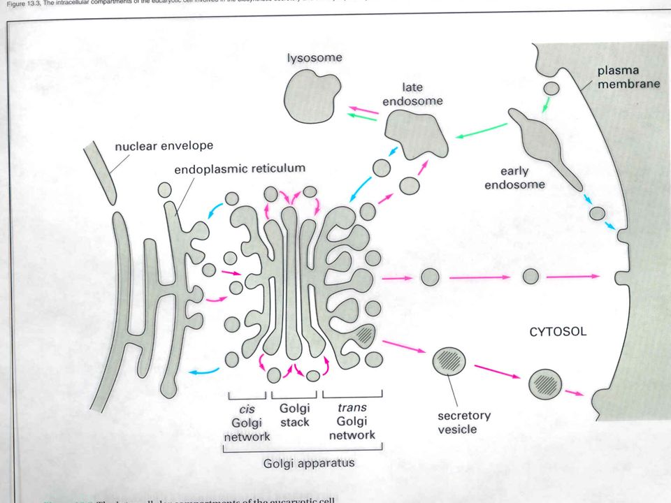
2.4 Membranes 2.4.7 Explain how vesicles are used to transport materials within a cell between the rough endoplasmic reticulum, Golgi apparatus and plasma membrane. (3) 2.4.8 Describe how the fluidity of the membrane allows it to change shape, break and reform during endocytosis and exocytosis. (2)
 Describe how the fluidity of the membrane allows it to change shape, break and reform during endocytosis and exocytosis. (2)")
28
2.4 Membranes Endocytosis – the mass movement INTO the cell by the membrane ‘pinching’ into a vacuole Exocytosis – the mass movement OUT of the cell by the fusion of a vacuole and the membrane This is possible because the of the fluid properties of the membrane (able to break and reform easily, phospholipids not attached just attracted)
")
29
Exocytosis

30
Endocytosis endo- and exo- -cytosis

31
Endocytosis can occur in three ways
Phagocytosis Pinocytosis Receptor-mediated endocytosis Pseudopodium of amoeba Food being ingested Phagocytosis Pinocytosis Receptor-mediated endocytosis Material bound to receptor proteins PIT Cytoplasm Plasma membrane TEM 54,000 TEM 96,500 LM 230 Figure 5.19C

32
CONNECTION Faulty membranes can overload the blood with cholesterol
Harmful levels of cholesterol Can accumulate in the blood if membranes lack cholesterol receptors LDL particle Protein Phospholipid outer layer Cytoplasm Receptor protein Plasma membrane Vesicle Cholesterol Figure 5.20

33
Endocytosis and Exocytosis

Similar presentations


 Cells. Figure 4.1x Cell Theory: - all organisms are composed of cells - all cells come from other cells.>")


