Download presentation
Presentation is loading. Please wait.

2
Dr. Jeevan Shetty MBBS MD Associate Professor Reference Book: Text book of Biochemistry By DM Vasudevan 5th edition

3
At the end of the topic student shall be able to Define and classify ELISA Principle of ELISA technique Instrumentation in ELISA Procedure of measurement Interpretation of results Advantages of ELISA over RIA Application of ELISA in medicine and pharmacy

4
Definition - Is a biochemical technique used mainly in immunology to detect the presence of an antibody or an antigen in a sample. The technique is divided into 1- Competitive ELISA - antigen detection or antibody detection 2- Sandwich ELISA (also called direct ELISA) 3- Indirect ELISA
 3- Indirect ELISA.")
5
Principle - Single antibody (competitive ELISA) The labelled antigen competes for primary antibody binding sites with the sample antigen (unlabelled). The more antigen in the sample, the less labelled antigen is retained in the well and the weaker is the signal.

6
Single antibody (competitive ELISA) Antibodies are coated on to the micotitre plate Serum (unlabeled antigen) and labeled antigen is added to the plate. Both antigen in the serum and labeled antigen binds to coated antibody and excess antigen is washed. Substrate for the enzyme is added and observe the color development. Intensity of the color developed is inversely proportional to the amount of antigen in the serum.

8
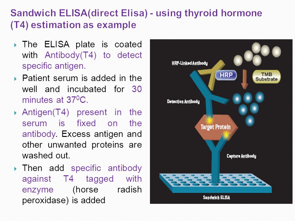
The ELISA plate is coated with Antibody(T4) to detect specific antigen. Patient serum is added in the well and incubated for 30 minutes at 37 0 C. Antigen(T4) present in the serum is fixed on the antibody. Excess antigen and other unwanted proteins are washed out. Then add specific antibody against T4 tagged with enzyme (horse radish peroxidase) is added
 present in the serum is fixed on the antibody. Excess antigen and other unwanted proteins are washed out. Then add specific antibody against T4 tagged with enzyme (horse radish peroxidase) is added.")
9
If the antigen is already fixed, antibody-HRP- conjugate will be fixed in the well, Then a color reagent, containing hydrogen peroxide (H 2 O 2 ) and diamino benzidine (DAB) are added. The reaction is as follows H 2 O 2 H 2 O + Nascent oxygen Diamino benzidine Oxidized DAB (Colorless) (Brown color) This is called sandwich ELISA. Development of brown color indicates the presence of antigen in the serum Color developed is proportional to the antigen in the serum.
 (Brown color) This is called sandwich ELISA. Development of brown color indicates the presence of antigen in the serum Color developed is proportional to the antigen in the serum..")
11
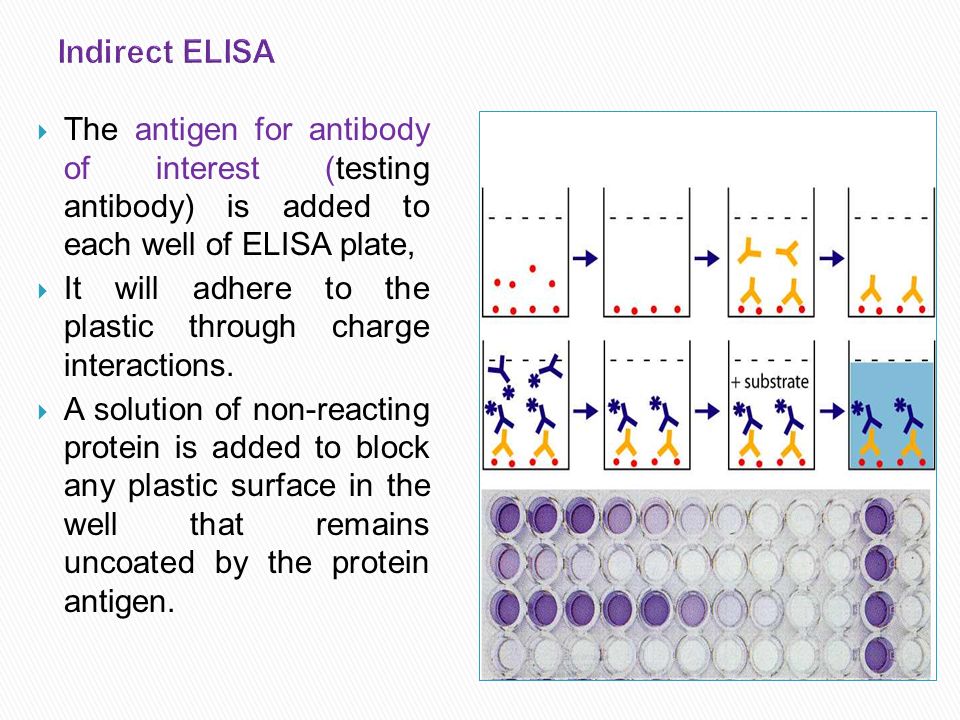
The antigen for antibody of interest (testing antibody) is added to each well of ELISA plate, It will adhere to the plastic through charge interactions. A solution of non-reacting protein is added to block any plastic surface in the well that remains uncoated by the protein antigen.

12
Then the serum is added, which contains a mixture of the serum antibodies, of unknown concentration, some of which may bind specifically to the test antigen that is coating the well. Afterwards, a secondary antibody is added, which will bind to the antibody bound to the test antigen in the well. This secondary antibody often has an enzyme attached to it

13
A substrate for this enzyme is then added. Often, this substrate changes colour upon reaction with the enzyme. The colour change shows that secondary antibody has bound to primary antibody, which strongly implies that the donor has had an immune reaction to the test antigen. The higher the concentration of the primary antibody that was present in the serum, the stronger the colour change. Often a spectrometer is used to give quantitative values for colour strength

14
Solid Phase : Plastic tubes / Micro titre plates Enzymes : Peroxidase (Hydrogen Peroxide) Alkaline Phosphatase (PNPP – para nitrophenyl phosphate)
 Alkaline Phosphatase (PNPP – para nitrophenyl phosphate)")
16
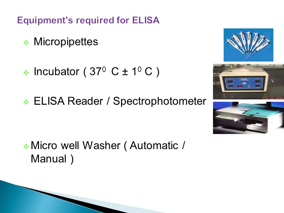
Micropipettes Incubator ( 37 0 C ± 1 0 C ) ELISA Reader / Spectrophotometer Micro well Washer ( Automatic / Manual )
 ELISA Reader / Spectrophotometer Micro well Washer ( Automatic / Manual )")
17
Before starting the work read kit instruction carefully 1- The 96 well plate is labeled carefully and the first wells are used to draw the standard curve

18
The sample is added to plate in duplicate or triplicate and then the mean result is calculated The quality control sample which is provided with the kit is treated as the test samples

19
After reading the results the standard curve is drawn were the concentration is blotted on the X-axis and the absorbance on the Y-axis Concentration ng/ml Absorption nm

20
The standards concentrations is specified on the x-axis and the reading of each standard is specified on the y-axis and the standard curve is drawn

21
This standard curve is used to determine the unknown concentration of each sample by finding the opposite concentration to the absorbance Concentration ng/ml Absorption nm

22
The quality control sample concentration is determined from the standard curve and if the result is in the range given by the kit manufacturer the results could be accepted.

23
Screening donated blood for evidence of viral contamination by ◦ HIV-1 and HIV-2 (presence of anti-HIV antibodies) ◦ hepatitis C (presence of antibodies) ◦ hepatitis B (testing for both antibodies and a viral antigen) Measuring hormone levels ◦ β-HCG (as a test for pregnancy) ◦ LH (determining the time of ovulation) ◦ TSH, T3 and T4 (for thyroid function) ◦ Hormones (e.g., anabolic steroids, HGH) that may have been used illicitly by athletes.
 ◦ hepatitis C (presence of antibodies) ◦ hepatitis B (testing for both antibodies and a viral antigen) Measuring hormone levels ◦ β-HCG (as a test for pregnancy) ◦ LH (determining the time of ovulation) ◦ TSH, T3 and T4 (for thyroid function) ◦ Hormones (e.g., anabolic steroids, HGH) that may have been used illicitly by athletes.")
24
Detecting infections ◦ sexually-transmitted agents like HIV, syphilis, and chlamydia ◦ hepatitis B and C ◦ Toxoplasma gondii Detecting allergens in food and house dust Measuring "rheumatoid factors" and other autoantibodies in autoimmune diseases like systemic lupus erythematous (SLE). Measuring toxins in contaminated food Detecting illicit drugs, e.g., ◦ cocaine ◦ opiates ◦ Δ-9-tetrahydrocannabinol, the active ingredient in marijuana

25
Serum Hormone estimation T 4, TSH, FSH, LH, Insulin etc Tumor Marker estimation AFP, PSA, HCG,CEA, CA-125 etc AFP, PSA, HCG,CEA, CA-125 etc Detection of Bacterial toxins, Viruses etc to Rubella virus Antiviral antibodies eg to Rubella virus to Salmonella Antibacterial antibodies eg to Salmonella Auto antibodies ANA, anti-DNA, anti Thyroglobulin

26
It is cheaper than RIA Lacks radiological hazards of RIA Reagents have more shelf-life compared to RIA It is equally sensitive and specific as RIA Suitable for use even in small laboratories

Similar presentations
>")


 are gamma globulin proteins that are.>")


>")


