Download presentation
Presentation is loading. Please wait.

2
به نام پروردگار

3
زیست شناسی سلولی و مولکولی DNA و همانندسازی آن

5
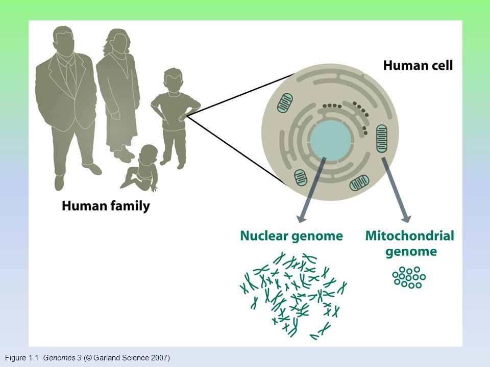
Figure 1.1 Genomes 3 (© Garland Science 2007)
")
6
Figure 1.2 Genomes 3 (© Garland Science 2007)
")
7
کشف موکول اطلاعاتی

8
فردریک مایشر ( ۱۸۹۵ - ۱۸۴۴ ) بررسی شیمیایی سلول های سفید خون گلبول های سفید را تحت تأثیر عصاره ی معده ی خوک هسته ی سلول ها را از سیتوپلاسم جدا کرد. هسته ها را تحت تأثیر هیدروکسید سدیم قرار داد تجزیه ی شیمیایی آن نشان داد، کربن، هیدروژن، اکسیژن، نیتروژن و درصد زیادی فسفر، عنصر های سازنده ی آن هستند نوکلئین

9
Figure 1.3a Genomes 3 (© Garland Science 2007) آسوالد آوری
 آسوالد آوری")
11
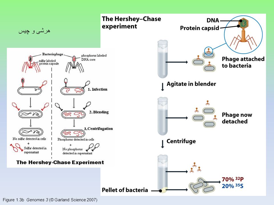
Figure 1.3b Genomes 3 (© Garland Science 2007) هرشی و چیس
 هرشی و چیس")
12
چگونگی همانندسازی

13
تئوریهای همانندسازی

14
Figure 15.2 Genomes 3 (© Garland Science 2007)
")
15
Figure 15.3a Genomes 3 (© Garland Science 2007)
")
16
Figure 15.3b Genomes 3 (© Garland Science 2007)
")
17
نیمه حفاظتی بودن همانند سازی آزمایش مسلسون و استال

18
ساختمان

19
Figure 1.4 Genomes 3 (© Garland Science 2007)
")
20
Figure 1.4a Genomes 3 (© Garland Science 2007)
")
21
Figure 1.4b Genomes 3 (© Garland Science 2007)
")
22
Figure 1.5 Genomes 3 (© Garland Science 2007)
")
23
Figure 1.6 Genomes 3 (© Garland Science 2007)
")
24
Figure 1.7 Genomes 3 (© Garland Science 2007)
")
25
Figure 1.8a Genomes 3 (© Garland Science 2007)
")
26
Figure 1.8b Genomes 3 (© Garland Science 2007)
")
27
DNA is a Double Helix Nucleotides – A, G, T, C Sugar and phosphate form the backbone Bases lie between the backbone Held together by H-bonds between the bases – A-T – 2 H bonds – G-C – 3 H bonds

28
DNA Template template Each strand of the parent DNA is used as a template to make the new daughter strand DNA replication makes 2 new complete double helices each with 1 old and 1 new strand

29
Watson and Crick 1953 article in Nature

31
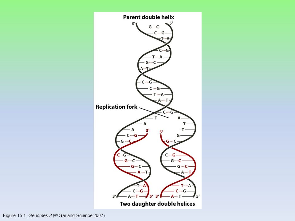
Figure 15.1 Genomes 3 (© Garland Science 2007)
")
32
Table 1.1 Genomes 3 (© Garland Science 2007)
")
33
Figure 1.9 Genomes 3 (© Garland Science 2007)
")
34
مراحل باز شدن تراکم باز شدن دو رشته تشکیل کمپلکس همانند سازی ( چنگال و پروتئین ها ) همانندسازی و پیشرفت
 همانندسازی و پیشرفت")
35
Figure 15.4 Genomes 3 (© Garland Science 2007)
")
36
Table 15.1 Genomes 3 (© Garland Science 2007)
")
37
Figure 15.5 Genomes 3 (© Garland Science 2007)
")
38
Double helix structure of DNA “It has not escaped our notice that the specific pairing we have postulated immediately suggests a possible copying mechanism for the genetic material.” Watson & Crick

39
Directionality of DNA You need to number the carbons! – it matters! OH CH 2 O 4 5 3 2 1 PO 4 N base ribose nucleotide This will be IMPORTANT!!

40
The DNA backbone Putting the DNA backbone together – refer to the 3 and 5 ends of the DNA the last trailing carbon OH O 3 PO 4 base CH 2 O base O P O C O –O–O CH 2 1 2 4 5 1 2 3 3 4 5 5 Sounds trivial, but … this will be IMPORTANT!!

41
Anti-parallel strands Nucleotides in DNA backbone are bonded from phosphate to sugar between 3 & 5 carbons – DNA molecule has “direction” – complementary strand runs in opposite direction 3 5 5 3

42
Bonding in DNA ….strong or weak bonds? How do the bonds fit the mechanism for copying DNA? 3 5 3 5 covalent phosphodiester bonds hydrogen bonds

43
Base pairing in DNA Purines – adenine (A) – guanine (G) Pyrimidines – thymine (T) – cytosine (C) Pairing – A : T 2 bonds – C : G 3 bonds
 – guanine (G) Pyrimidines – thymine (T) – cytosine (C) Pairing – A : T 2 bonds – C : G 3 bonds")
44
Copying DNA Replication of DNA – base pairing allows each strand to serve as a template for a new strand – new strand is 1/2 parent template & 1/2 new DNA

45
DNA Replication Large team of enzymes coordinates replication Let ’ s meet the team …

46
Replication: 1st step Unwind DNA – helicase enzyme unwinds part of DNA helix stabilized by single-stranded binding proteins single-stranded binding proteins replication fork helicase

47
DNA Polymerase III Replication: 2nd step But … We ’ re missing something! What? Where ’ s the ENERGY for the bonding! Build daughter DNA strand add new complementary bases DNA polymerase III

48
energy ATP GTPTTPCTP Energy of Replication Where does energy for bonding usually come from? ADPAMPGMPTMPCMP modified nucleotide energy We come with our own energy! And we leave behind a nucleotide! You remember ATP! Are there other ways to get energy out of it? Are there other energy nucleotides? You bet!

49
Energy of Replication The nucleotides arrive as nucleosides – DNA bases with P–P–P P-P-P = energy for bonding – DNA bases arrive with their own energy source for bonding – bonded by enzyme: DNA polymerase III ATPGTPTTPCTP

50
Adding bases – can only add nucleotides to 3 end of a growing DNA strand need a “starter” nucleotide to bond to – strand only grows 5 3 DNA Polymerase III DNA Polymerase III DNA Polymerase III DNA Polymerase III energy Replication energy 3 3 5 B.Y.O. ENERGY! The energy rules the process 5

51
energy 35 5 5 3 need “primer” bases to add on to energy 3 no energy to bond energy ligase 35

52
Limits of DNA polymerase III can only build onto 3 end of an existing DNA strand Leading & Lagging strands 5 5 5 5 3 3 3 5 3 5 3 3 Leading strand Lagging strand Okazaki fragments ligase Okazaki Leading strand continuous synthesis Lagging strand Okazaki fragments joined by ligase “spot welder” enzyme DNA polymerase III 3 5 growing replication fork

53
DNA polymerase III Replication fork / Replication bubble 5 3 5 3 leading strand lagging strand leading strand lagging strand leading strand 5 3 3 5 5 3 5 3 5 3 5 3 growing replication fork growing replication fork 5 5 5 5 5 3 3 5 5 lagging strand 5 3

54
DNA polymerase III RNA primer built by primase serves as starter sequence for DNA polymerase III Limits of DNA polymerase III can only build onto 3 end of an existing DNA strand Starting DNA synthesis: RNA primers 5 5 5 3 3 3 5 3 5 3 5 3 growing replication fork primase RNA

55
DNA polymerase I removes sections of RNA primer and replaces with DNA nucleotides But DNA polymerase I still can only build onto 3 end of an existing DNA strand Replacing RNA primers with DNA 5 5 5 5 3 3 3 3 growing replication fork DNA polymerase I RNA ligase

56
Loss of bases at 5 ends in every replication chromosomes get shorter with each replication limit to number of cell divisions? DNA polymerase III All DNA polymerases can only add to 3 end of an existing DNA strand Chromosome erosion 5 5 5 5 3 3 3 3 growing replication fork DNA polymerase I RNA Houston, we have a problem!

57
Repeating, non-coding sequences at the end of chromosomes = protective cap limit to ~50 cell divisions Telomerase enzyme extends telomeres can add DNA bases at 5 end different level of activity in different cells high in stem cells & cancers -- Why? telomerase Telomeres 5 5 5 5 3 3 3 3 growing replication fork TTAAGGG

58
Replication fork 3’ 5’ 3’ 5’ 3’ 5’ helicase direction of replication SSB = single-stranded binding proteins primase DNA polymerase III DNA polymerase I ligase Okazaki fragments leading strand lagging strand SSB

59
DNA polymerases DNA polymerase III – 1000 bases/second! – main DNA builder DNA polymerase I – 20 bases/second – editing, repair & primer removal DNA polymerase III enzyme Arthur Kornberg 1959 Roger Kornberg 2006

60
Editing & proofreading DNA 1000 bases/second = lots of typos! DNA polymerase I – proofreads & corrects typos – repairs mismatched bases – removes abnormal bases repairs damage throughout life – reduces error rate from 1 in 10,000 to 1 in 100 million bases

61
Fast & accurate! It takes E. coli <1 hour to copy 5 million base pairs in its single chromosome – divide to form 2 identical daughter cells Human cell copies its 6 billion bases & divide into daughter cells in only few hours – remarkably accurate – only ~1 error per 100 million bases – ~30 errors per cell cycle

62
1 2 3 4 What does it really look like?

64
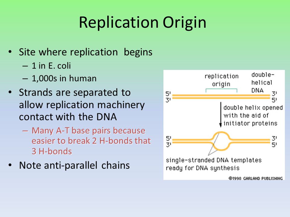
Replication Origin Site where replication begins – 1 in E. coli – 1,000s in human Strands are separated to allow replication machinery contact with the DNA – Many A-T base pairs because easier to break 2 H-bonds that 3 H-bonds Note anti-parallel chains

65
Replication Fork Bidirectional movement of the DNA replication machinery

66
Figure 15.6 Genomes 3 (© Garland Science 2007)
")
67
Figure 15.7 Genomes 3 (© Garland Science 2007)
")
68
Figure 15.8a Genomes 3 (© Garland Science 2007)
")
69
Figure 15.8b Genomes 3 (© Garland Science 2007)
")
70
Figure 15.9a Genomes 3 (© Garland Science 2007)
")
71
Figure 15.9b Genomes 3 (© Garland Science 2007)
")
72
Figure 15.10 Genomes 3 (© Garland Science 2007)
")
73
Figure 15.11 Genomes 3 (© Garland Science 2007)
")
74
Figure 15.12 Genomes 3 (© Garland Science 2007)
")
75
Table 15.2 Genomes 3 (© Garland Science 2007)
")
76
Figure 15.13 Genomes 3 (© Garland Science 2007)
")
77
Figure 15.13a Genomes 3 (© Garland Science 2007)
")
78
Figure 15.13b Genomes 3 (© Garland Science 2007)
")
79
Figure 15.14 Genomes 3 (© Garland Science 2007)
")
80
Figure 15.15a Genomes 3 (© Garland Science 2007)
")
81
Figure 15.15b Genomes 3 (© Garland Science 2007)
")
82
Figure 15.16 Genomes 3 (© Garland Science 2007)
")
83
Figure 15.17 Genomes 3 (© Garland Science 2007)
")
84
Figure 15.18 Genomes 3 (© Garland Science 2007)
")
85
Figure 15.19 Genomes 3 (© Garland Science 2007)
")
86
Figure 15.20 Genomes 3 (© Garland Science 2007)
")
87
Figure 15.20a Genomes 3 (© Garland Science 2007)
")
88
Figure 15.20b Genomes 3 (© Garland Science 2007)
")
89
Figure 15.21 Genomes 3 (© Garland Science 2007)
")
90
Figure 15.22a Genomes 3 (© Garland Science 2007)
")
91
Figure 15.22b Genomes 3 (© Garland Science 2007)
")
92
Figure 15.23 Genomes 3 (© Garland Science 2007)
")
93
Figure 15.24a Genomes 3 (© Garland Science 2007)
")
94
Figure 15.24b Genomes 3 (© Garland Science 2007)
")
Similar presentations


 for growth & development From.>")


 RIBOSE (RNA) Built from NUCLEOTIDE SUBUNITS NITROGEN BASES.>")


>")


