Download presentation
Presentation is loading. Please wait.

1
THE SKELETAL SYSTEM

2
I. BONE STRUCTURE & FUNCTION A.FUNCTION 1.Support 2.Protection 3.Movement 4.Blood formation 5.Electrolyte Balance 6.Acid Base Balance

3
I. BONE STRUCTURE & FUNCTION B.Structure 1.Shape –Long bones –Short bones –Flat bones –Irregular bones

4
Long bonesShort bones

5
Flat bones Irregular bones

6
Classify the bone types to the left

7
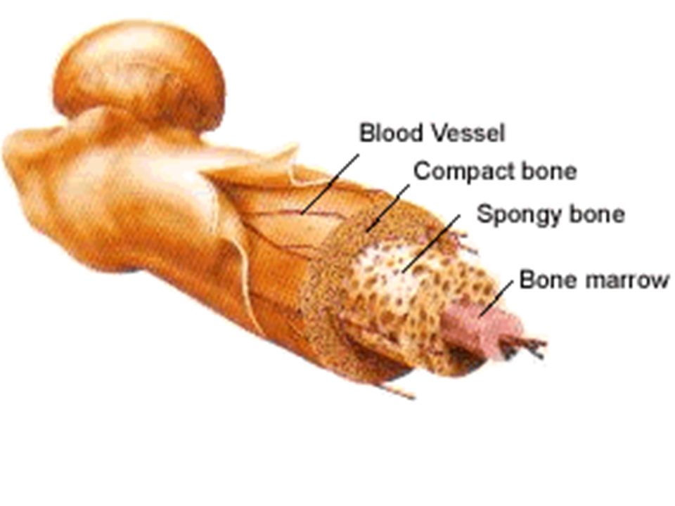
B. Structure 2. Parts of Bone –Epiphyses Contains red bone marrow Spongy bone and compact bone

8
B.Structure 2.Parts of Bone –Diaphysis Shaft of bone Contains yellow bone marrow (stores fat) Spongy and compact bone
 Spongy and compact bone")
10
B.Structure 2.Parts of Bone –Periosteum –Epiphyseal plate Allows for growth in bone Found only in children

11
3.Mature Bone Osseous Tissue B. Structure

12
Lamellar bone Called Lamellar bone Two kinds Compact Spongy (cancellous) 3. Mature Bone
 3. Mature Bone")
13
Spongy (cancellous) contains trabeculae contains trabeculae contains spaces contains spaces 3. Mature Bone

14
Dense, few spaces Dense, few spaces Haversian canals Haversian canals Concentric Lamellae Concentric Lamellae Compact Bone 3. Mature Bone

15
I. BONE STRUCTURE & FUNCTION Background minerals C.Histology 1.Matrix

17
C. Histology 2.Bone cells

18
Osteoblasts

19
Osteocytes Osteoclasts

20
Osteoblasts Osteoclasts Osteocytes

21
Name C, D & E D = Osteoblast E = Osteocytes C = Osteoclast

22
D. Membranes 1.Periosteum 1.Periosteum: a.The external covering of bone

23
D. Membranes 2.endosteum: a.Found on internal bone surface b.covers trabeculae of spongy bone c.in marrow cavities

25
II. BONE GROWTH & DEVELOPMENT A.Two Patterns of Bone Formation 1.Intramembranous bones –originate between sheet-like layers of connective tissues

26
II. BONE GROWTH & DEVELOPMENT A.Two Patterns of Bone Formation 2.Endochondral bones –begin as masses of hyaline cartilage that bone tissue later replaces.

27
II. BONE GROWTH & DEVELOPMENT B.Growth in Long Bones 1.grow by interstitial growth at epiphyseal plates a.rate of cartilage growth is balanced by replacement with bone b.end of growth as cartilage cells slow down division

28
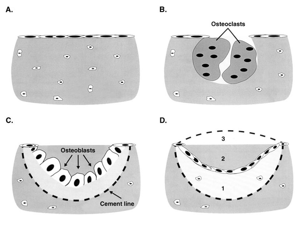
II. BONE GROWTH & DEVELOPMENT B.Growth in Long Bones 2. bones grow in width by appositional growth a.Osteoblasts in periosteum: secretes bone matrix b.Osteoclasts in endosteum: removes bone matrix (a little slower)
.")
29
Appositional Growth –New bone forms at ridges around blood vessels –Periosteum becomes endosteum

30
– New lamella formed – More bone added forming osteon Appositional Growth

31
II. BONE GROWTH & DEVELOPMENT C.Hormones 1. growth hormone from pituitary: stimulates growth in childhood a.Gigantism: excessive growth hormone b.dwarfism: not enough growth hormone or thyroid hormones

32
II. BONE GROWTH & DEVELOPMENT C.Hormones 2. thyroid: regulates activity of growth hormone 3. sex hormones: promote growth spurt, induce epiphyseal plate closure estrogen: maintains bone density

33
III. Bone Maintenance & Repair A.Bone remodeling: 1.Life long process 2. Local areas of bone are destroyed and rebuilt 3. Repairs microdamage caused by normal wear and tear

35
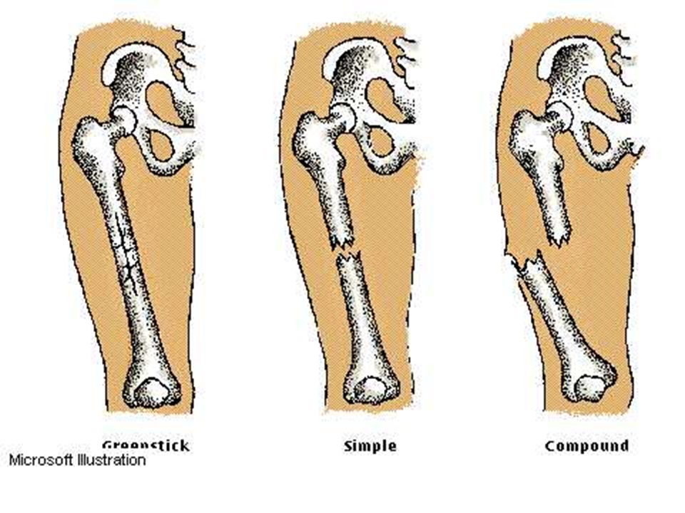
B.Fractures 1. Simple 1. Simple bone breaks cleanly, does not break through skin

36
B.Fractures 2. Compound 2. Compound broken ends protrude through the skin, risk of bone infection

37
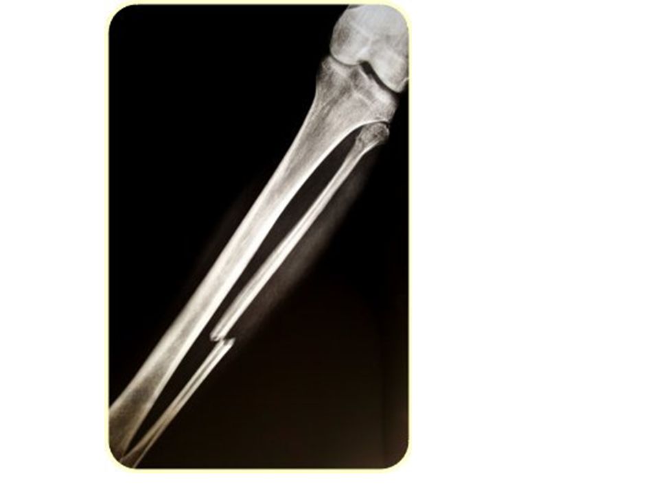
B.Fractures 3. Greenstick 3. Greenstick Greenstick: (children) Does not break completely
 Does not break completely")
44
C. Repair Hematoma forms 1. Hematoma forms 2. Spongy bone forms in area of hematoma 2. Spongy bone forms in area of hematoma III. Remodeling and Repair

45
C. Repair Osteoblasts lay down new bone matrix 3. Osteoblasts lay down new bone matrix 4. Remodeling 4. Remodeling

46
III. Calcium Homeostasis A.Blood Ca 2+ Level 1.Has a very narrow range 2. Ca 2+ Required for –For normal muscle contraction –Nerve impulses

47
III. Calcium Homeostasis A.Blood Ca 2+ Level 3.Abnormal levels –Hypocalcemia causes marked jitteriness and convulsive seizures

48
III. Calcium Homeostasis A.Blood Ca 2+ Level 3.Abnormal levels –Hypercalcemia the most common life- threatening metabolic disorder associated with cancer

49
III. Calcium Homeostasis B.Bone’s Role 1.Major storage site for calcium 2.Calcium moves –Into bone as osteoblasts build new bone –Out of bone as osteoclasts break down bone

50
III. Calcium Homeostasis C.Bone, Calcium and Hormones 1.Parathyroid Hormone – Increases blood Ca 2+ levels 2.Calcitonin Decreases blood Ca 2+ levels

51
D. Homeostatic Imbalances OsteopeniaOsteopenia – Inadequate ossification OsteoporosisOsteoporosis – Bone absorption outpaces deposition – Fractures common – More common in elderly women III. Calcium Homeostasis

52
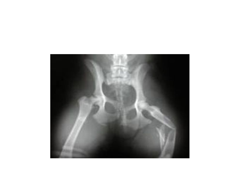
RicketsRickets – Lack of vitamin D or calcium during growth – Bowed legs – Deformed pelvis D. Homeostatic Imbalances Rickets

53
Osteosarcoma – Bone cancer – Usually between ages 10-25 – Survival rate is about 50% with amputation D. Homeostatic Imbalances

54
Bone spurBone spur – Abnormal projection at one site of bone due to overgrowth – Common in aging bones D. Homeostatic Imbalances

55
IV. The Skeleton There are 206 bone

56
IV. The Skeleton A.Organization 1.Axial Skeleton –Skull –Vertebral Column –Vertebrae –Ribs 2.Appendicular Skeleton –limbs –girdles

57
B. Male vs. Female Skeleton - Pelvis a.spines farther apart in male b.hole in ischium: smaller and triangular in female c.angle across pubic symphysis = pubic arch: less than 90° and more sharply angled in male d.distance between ischia larger in female

58
C.Bone Markings – 1. Kinds

59
For muscle attachment For formation of a joint To allow blood vessels or nerves to pass through Articulating Surfaces Openings Depressions & Enlargements 1. Kinds

60
C.Bone Markings – 2. Articulating surfaces A rounded projection set off from the body of a bone by a constriction (the neck) ex. head of femur Head
 ex. head of femur Head.")
61
C.Bone Markings – 2. Articulating surfaces Condyle Any large articulating surface, may be concave or convex

62
C.Bone Markings – 2. Articulating surfaces A smooth, flat surface, generally small Facet

63
C.Bone Markings – 2. Articulating surfaces A shallow depression Fossa

64
C.Bone Markings – 3. Enlargements & Processes generic term for bone projection that serves as a point for attachment of other structures Process

65
C.Bone Markings – 3. Enlargements projection or swelling to the side of or above a condyle Epicondyle

66
C.Bone Markings – Spine a sharp, slender projecting process 3. Enlargements

67
C.Bone Markings – a small rounded projection Tubercle 3. Enlargements

68
C.Bone Markings – a large rounded roughened projection 3. Enlargements Turberosity

69
C.Bone Markings – 3. Enlargements Trochanter a large blunt projection

70
C.Bone Markings – 3. Enlargements a prominent border or ridge Crest

71
C.Bone Markings – 3. Enlargements A major branch or division off of the main body of a bone Ramus

72
C.Bone Markings – 4. Openings

73
C.Bone Markings – 4. Openings Canal like opening Meatus

74
C.Bone Markings – 5. Depressions A shallow groove Sulcus

75
C.Bone Markings – 5. Depressions A very shallow groove Fovea

76
C.Bone Markings – 5. Depressions A deep groove Fissure

77
That’s All Folks!

Similar presentations


 - Tendons (muscle to bone)>")
 2.Protection: skull, vertebrae,>")


 Joints ► Cartilages Ligaments ► Divided.>")


 Joints Cartilages Ligaments Divided into two divisions Axial skeleton –>")

