Download presentation
Presentation is loading. Please wait.

1
به نام خدا

2
دكتر محمد امامي فوق تخصص ريه عضو هيات علمي دانشگاه

4
VTE

5
CLINICAL PRESENTATIONS OF DVT swelling pain erythema warmth Homan's sign (calf pain with flexion of the knee and dorsiflexion of the ankle) Moses’ sign (pain with calf compression against the tibia) palpable cord
 Moses’ sign (pain with calf compression against the tibia) palpable cord")
6
Of DVT Differential Diagnosis cellulitis arthritis muscular injury or tear neuropathy arterial insufficiency lymphedema ruptured Baker's cyst superficial thrombophlebitis chronic venous insufficiency

7
PE CLINICAL PRESENTATION OF PE CLINICAL PRESENTATION OF sudden onset of dyspnea Pleuritic chest pain Hemoptysis Cough A sense of impending doom Angina Syncope Fever

8
8 Clinical Features Signs with Angiographically Proven PE SignPercent Tachypnea > 20/min92 Rales58 Accentuated S253 Tachycardia >100/min44 Fever > 37.843 Diaphoresis36 S3 or S4 gallop34 Thrombophebitis32 Lower extremity edema24

9
Differential Diagnosis of PE Pneumonia, asthma, chronic obstructive pulmonary disease Congestive heart failure Pericarditis Pleurisy: "viral syndrome," costochondritis, musculoskeletal discomfort Rib fracture, pneumothorax Acute coronary syndrome Anxiety

10
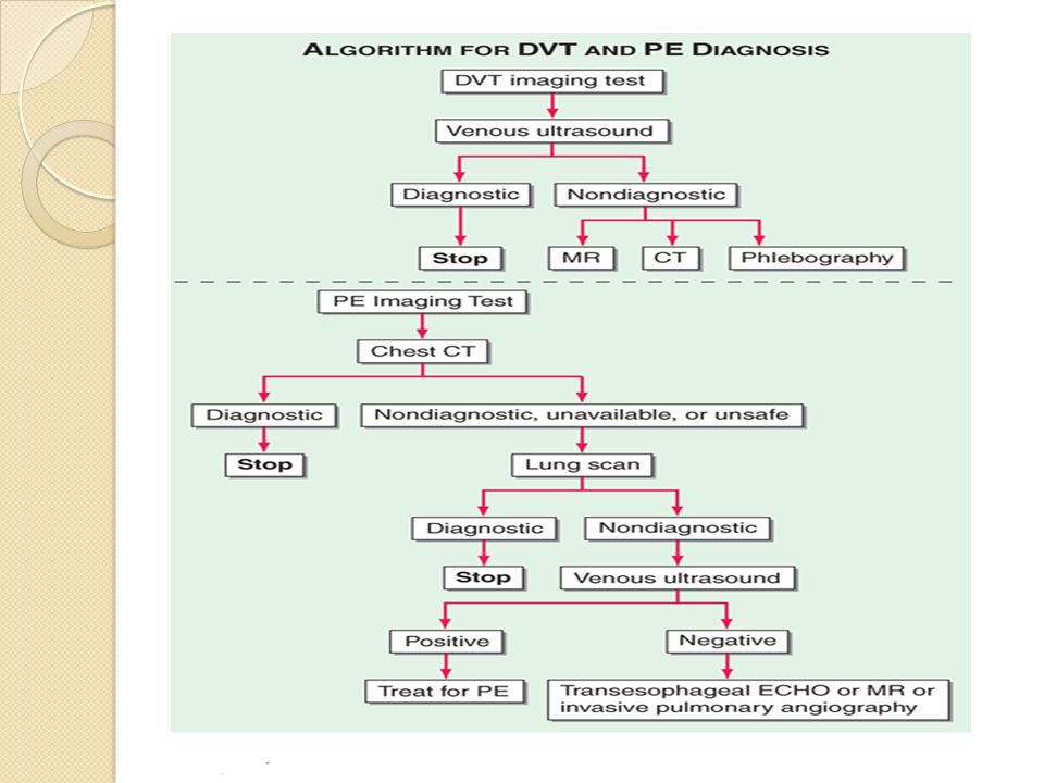
DIAGNOSIS OF DVT Multiple investigations have established that the clinical diagnosis of venous thrombosis is imprecise. In patients with clinical signs and symptoms suggestive of venous thrombosis, 60% to 80% will not have the diagnosis established by objective testing.

11
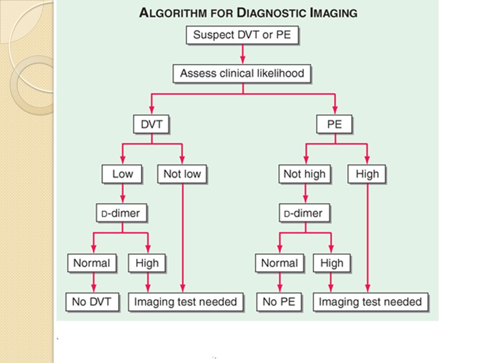
Clinical Prediction Rules Several clinical prediction rules for venous thrombosis have been developed and validated.The Wells rule, initially described in 1995 and revised in 1997.

12
Active cancer (patient receiving treatment for cancer within the previous 6 mo or currently receiving palliative treatment) 1 Paralysis, paresis, or recent plaster immobilization of the lower extremities 1 Recently bedridden for 3 days or more, or major surgery within the previous 12 wk requiring general or regional anesthesia 1 Localized tenderness along the distribution of the deep venous system 1 Entire leg swollen 1 Calf swelling at least 3 cm larger than that on the asymptomatic side (measured 10 cm below the tibial tuberosity) 1 Pitting edema confined to the symptomatic leg 1 Collateral superficial veins (nonvaricose) 1 Previously documented deep venous thrombosis 1 Alternate diagnosis at least as likely as deep venous thrombosis − 2
 1 Paralysis, paresis, or recent plaster immobilization of the lower extremities 1 Recently bedridden for 3 days or more, or major surgery within the previous 12 wk requiring general or regional anesthesia 1 Localized tenderness along the distribution of the deep venous system 1 Entire leg swollen 1 Calf swelling at least 3 cm larger than that on the asymptomatic side (measured 10 cm below the tibial tuberosity) 1 Pitting edema confined to the symptomatic leg 1 Collateral superficial veins (nonvaricose) 1 Previously documented deep venous thrombosis 1 Alternate diagnosis at least as likely as deep venous thrombosis − 2")
13
Low Clinical Likelihood of DVT if Point Score Is Zero or Less Moderate-Likelihood Score Is 1 to 2 High-Likelihood Score Is 3 or Greater.

17
the diagnosis of PE cannot be confirmed or excluded solely on clinical grounds.

18
Wells Clinical Model for Predicting the Pretest Probability of Pulmonary Embolism Wells Clinical Model for Predicting the Pretest Probability of Pulmonary Embolism

19
Clinical signs and symptoms of deep venous thrombosis 3.0 An alternative diagnosis is less likely than pulmonary embolism 3.0 Heart rate > 100 beats/min 1.5 Immobilization or surgery in the previous 4 wk 1.5 Previous deep venous thrombosis or pulmonary embolism 1.5 Hemoptysis 1.0 Malignancy (on treatment, treated in the last 6 mo, or palliative) 1.0
 1.0")
20
<2 points Low probability 2–6 points Intermediate probability >6 points High probability

22
D-Dimer TEST A plasmin-derived degradation product of cross-linked fibrin. D-dimer can be measured in whole blood or plasma to provide an indirect index of ongoing activation of the coagulation system. The quantitative plasma d-dimer enzyme- linked immunosorbent assay (ELISA) rises in the presence of DVT or PE because of the breakdown of fibrin by plasmin.
 rises in the presence of DVT or PE because of the breakdown of fibrin by plasmin..")
23
Enzyme-linked immunosorbent assay (ELISA) (results in >8 hrs) Quantitative rapid ELISA (results in 30 min) Semi-quantitative rapid ELISA (results in 10 min) Qualitative rapid ELISA (results in 10 min) Quantitative latex agglutination assay (results in 10 to 15 min) Semi-quantitative latex agglutination assay (results in 5 min) Erythrocyte agglutination assay (SimpliRED) (results in 2 min)
 (results in >8 hrs) Quantitative rapid ELISA (results in 30 min) Semi-quantitative rapid ELISA (results in 10 min) Qualitative rapid ELISA (results in 10 min) Quantitative latex agglutination assay (results in 10 to 15 min) Semi-quantitative latex agglutination assay (results in 5 min) Erythrocyte agglutination assay (SimpliRED) (results in 2 min)")
24
For the quantitative assays, a level >500 ng/mL is usually considered abnormal

25
D -Dimer testing has proved to be highly sensitive but not specific. Elevation of d-dimer indicates endogenous although often clinically ineffective thrombolysis. The sensitivity of the d-dimer is >80% for DVT (including isolated calf DVT) and >95% for PE.
 and >95% for PE..")
26
The d-dimer is less sensitive for DVT than for PE because the DVT thrombus size is smaller. The d-dimer is a useful "rule out" test. More than 95% of patients with a normal (<500 ng/mL) d-dimer do not have PE. elevated levels are present in nearly all patients with thromboembolism.
 d-dimer do not have PE. elevated levels are present in nearly all patients with thromboembolism..")
27
False-positive D-dimer elevations advancing age pregnancy trauma infections the postoperative period inflammatory states malignancy myocardial infarction pneumonia

28
hospitalized patients are more likely to have an elevated D-dimer level than outpatients. highly sensitive assays have proved capable of safely excluding venous thrombosis in outpatients presenting with a low or intermediate clinical likelihood of the disease.

29
a normal D -dimer result can safely exclude embolism in patients with a low clinical probability of disease.

Similar presentations


 The Patient Journey>")


 in a deep vein, usually in the legs, which partially or completely.>")


 around valves propagation Virchow’s triad.>")


