Download presentation
Presentation is loading. Please wait.

1
Medical School Histology Basics Epithelium and junctions
VIBS 289 lab Larry Johnson Texas A&M University

2
Epithelium - function Epithelium forms the outer protective surface of the body and all the glands. It lines the walls of most closed cavities of the body and all passages leading to the exterior, and it lines blood and lymphatic vessels. skin gland kidney cavities bladder vessels

3
Epithelium - function Epithelial tissues participate in the metabolism of the body through: i) the absorption of substances from the exterior, e.g., from the lumen of the intestines; and ii) the elimination of other substances to the outside. All substances, including gases, normally received and given off from the body must pass through epithelia. For the performance of secretory functions, the epithelial tissues form specialized structures called glands. Sweat gland Intestine
 the absorption of substances from the exterior, e.g., from the lumen of the intestines; and ii) the elimination of other substances to the outside. All substances, including gases, normally received and given off from the body must pass through epithelia. For the performance of secretory functions, the epithelial tissues form specialized structures called glands. Sweat. gland. Intestine.")
4
Origin and Distribution of Epithelium
Ectoderm - epidermis of skin and epithelium of cornea together covers the entire surface of the body; sebaceous and mammary glands Endoderm - alimentary tract, liver, pancreas, gastric glands, intestinal glands Endocrine glands - lose connection with surface Mesoderm Endothelium - lining of blood vessels Mesothelium - lining serous cavities ECTODERM MESODERM ENDODERM

5
CLASSIFICATION OF EPITHELIA
NUMBER OF LAYERS SIMPLE - ONE LAYER STRATIFIED - MORE THAN ONE LAYER SHAPE OF SUPERFICIAL CELLS SQUAMOUS - FLAT CUBOIDAL - CUBE COLUMNAR – COLUMN OTHER PSEUDOSTRATIFIED - NOT TRULY STRATIFIED TRANSITIONAL - URINARY TRACT

6
Slide 109: Skin, hand, monkey
dermis Simple squamous endothelial cells lining blood lymph vessels Stratified squamous epithelium of skin Stratified cuboidal of sweat ducts dermis epidermis Sweat gland Stratified cuboidal

8
32409 Rat intestine Smooth muscle Simple columnar epithelium 148
Goblet cells Small intestinal villi

9
32409 EM 3 Brush border Intestinal absorptive cell nuclei Goblet cell
EM 3: region of basal lamina and connective tissue beneath intestinal epithelial absorptive cells Plasma membrane Basal lamina Connective tissue Brush border Intestinal absorptive cell nuclei Goblet cell

10
EM 3: Intestine (Basal part of cell)
Basal lamina Intestinal absorptive cells Connective tissue 148

12
Slide 258: Kidney (PAS) Basement membrane of epithelium Simple squamous epithelium Simple cuboidal in medulla and cortex
 Basement membrane of epithelium Simple squamous epithelium Simple cuboidal in medulla and cortex")
13
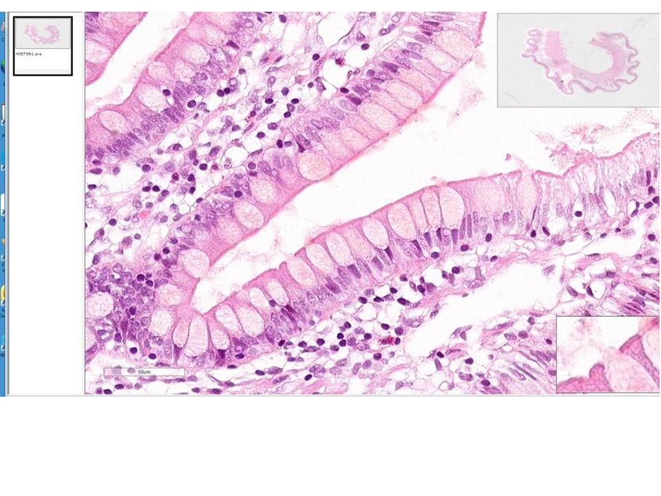
Ileum 148 Intestinal absorptive cells and goblet cells Simple columnar
epithelium Brush border Goblet cells

14
249 Ileum, monkey (PAS) Basement membrane of epithelium
Simple columnar epithelium Connective tissue Brush border Goblet cells Basement membrane, brush border and goblet cells are PAS positive for sugars Cell nuclei

16
The basal lamina of epithelial cells usually is thin (EM 10f); however, under pathological conditions, it is thickened as in the lymphatic vessel and seminiferous epithelium (Sertoli cell) of an aged-rat testis (EM 8h). EM 10f EM 8h basal lamina

17
SPECIALIZATION OF EPITHELIA
MAINTAIN EXTENSIVE CONTACTS AMONG CELLS STRUCTURALLY AND FUNCTIONALLY POLARIZED JUNCTIONS ZONULA OCCLUDENS - TIGHT JUNCTION (BELT) ZONULA ADHERENS – ADHERING BELT DESMOSOME (MACULA ADHERENS) - SPOT ATTACHMENT GAP JUNCTIONS - COMMUNICATION
 ZONULA ADHERENS – ADHERING BELT. DESMOSOME (MACULA. ADHERENS) - SPOT ATTACHMENT. GAP JUNCTIONS - COMMUNICATION.")
18
Terminal bars 32409

19
Terminal bars belt Terminal bars are part of a belt (zonula)
148 250 Intestinal absorptive cells Terminal bars Goblet cells 32409 belt 250 Terminal bars are part of a belt (zonula) around the cells.
 around the cells.")
20
EM 4: Intestine (Apical)
Tight junction, zonula adherens, Goblet cells terminal web 148

21
EM 2: Liver Gap junction, desmosome, tight junction

22
EM 2a: Liver- Gap junctions

23
133 133 vein valve Simple squamous epithelium cells viewed “face on” venule Lumen of lymphatic vessel Simple squamous epithelium cells viewed on their sides capillary valve 250

24
109 valve arteriole capillary 19680 testis Lumen of lymphatic vessels 196 Simple squamous epithelium valve vein

25
EM 10A showing capillary endothelial cells
Simple squamous epithelium : cells viewed on the side 109

27
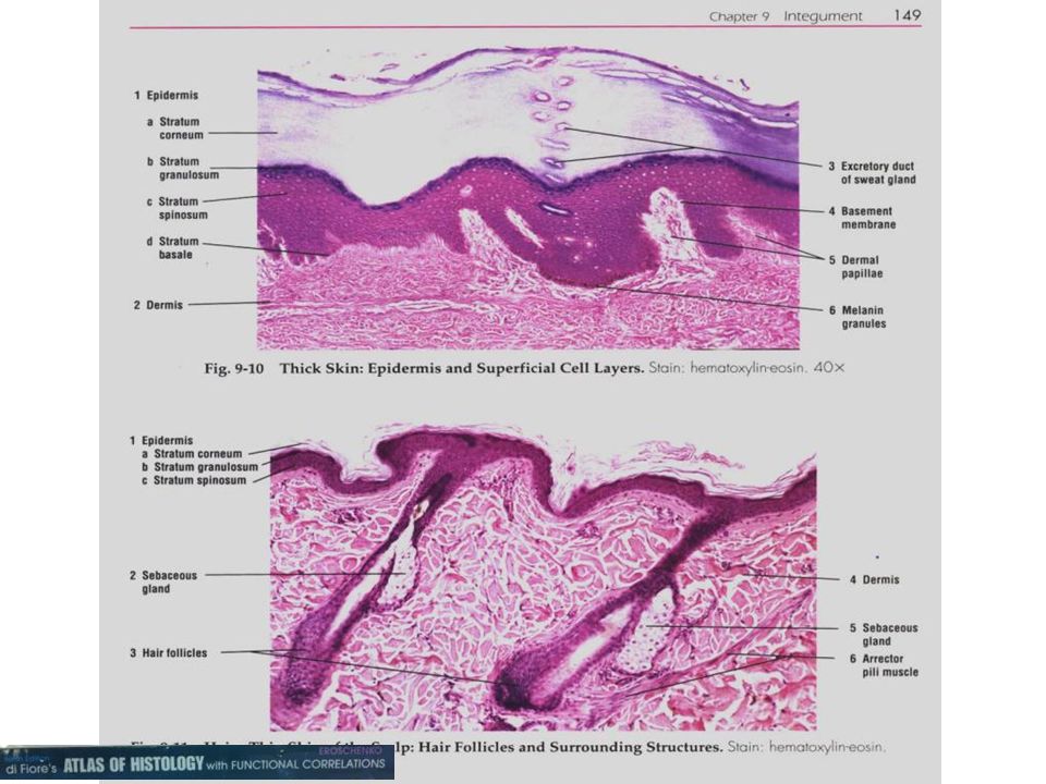
Stratified squamous epithelium
109 Keratinized dead flattered cells at surface

28
Slide 109: Skin, hand, monkey
prickle cell" layer (desmosomes) epidermis dermis
 epidermis. dermis.")
29
Slide 109 : Skin, hand, monkey
Fat cells dermis epidermis Stratified cuboidal is only found in sweat ducts of skin (Slide 109).
.")
31
Nuclei present in flat cells at surface
stratified squamous epithelium non-keratinized HISTO052 tongue Vagina (Slide 178)
")
33
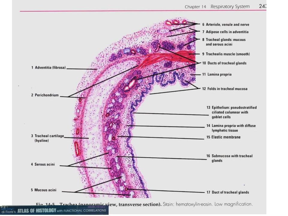
Lung air space Slide 133: Trachea, monkey 133 432 Simple squamous lining the air spaces of lungs Pseudostratified epithelium lining tracheal lumen Ciliated epithelium of trachea, goblet cells, thick basement membrane

34
tracheal lumen (Slide 133),
transition of the luminal epithelium from stratified squamous of vocal cords to pseudostratified, ciliated epithelium Plasma cells 133 pseudostratified, ciliated epithelium called respiratory epithelium human larynx, Slide 429 stratified squamous Note thickened basement membrane typical of this epithelium

36
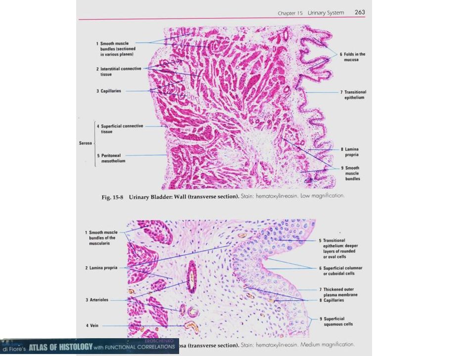
Transitional epithelium
urinary bladder (Slide 160) ureter (Slide 262)
 ureter (Slide 262)")
37
Slide 160 Image of the Urinary bladder
Transitional epithelium Mesothelium Lumen of bladder

38
HISTO052 (tongue) to observe mucus and serous glands
 to observe mucus and serous glands")
39
In summary All substances, including gases, normally received and given off from the body must pass through epithelia.

40
Questions on Epithelia
Which item(s) is/are characteristic of epithelia? a. secretory cells of glands b. covers organs c. line urinary tract d. a & b e. a, b, & c Which junction - description pair match? a. Zonula occludes – tight junction around the cell b. Zonula adherens – adhering junction around the cell c. Hemidesmosome – spot attachment of cells to the CT below d. a & b e. a, b, & c Which embryonic origin - distribution of epithelium do not match? a. endoderm - endothelium b. endoderm - alimentary tract c. mesoderm - mesothelium d. ectoderm - mammary gland e. ectoderm - epidermis
 is/are characteristic of epithelia a. secretory cells of glands. b. covers organs. c. line urinary tract. d. a & b. e. a, b, & c. Which junction - description pair match a. Zonula occludes – tight junction around the cell. b. Zonula adherens – adhering junction around the cell. c. Hemidesmosome – spot attachment of cells to the CT below. d. a & b. e. a, b, & c. Which embryonic origin - distribution of epithelium do not match a. endoderm - endothelium. b. endoderm - alimentary tract. c. mesoderm - mesothelium. d. ectoderm - mammary gland. e. ectoderm - epidermis.")
41
Many illustrations in these VIBS Histology YouTube videos were modified from the following books and sources: Many thanks to original sources! Bruce Alberts, et al Molecular Biology of the Cell. Garland Publishing, Inc., New York, NY. Bruce Alberts, et al Molecular Biology of the Cell. Garland Publishing, Inc., New York, NY. William J. Banks, Applied Veterinary Histology. Williams and Wilkins, Los Angeles, CA. Hans Elias, et al Histology and Human Microanatomy. John Wiley and Sons, New York, NY. Don W. Fawcett Bloom and Fawcett. A textbook of histology. W. B. Saunders Company, Philadelphia, PA. Don W. Fawcett Bloom and Fawcett. A textbook of histology. Chapman and Hall, New York, NY. Arthur W. Ham and David H. Cormack Histology. J. S. Lippincott Company, Philadelphia, PA. Luis C. Junqueira, et al Basic Histology. Lange Medical Publications, Los Altos, CA. L. Carlos Junqueira, et al Basic Histology. Appleton and Lange, Norwalk, CT. L.L. Langley, et al Dynamic Anatomy and Physiology. McGraw-Hill Book Company, New York, NY. W.W. Tuttle and Byron A. Schottelius Textbook of Physiology. The C. V. Mosby Company, St. Louis, MO. Leon Weiss Histology Cell and Tissue Biology. Elsevier Biomedical, New York, NY. Leon Weiss and Roy O. Greep Histology. McGraw-Hill Book Company, New York, NY. Nature ( Vol. 414:88,2001. Arthur C. Guyton,1971.Textbook of Medical Physiology W.B. Saunders company, Philadelphia, PA WW Tuttle and BA Schottelius Textbook of Physiology C.V. Mosby Co. A.L. Mescher Junqueira’s Basis Histology text and atlas, 13th ed. McGraw

42
On road from Fort Stockton
to Big Bend National Park, TX Ranch just south of Fort Stockton, TX

43
The end of

Similar presentations


 – 2.Connective (CT) – 3.Muscle – 4.Nervous.>")

 types of tissue: – 1. Epithelial – 2. Connective – 3. Muscle – 4. Nervous.>")

 Bram Welch-Horan (tbw5) October 11, 2005.>")


 Dr. Abdullah Aldahmash.>")


