Download presentation
Presentation is loading. Please wait.

1
Plastic Surgery Dr. Jalal Ali Hassan Lect. 3

2
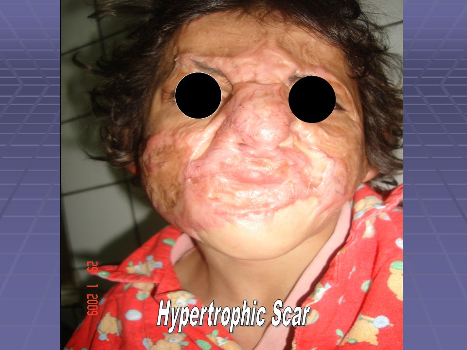
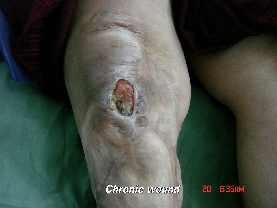
Wound Healing Wound Healing is a mechanism by which the body attempts to restore the integrity of the injured part. During repair,a complex chain of events eventually leads to the formation of a scar leads to the formation of a scar In certain circumstances, the cellular processes that contribute to repair become unregulated, leading to excessive scaring in the form of hypertrophic scars and keloids,at other times, abnormalities in repair occur, leading to deficiencies in wound healing such as are seen in chronic, non healig wound.

6
Chronic scar & Marjolins ulcer

7
Factors Influencing Healing of a wound 1-Site of the wound 2-Structures involved 3-Mechanism of wounding: Incision, Crush, Crush avulsion 4-Contamination(foreign bodies,bacteria) 5-Loss of tissue 6-Other local factors:vascular insufficiency (arterial or venous),previous radation,pressure 7-Systemic factors: malnutrition or vitamin & mineral deficiencies Disease(D.M) Medications(e.g. steroids) Medications(e.g. steroids) Immune deficiencies(e.g. chemotherapy,AIDS) Smoking
 Medications(e.g. steroids) Immune deficiencies(e.g. chemotherapy,AIDS) Smoking.")
8
Four Types of Wound Healing 1- Primary healing: Occurs when the wound is closed surgically within hours of its creation. The wound edges are reapproximated directly using sutures or by some other mechanical means,collagen metabolism provides long-term strength to the wound, when normal, synthesis, deposition & cross - linking. Epithelization, provides coverage of the wound surface & acts as a barrier from bacterial invasion.

11
2- Delayed Primary Healing: Contaminated or poorly delineated wound is left open to prevent wound infection. After 3-4 days local phagocytic cell recruitment into the wound has occurred & angiogenesis has begun. After 3-4 days local phagocytic cell recruitment into the wound has occurred & angiogenesis has begun. Inflamatory cells are present that destroy contaminating bacteria. Decreases the risk Inflamatory cells are present that destroy contaminating bacteria. Decreases the risk of infection in contaminated wounds of infection in contaminated wounds

13
3-Secondary Healing: An open full-thickness wound is allowed to close by both wound contraction & epithelization. Appropriate for infected or contaminated wounds.Allows drainage of fluid. Allows debidement with dressing chandes. Prolonged inflammatory phase leading to increased scaring& wound contracture. Contracture occur by myofibroblasts.

15
4-Healing of Partial-thickness wounds: Partial-thickness wounds which involve the epithelium & the superficial portion of the dermis, heal mainly by epithelization. There is minimal collagen deposition & an absence of wound contraction.

19
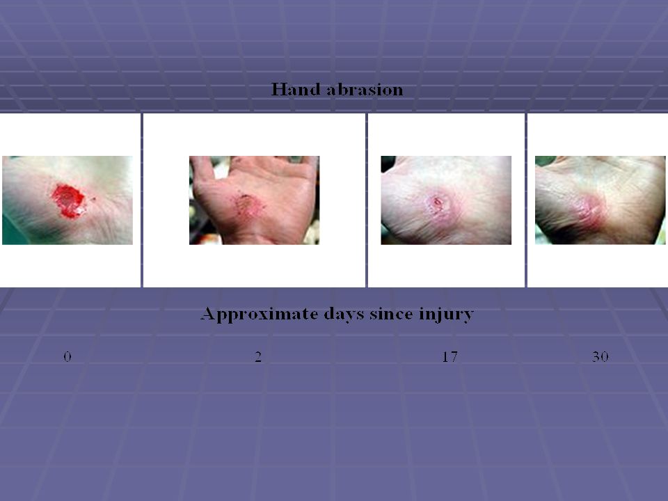
Phases of Wound Healing A- Inflammatory Phase : 1-Begins at the time of injury,lasts 2-3days 2-Begins with vasoconstruction to achieve hemostasis(epinephrine & thromboxane) 3-Platelet pluge forms & clotting cascade is activated, resulting in fibrin deposition 4-Platelets release - platelet-derived growth factor(PDGF)& transforming growth factor – B (TGF-B) from their alpha granules,attracting inflammatory cells,particularly Macrophages. 5-After hemostasis is achieved,vasodilatation occurs & vascular permability increases (due to histamine, platelet-activating factor, bradykinin,prostaglandin I- 2, prostaglandin E 2 & nitric oxide) aiding the infiltration of inflammatoty cells in to the wound. 6- Neutrophils peak at 24 hours & help with debridement 7-Monocytes enter the wound,becoming macrophages,& peak within 2-3 days 8-Macrophages produce PDGF&TGF-B,attracting fibroblasts & stimulating collagen production production
 aiding the infiltration of inflammatoty cells in to the wound. 6- Neutrophils peak at 24 hours & help with debridement 7-Monocytes enter the wound,becoming macrophages,& peak within 2-3 days 8-Macrophages produce PDGF&TGF-B,attracting fibroblasts & stimulating collagen production production.")
20
Phase I: Inflammation (Day 1-5)
")
21
B- Proliferative Phase: Lasts from the 3 rd day to the 3 rd week 1- Fibroblasts: attracted & activated by PDGF& TGF-B, arrive day 3, reach peak numbers by day 7 reach peak numbers by day 7 2-Collagen synthesis mainly type III( blood vessels &immature scar), angiogenesis & epithelization occur. angiogenesis & epithelization occur. 3-Total collagen content increases for 3weeks until collagen production & breakdown become equal &the remodeling phase begins. Fibroblasts require vitamin- C to produce collagen.

22
Phase II: Migration and Proliferation (Day 5-14)
")
23
C- Remodeling Phase : Increased collagen production & breakdown continue for 6 months Increased collagen production & breakdown continue for 6 months to 1 year. to 1 year. 1-Type I collagen replaces type III until it reaches a 4 :1 ratio of type I to type III (that of normal skin & mature scar tissue ) 2-Wound strength increases as collagen reorganizes along lines of tension & is cross- linked. tension & is cross- linked. 3-Vasculaity decreases 4-Fibroblast & Myofibroblasts cause wound contraction during the remodeling phase. remodeling phase.
 2-Wound strength increases as collagen reorganizes along lines of tension & is cross- linked. tension & is cross- linked. 3-Vasculaity decreases 4-Fibroblast & Myofibroblasts cause wound contraction during the remodeling phase. remodeling phase..")
25
Causes of abnormal wound healing -Hyperglycemia -Arterial disease –ischemia leads to inhibited collagen production & infection. -Venous insufficincy, increase venous pressure lead to edema &decrease O2 diffusion collagen production & infection. -Venous insufficincy, increase venous pressure lead to edema &decrease O2 diffusion -Abnormal pressure distribution -Nutritional deficiencies -Infection increases collagen breakdown & decrease epithelization

26
Scar & Scar Revision Scar :is a mark remaining after the healing of a wound or other morbid process. of a wound or other morbid process. Features of a good scars : 1- Fine line or series of lines to RSTL, contour 1- Fine line or series of lines to RSTL, contour junction & skin wrinkles. junction & skin wrinkles. 2-No contour irregularities. 2-No contour irregularities. 3-No pigmentation abnormalities. 3-No pigmentation abnormalities. 4-No contractures or distortion of adjacent 4-No contractures or distortion of adjacent structures. structures.

27
Types of Scars : - Immature scar - Immature scar - Mature scar - Mature scar

28
Objectives of Scar Revision: 1-To improve scar direction 2-To decrease scar width 3-To divide a long scar into smaller components. 4-To correct mal alignment or distortion of anatomical units. anatomical units. 5-To improve any surface irregularities. 6-To correct any pigmentation irregularities.

29
How to obtaining a fine line scar: A- Controllable Factors; 1- A traumatic technique. 1- A traumatic technique. 2-Eversion of wound edges. 2-Eversion of wound edges. 3-Placement of the scar in the same 3-Placement of the scar in the same direction of the skin lines. direction of the skin lines.

30
B – Non controllable Factors: 1-Age 1-Age 2-Site 2-Site 3-Type of the skin 3-Type of the skin 4-Length of the scar 4-Length of the scar 5-U –shaped scar 5-U –shaped scar

31
C- Complicating Factors: 1-Skin disorders e.g;Ehler –Danlos syndro ( genitically transmited disease,hyperextensible &laxity occur prematurely -abnormal collagen maturation & tissue fragility - surgery ass. With prolonged healing, haemorrhage, and darkly pigmented or telangiectatic haemorrhage, and darkly pigmented or telangiectatic hypertrophic scars.-elective surgery is usually not adviced hypertrophic scars.-elective surgery is usually not adviced 2-Infection 2-Infection 3-Individual healing mechnism. 3-Individual healing mechnism.

32
Factors to be considered before performing Scar Revision: 1-Time since injury. 2-Nature of the injuring agent. 3-Age4-Location 5- Ethnic back ground e.g; hyperpigmentation less in lighter skin 6- Skin tone & light effect; scars are visible by different color than the surrounding light reflection from the scar surface. surface. 7- Healing of previous scar. 8- Nature of the scar: spread wide scar,hypertrophic scar true keloid. true keloid. 9-Whether any skin lost. 10-Perception & expectations of the patient & family.

33
Treatment : Medical ; 1- Ionizing Radiation 1- Ionizing Radiation 2- Steroid injection 2- Steroid injection 3- Pressure; a -pressure splint. 3- Pressure; a -pressure splint. b –Silicon gel sheet. b –Silicon gel sheet. Surgical : 1-Excision 6-Skin flap 2-Excision &underminig 7-Dermabrasion 2-Excision &underminig 7-Dermabrasion 3-Z- Plasty 8-Tissue expantion 3-Z- Plasty 8-Tissue expantion 4- W- Plasty 9-Laser 4- W- Plasty 9-Laser 5-Skin graft 5-Skin graft

34
Undesirable Results from Scar Revision: 1- Haematoma,if tissue dead space not closed by suturing or compression dressing 1- Haematoma,if tissue dead space not closed by suturing or compression dressing 2-Infection : rare in scar revision of face, 2-Infection : rare in scar revision of face, occur on the trunk& extremities, retained occur on the trunk& extremities, retained foreign bodies lead to infection foreign bodies lead to infection 3-Hyperpigmentation : use of SPF15%, Tretinoin 3-Hyperpigmentation : use of SPF15%, Tretinoin & Hydroquinone & Hydroquinone 4-Milia are small sebaceous inclusion cysts. 4-Milia are small sebaceous inclusion cysts. 5-Dehisence: due to excess tension or direct 5-Dehisence: due to excess tension or direct trauma after sutures removed. trauma after sutures removed. - Use skin tapes after suture removal. - Use skin tapes after suture removal.

Similar presentations


 Immediate threat: –Dehydration and electrolyte.>")


