Download presentation
Presentation is loading. Please wait.

1
The Integumentary System Human Anatomy and Physiology

2
Types of Membranes

3
Functions of the Integumentary System Protective covering Prevents harmful substances and organisms from entering the body Reduces water loss from deeper tissues Regulation of body temperature Houses sensory receptors Contains immune system cells Synthesizes vitamin D Excretes small quantities of waste Absorption of drugs and other agents

5
Components of the Integumentary System Skin Hair Nails Sebaceous glands Sweat glands

6
Layers of the Skin Epidermis Dermis Subcutaneous layer (hypodermis)
")
8
Thick versus Thin Skin Thick Skin Palms of hands and soles of feet Hairless Subject to much abrasion Thicker epidermis (has an extra layer) Thin Skin Found everywhere else on the body Has hair Lacks one layer of the epidermis “Thick” and “thin” are not describing actual depth of tissue!!! Thickest skin = upper back Thinnest skin = eyelids

9
Epidermis Stratified squamous epithelium Lacks blood vessels Grows from the bottom layer (stratum basale) Keratinization
 Keratinization")
10
Layers of Epidermis Stratum corneum Stratum lucidum Stratum granulosum Stratum spinosum Stratum basale Basement membrane

11
Epidermal Layers Stratum corneum – flattened cells, desicated, anucleate cells, keratinized Water barrier Varies in thickness Thickens with unusual amounts of friction calluses Stratum lucidum – in thick skin only, cells in process of keratinization

12
Epidermal Layers continued… Stratum granulosum – only a few cells thick, appears granular Cells contain numerous keratin granules Stratum spinosum – several cells thick, numerous cytoplasmic processes called spines, nuclei elongated Stratum basale – single layer of cells on bottom, contains skin stem cells Cells appear cuboidal or low columnar

14
Dermis Epidermal ridges and dermal papillae Irregular dense connective tissue Thicker than epidermis Muscle and nerve fibers, blood vessels, hair follicles, sebaceous glands, and sweat glands 2 layers: papillary and reticular

15
Layers of Dermis Papillary layer Thinner, superficial layer Loose CT Contains blood vessels that serve the epidermis Contains nerve processes Reticular layer Varies in thickness, but generally thicker than papillary layer Contains thicker collagen and elastic fibers May contain smooth muscle cells

16
Subcutaneous Layer/Hypodermis Loose connective tissue and adipose tissue Connective tissue fibers are continuous with dermis Panniculus adiposus – layer of adipose tissue that insulates and stores energy Arrector pili muscles originate here

17
Cells of the Epidermis Keratinocytes Main cell type Produce keratin Melanocytes In stratum basale Contain melanin granules Langerhans cells Involved in contact dermatitis reactions Merkel cells In stratum basale Most abundant in fingertips Sense light touch

18
Skin Color There are 3 pigments involved in skin color: melanin, carotene, and hemoglobin. Melanin is the only pigment made by the skin (from tyrosine) and ranges in color from yellow red-brown black. Skin color differences result from the kind and amount of melanin made and retained by the skin cells. All humans have relatively the same number of melanocytes.
 and ranges in color from yellow red-brown black. Skin color differences result from the kind and amount of melanin made and retained by the skin cells. All humans have relatively the same number of melanocytes..")
19
Skin Color continued… Freckles and moles are local accumulations of melanin. A tan is darkening of the skin as a result of increased melanin production, usually in response to prolonged exposure to UV radiation. Carotene is a yellow orange pigment found in certain plant products. It tends to accumulate in the stratum corneum and in the fat of the hypodermis, and it is more obvious when large amounts of carotene-rich foods are eaten.

20
Skin Color continued… Hemoglobin gives a pinkish hue to fair skin which is most noticeable in Caucasian skin. A more crimson pigment results when the hemoglobin is highly oxygenated. Hemoglobin is found in the RBCs of the dermal capillaries. Hair color is genetically determined and results from the amount and type of pigment secreted by melanocytes near hair follicles. Dark hair has more melanin than light hair. Red hair contains an iron pigment called trichosiderin, and gray hair is a mixture of pigmented and unpigmented hair.

21
Nerve Supply to the Skin Free nerve endings found in the epidermis and papillary dermis sense temperature, vibration, pain, etc. Encapsulated nerve endings: Pacinian corpuscles – deep dermis and hypodermis; sense deep pressure Meissner’s corpuscles – in papillary region of dermis; sense light touch

22
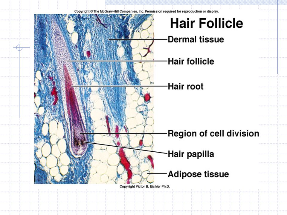
Hair Present on all surfaces except for palms, soles, lips, nipples, and parts of external reproductive organs Made of keratinized cells Hair follicle Hair papilla Hair shaft Hair color Arrector pili

26
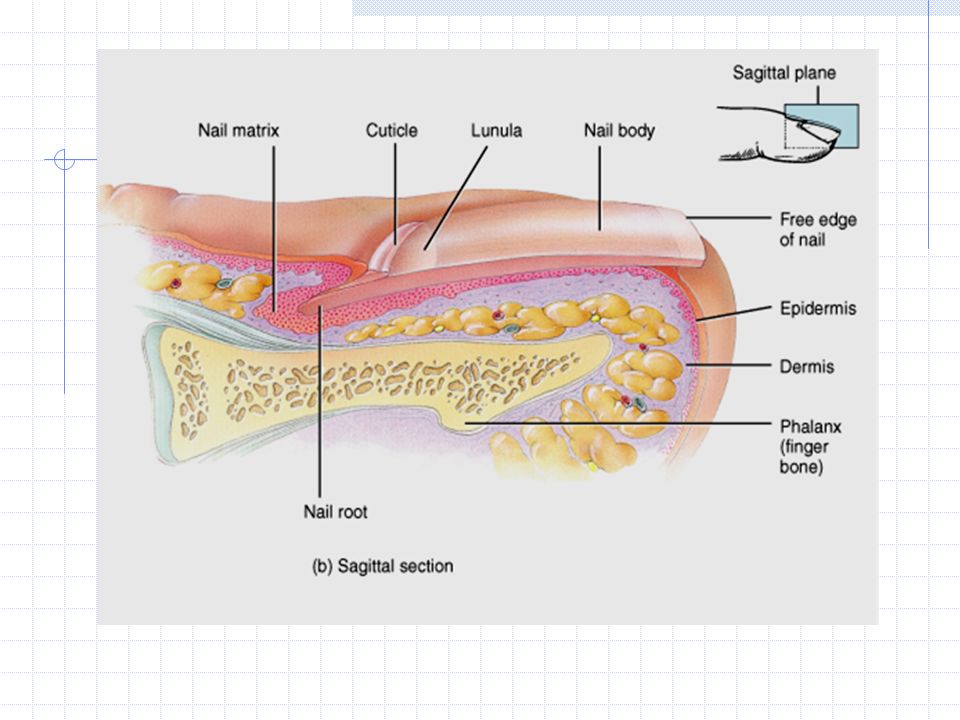
Nails Protective coverings on the ends of fingers and toes Nail plate Nail bed Lunula

28
Sebaceous Glands Sebaceous glands are associated with hair follicles Sebum Found everywhere except palms and soles Acne

30
Sweat Glands In dermis or superficial subcutaneous layer Eccrine glands Most numerous Produce sweat on hot days and during exercise Apocrine glands Become active at puberty Secretions smell because of bacterial activity Active during emotional upset, fright, pain, sexual arousal Ceruminous glands and mammary glands

32
Healing of Wounds Inflammation = normal response to injury or stress Epidermal cuts Deep cuts Blood clots Scabs Scars

33
Healing of Burns First degree burns Superficial partial- thickness burn Second degree burns Deep partial- thickness burn Third degree burns Full-thickness burn Rule of 9s

34
Aging and Skin Epidermal cells reproduce slower larger and more irregular shape Age spots – sites of oxidation of fats in secretory cells of apocrine and eccrine glands Dermis reduces wrinkling and sagging Drier skin because of less oil from sebaceous glands Gray or white hair from decreased melanin production

35
Aging – continued… Slower hair growth and fewer hair follicles thinner hair and/or hair loss Less blood supply to nail beds impaired growth Diminished sensitivity to pain and pressure because of fewer receptors Fewer sweat glands, fewer dermal blood vessels, and declined ability to shiver decreased ability to control temperature Diminished ability to activate vitamin D reduced skeletal health

Similar presentations


 Largest organ of the body (15% of body weight) Skin thickness variable, normally 1-2 mm Protection –chemical barrier (waterproof)>")
