Download presentation
Presentation is loading. Please wait.

1
Synaptic transmission: communication between neurons

2
Two principal kinds of synapses: electrical and chemical

3
Gap junctions are formed where hexameric pores called connexons connect with one between cells

4
Electrical synapses are built for speed

5
Contrast with chemical synapse Delay of about 1 ms

6
Electrical coupling is a way to synchronize neurons with one another

7
Electrical synapses are not presently considered to be the primary means of communication between neurons in the mammalian nervous system, but they may prove to be more important than presently recognized

8
Rectification and uni-directionality of electrical synapses …not just simple bidirectional bridges between cells Conductance through gap junctions may be sensitive to the junctional potential (i.e the voltage drop between the two coupled cells), or sensitive to the membrane potential of either of the coupled cells Glial cells can also be connected by gap junctions, which allows synchronous oscillations of intracellular calcium http://users.umassmed.edu/michael.sanderson/mjslab/MOVIE.HTM
, or sensitive to the membrane potential of either of the coupled cells Glial cells can also be connected by gap junctions, which allows synchronous oscillations of intracellular calcium")
9
Chemical synapses: the predominant means of communication between neurons

13
An early experiment to support the neurotransmitter hypothesis

14
Criteria that define a neurotransmitter: 1.Must be present at presynaptic terminal 2.Must be released by depolarization, Ca ++ -dependent 3.Specific receptors must be present

15
Neurotransmitters may be either small molecules or peptides Mechanisms and sites of synthesis are different Small molecule transmitters are synthesized at terminals, packaged into small clear-core vesicles (often referred to as ‘synaptic vesicles’ Peptides, or neuropeptides are synthesized in the endoplasmic reticulum and transported to the synapse, sometimes they are processed along the way. Neuropeptides are packaged in large dense-core vesicles

16
Neurotransmitter is released in discrete packages, or quanta

17
Failure analysis reveals that neurons release many quanta of neurotransmitter when stimulated, that all contribute to the response Quantal content: The number of quanta released by stimulation of the neuron Quantal size: How size of the individual quanta

18
Quanta correspond to release of individual synaptic vesicles EM images and biochemistry suggest that a MEPP could be caused by a single vesicle EM studies revealed correlation between fusion of vesicles with plasma membrane and size of postsynaptic response

19
4-AP was used to vary the efficiency of release

20
Calcium influx is necessary for neurotransmitter release Voltage-gated calcium channels

21
Calcium influx is sufficient for neurotransmitter release

22
Synaptic release II The synaptic vesicle release cycle 1.Tools and Pools 2.Molecular biology and biochemistry of vesicle release: 1.Docking 2.Priming 3.Fusion 3.Recovery and recycling of synaptic vesicles

23
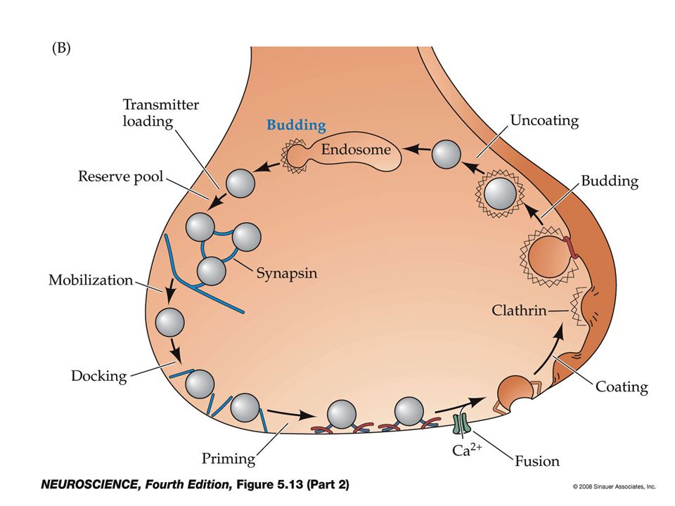
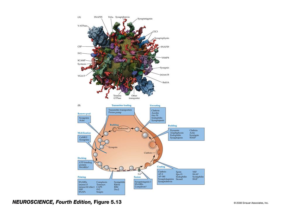
The synaptic vesicle cycle

24
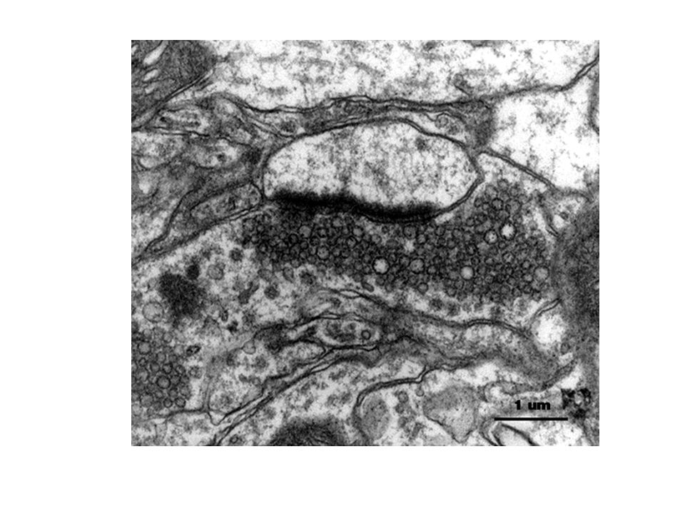
How do we study vesicle dynamics? Morphological techniques Electron microscopy to obtain static pictures of vesicle distribution; TIRFM (total internal reflection fluorescence microscopy) to visualize movement of vesicles close to the membrane Physiological studies Chromaffin cells Neuroendocrine cells derived from adrenal medulla with large dense-core vesicles. Can measure membrane fusion (capacitance measurements), or direct release of catecholamine transmitters using carbon fiber electrodes (amperometry) Neurons Measure release of neurotransmitter from a presynaptic cell by quantifying the response of a postsynaptic cell Genetics Delete or overexpress proteins in mice, worms, or flies, and analyze phenotype using the above techniques
 to visualize movement of vesicles close to the membrane Physiological studies Chromaffin cells Neuroendocrine cells derived from adrenal medulla with large dense-core vesicles. Can measure membrane fusion (capacitance measurements), or direct release of catecholamine transmitters using carbon fiber electrodes (amperometry) Neurons Measure release of neurotransmitter from a presynaptic cell by quantifying the response of a postsynaptic cell Genetics Delete or overexpress proteins in mice, worms, or flies, and analyze phenotype using the above techniques.")
25
Synaptic vesicle release consists of three principal steps: 1.Docking Docked vesicles lie close to plasma membrane (within 30 nm) 1.Priming Primed vesicles can be induced to fuse with the plasma membrane by sustained depolarization, high K +, elevated Ca ++, hypertonic sucrose treatment 2.Fusion Vesicles fuse with the plasma membrane to release transmitter. Physiologically this occurs near calcium channels, but can be induced experimentally over larger area (see ‘priming’). The ‘active zone’ is the site of physiological release, and can sometimes be recognized as an electron-dense structure..
. The ‘active zone’ is the site of physiological release, and can sometimes be recognized as an electron-dense structure...")
26
Becherer, U, Rettig, J. Cell Tissue Res (2006) 326:393 Morphologically, vesicles are classified as docked or undocked. Docked vesicles are further subdivided into primed and unprimed pools depending on whether they are competent to fuse when cells are treated with high K +, elevated Ca ++, sustained depolarization, or hypertonic sucrose treatment. Synaptic vesicles exist in multiple pools within the nerve terminal (reserve pool) (Release stimulated by flash-photolysis of caged calcium)
 326:393 Morphologically, vesicles are classified as docked or undocked. Docked vesicles are further subdivided into primed and unprimed pools depending on whether they are competent to fuse when cells are treated with high K +, elevated Ca ++, sustained depolarization, or hypertonic sucrose treatment. Synaptic vesicles exist in multiple pools within the nerve terminal (reserve pool) (Release stimulated by flash-photolysis of caged calcium).")
27
In CNS neurons, vesicles are divided into Reserve pool (80-95%) Recycling pool (5-20%) Readily-releasable pool (0.1-2%; 5-10 synapses per active zone) Rizzoli, Betz (2005). Nature Reviews Neuroscience 6:57-69) A small fraction of vesicles (the recycling pool) replenishes the RRP upon mild stimulation. Strong stimulation causes the reserve pool to mobilize and be released
 A small fraction of vesicles (the recycling pool) replenishes the RRP upon mild stimulation. Strong stimulation causes the reserve pool to mobilize and be released.")
28
Vesicle release requires many proteins on vesicle and plasma membrane

29
Docking: UNC-18 (or munc-18) is necessary for vesicle docking (Weimer et al. 2003, Nature Neuroscience 6:1023) 1.unc-18 mutant C. elegans have neurotransmitter release defect 2.unc-18 mutant C. elegans have reduction of docked vesicles
 1.unc-18 mutant C. elegans have neurotransmitter release defect 2.unc-18 mutant C. elegans have reduction of docked vesicles.")
30
Unc-18 mutants are defective for evoked and spontaneous release

31
Unc-18 mutants are defective for calcium-independent release primed vesicles occasionally fuse in the absence of calcium; a calcium-independent fusion defect suggests a lack of primed vesicles

32
UNC-18 (munc18) is required for docking: unc-18 mutants have fewer docked vesicles
 is required for docking: unc-18 mutants have fewer docked vesicles")
33
Summary: Unc-18 mutants are unable to dock vesicles efficiently. Impaired docking leads to fewer primed vesicles; fewer primed vesicles leads to reduced overall neurotransmitter release.

35
Priming Vesicles in the reserve pool undergo priming to enter the readily- releasable pool At a molecular level, priming corresponds to the assembly of the SNARE complex

36
The SNARE complex

37
UNC-13 is a critical priming factor Richmond and Jorgensen (1999) Nature Neuroscience 2:959 normalunc-13 mutants unc-13 mutants have higher levels of synaptic vesicles than normal No docking defect was observed
 Nature Neuroscience 2:959 normalunc-13 mutants unc-13 mutants have higher levels of synaptic vesicles than normal No docking defect was observed")
38
unc-13 mutants have evoked release defect

39
Calcium-indepenent release is also defective, indicating that the defect is in priming

40
Munc-13 function in priming

41
Inhibitory domain, folds back on itself “open” syntaxin doesn’t fold properly

42
unc-13 defect can be bypassed by providing an “open” form of syntaxin

43
Model for unc-13, unc-18, syntaxin interaction in priming

44
Synaptotagmin functions as a calcium sensor, promoting vesicle fusion

45
Synaptic vesicles recycle post-fusion

46
Modern methods to track recycling membrane

47
The ATP-ase NSF disassembles the SNARE complex Endocytosis retrieves synaptic vesicle membrane and protein from the plasma membrane following fusion

Similar presentations


 and inspiratory neuron (bottom trace) were labeled with dye during intracellular.>")


 Ian Parker Lecture # 18 - Quantal release of neurotransmitter.>")


 Nervous system functions Structure of a neuron Sensory, motor, inter- neurons Membrane potential Sodium.>")


