Download presentation
Presentation is loading. Please wait.

1
Chapter 43 The Body’s Defenses

3
Lines of Defense Nonspecific Defense Mechanisms……

4
Phagocytic and Natural Killer Cells Neutrophils –60-70% WBCs; engulf and destroy microbes at infected tissue Monocytes: 5% WBCs; develop into…. –Macrophages( largest,long-lived) enzymatically destroy microbes Eosinophils 1.5% WBCs; destroy large parasitic invaders (blood flukes) Natural killer (NK) cells destroy virus-infected body cells & abnormal cells WBC=leukocytes
 enzymatically destroy microbes Eosinophils 1.5% WBCs; destroy large parasitic invaders (blood flukes) Natural killer (NK) cells destroy virus-infected body cells & abnormal cells WBC=leukocytes.")
5
The Inflammatory Response 1- Tissue injury; release of chemical signals~ basophils/mast cells (in connective tissue) release histamine (causes Step 2... –prostaglandins: increases blood flow & vessel permeability 2/3- Dilation and increased permeability of capillary~ chemokines: secreted by blood vessel endothelial cells and monocytes attrack phagocytes to area 4- Phagocytosis of pathogens~ fever & pyrogens: leukocyte-released molecules increase body temperature

6
Specific Immunity Lymphocyctes pluripotent stem cells... B Cells (bone marrow) T Cells (thymus) Antigen: a foreign molecule that elicits a response by lymphocytes (virus, bacteria, fungus, protozoa, parasitic worms) Antibodies: antigen-binding immunoglobulin, produced by B cells Antigen receptors: plasma membrane receptors on b and T cells
 T Cells (thymus) Antigen: a foreign molecule that elicits a response by lymphocytes (virus, bacteria, fungus, protozoa, parasitic worms) Antibodies: antigen-binding immunoglobulin, produced by B cells Antigen receptors: plasma membrane receptors on b and T cells.")
7
Clonal selection Effector cells: short-lived cells that combat the antigen Memory cells: long-lived cells that bear receptors for the antigen Clonal selection: antigen-driven cloning of lymphocytes “Each antigen, by binding to specific receptors, selectively activates a tiny fraction of cells from the body’s diverse pool of lymphocytes; this relatively small number of selected cells gives rise to clones of thousands of cells, all specific for and dedicated to eliminating the antigen.”

8
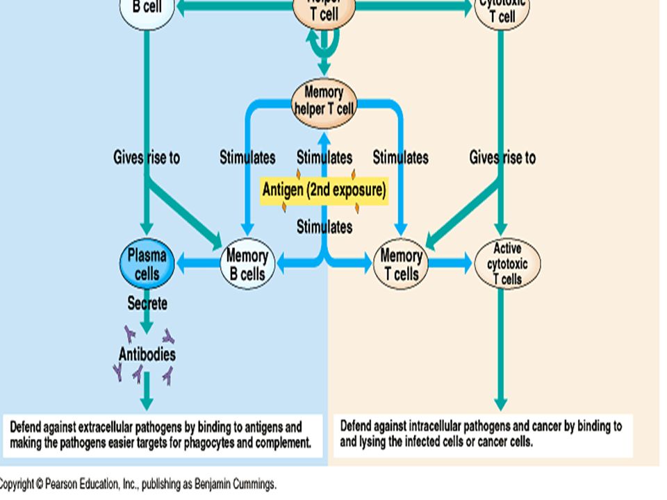
Immune Responses Primary immune response: lymphocyte proliferation and differentiation the 1st time the body is exposed to an antigen Plasma cells: antibody-producing effector B-cells Secondary immune response: immune response if the individual is exposed to the same antigen at some later time ~ Immunological memory

9
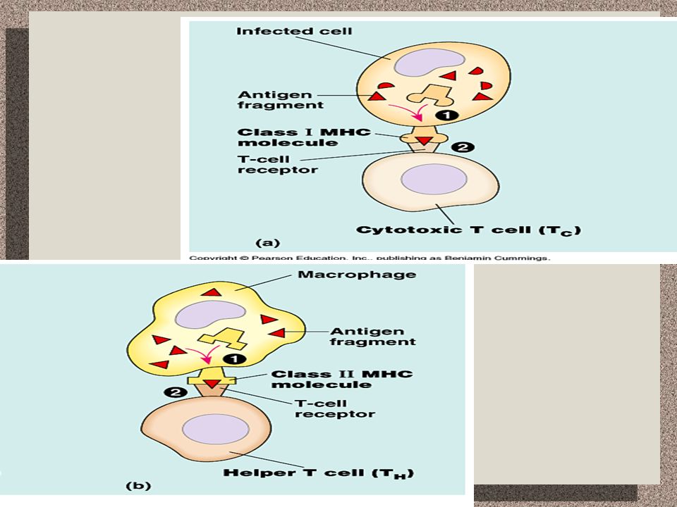
Self/Nonself Recognition Self-tolerance: capacity to distinguish self from non-self Autoimmune diseases: failure of self-tolerance; multiple sclerosis, lupus, rheumatoid arthritis, insulin-dependent diabetes mellitus Major Histocompatability Complex (MHC): body cell surface antigens coded by a family of genes Class I MHC molecules: found on all nucleated cells Class II MHC molecules: found on macrophages, B cells, and activated T cells Antigen presentation: process by which an MHC molecule “presents’ an intracellular protein to an antigen receptor on a nearby T cell Cytotoxic (killer)T cells (T C ): bind to protein fragments displayed on class I MHC molecules Helper T cells (T H ): bind to proteins displayed by class II MHC molecules
: body cell surface antigens coded by a family of genes Class I MHC molecules: found on all nucleated cells Class II MHC molecules: found on macrophages, B cells, and activated T cells Antigen presentation: process by which an MHC molecule presents’ an intracellular protein to an antigen receptor on a nearby T cell Cytotoxic (killer)T cells (T C ): bind to protein fragments displayed on class I MHC molecules Helper T cells (T H ): bind to proteins displayed by class II MHC molecules")
11
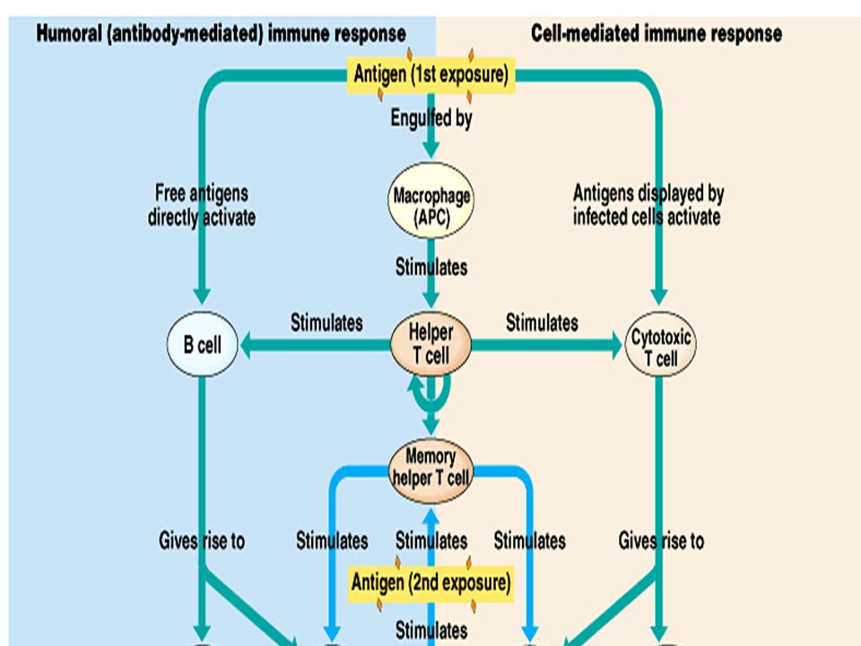
Types of immune responses Humoral immunity B cell activation Production of antibodies Defend against bacteria, toxins, and viruses free in the lymph and blood plasma Cell-mediated immunity T cell activation Binds to and/or lyses cells Defend against cells infected with bacteria, viruses, fungi, protozoa, and parasites; nonself interaction

14
Helper T lymphocytes Function in both humoral & cell-mediated immunity Stimulated by antigen presenting cells (APCs) T cell surface protein CD4 enhances activation Cytokines secreted (stimulate other lymphocytes): a) interleukin-2 (IL-2): activates B cells and cytotoxic T cells b) interleukin-1 (IL-1): activates helper T cell to produce IL-2
 T cell surface protein CD4 enhances activation Cytokines secreted (stimulate other lymphocytes): a) interleukin-2 (IL-2): activates B cells and cytotoxic T cells b) interleukin-1 (IL-1): activates helper T cell to produce IL-2")
15
Cell-mediated: cytotoxic T cells Destroy cells infected by intracellular pathogens and cancer cells Class I MHC molecules (nucleated body cells) expose foreign proteins Activity enhanced by CD8 surface protein present on most cytotoxic T cells (similar to CD4 and class II MHC) T C cell releases perforin, a protein that forms pores in the target cell membrane; cell lysis and pathogen exposure to circulating antibodies
 expose foreign proteins Activity enhanced by CD8 surface protein present on most cytotoxic T cells (similar to CD4 and class II MHC) T C cell releases perforin, a protein that forms pores in the target cell membrane; cell lysis and pathogen exposure to circulating antibodies")
16
Humoral response: B cells Stimulated by T-dependent antigens (help from TH cells) Macrophage (APCs) with class II MHC proteins Helper T cell (CD4 protein) Activated T cell secretes IL-2 (cytokines) that activate B cell B cell differentiates into memory and plasma cells (antibodies)
 Macrophage (APCs) with class II MHC proteins Helper T cell (CD4 protein) Activated T cell secretes IL-2 (cytokines) that activate B cell B cell differentiates into memory and plasma cells (antibodies)")
17
Antibody Structure & Function Epitope: region on antigen surface recognized by antibodies 2 heavy chains and 2 light chains joined by disulfide bridges Antigen-binding site (variable region)
")
18
5 classes of Immunoglobins:antibodies IgM: 1st to circulate; indicates infection; too large to cross placenta IgG: most abundant; crosses walls of blood vessels and placenta; protects against bacteria, viruses, & toxins; activates complement IgA: produced by cells in mucous membranes; prevent attachment of viruses/bacteria to epithelial surfaces; also found in saliva, tears, and perspiration IgD: do not activate complement and cannot cross placenta; found on surfaces of B cells; probably help differentiation of B cells into plasma and memory cells IgE: very large; small quantity; releases histamines-allergic reaction

21
Antibody-mediated Antigen Disposal Neutralization (opsonization): antibody binds to and blocks antigen activity Agglutination: antigen clumping Precipitation: cross-linking of soluble antigens Complement fixation: activation of 20 serum proteins, through cascading action, lyse viruses and pathogenic cells
: antibody binds to and blocks antigen activity Agglutination: antigen clumping Precipitation: cross-linking of soluble antigens Complement fixation: activation of 20 serum proteins, through cascading action, lyse viruses and pathogenic cells")
23
Immunity in Health & Disease Active immunity: long term/ natural: conferred immunity by recovering from disease artificial: immunization and vaccination; produces a primary response Passive immunity: short term transfer of immunity from one individual to another natural: mother to fetus; breast milk artificial: rabies antibodies ABO blood groups (antigen presence) Rh factor (blood cell antigen); Rh- mother vs. an Rh+ fetus (inherited from father)
.")
24
Abnormal immune function I Allergies hypersensitive responses to environmental antigens (allergens); mast cells release histamine causes dilation and blood vessel permeability, epinephrine Antihistamines can relieve symptoms anaphylactic shock: life threatening reaction to injected or ingested allergens.
; mast cells release histamine causes dilation and blood vessel permeability, epinephrine Antihistamines can relieve symptoms anaphylactic shock: life threatening reaction to injected or ingested allergens.")
25
Abnormal immune function II Autoimmune disease: –The system turns against the body’s own molecules –Examples: multiple sclerosis, lupus, rheumatoid arthritis, insulin-dependent diabetes mellitus Rheumatoid arthritis

26
Abnormal immune function III Immunodeficiency disease: Immune components are lacking, and infections recur Ex: SCIDS (bubble-boy); A.I.D.S.
; A.I.D.S.")
Similar presentations