Download presentation
Presentation is loading. Please wait.

1
Cardiovascular System
Chapter 11 Cardiovascular System

2
The major function of the cardiovascular system is transportation.
The blood is the transport vehicle. It carries oxygen, nutrients, cell wastes, hormones, and many other substances vital for body homeostasis to and from cells.

3
The force to move the blood throughout the body is provided by the beating heart.

4
The Cardiovascular System consists of:
Blood (transport vehicle) Heart (pump) Blood vessels (network of tubes)
 Heart (pump) Blood vessels (network of tubes)")
5
Location and Size of the Heart
The heart is the size of a fist The heart is located between the two lungs A double sac called the pericardium surrounds the heart.

6
Lubricating fluid is found in between the two layers of the sac
Lubricating fluid is found in between the two layers of the sac. This fluid allows the heart to beat easily in a frictionless environment.

7
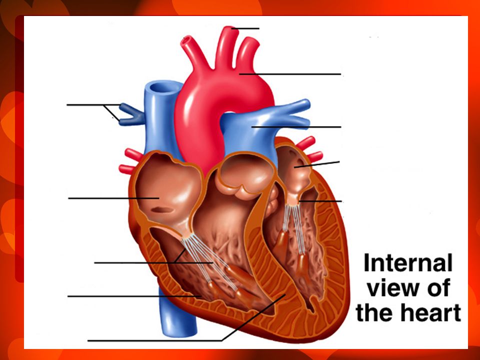
The walls of the heart are made of 3 layers:
Epicardium – outer layer Myocardium – layer that contracts Endocardium – lines the chambers and goes into the blood vessels

8
The heart has four chambers or cavities.
The two atria are located at the superior or top of the heart and the two ventricles are located at the inferior or bottom of the heart.

10
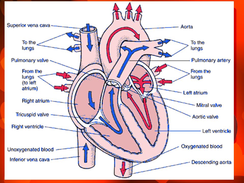
Blood flow through the heart

11
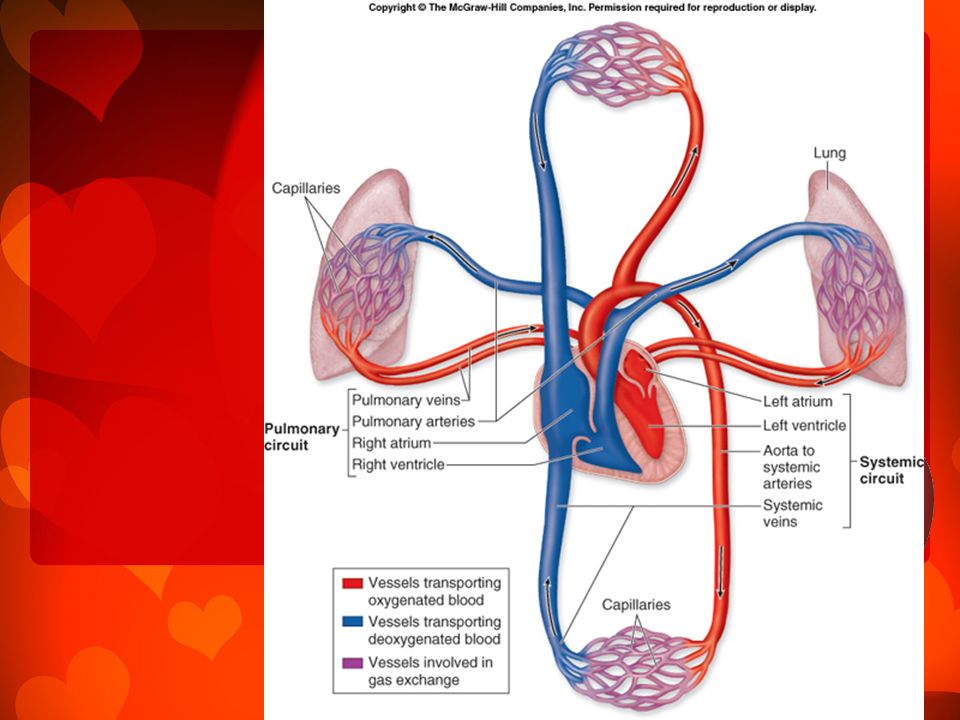
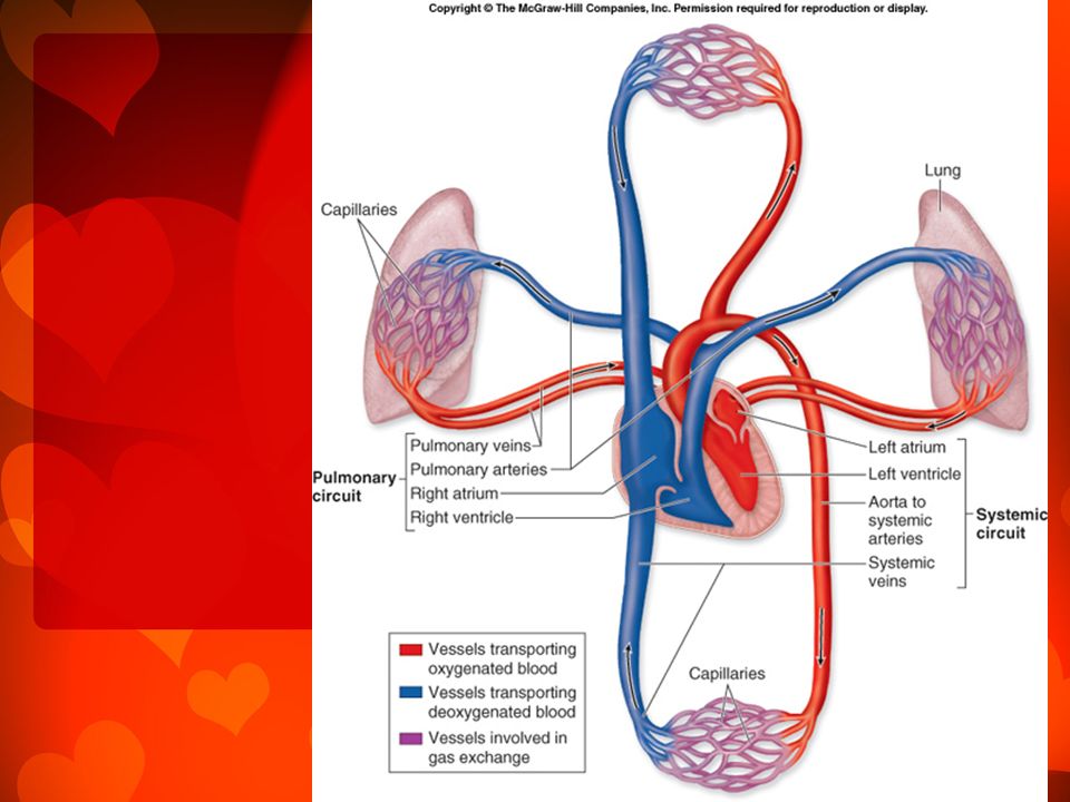
Pulmonary Circulation
Right side: pumps to lungs oxygen poor on its way to the lungs – picks up oxygen – becomes oxygen rich

13
The Steps: Oxygen poor blood enter the superior and inferior vena cava
Blood goes into the right atrium then right ventricle Blood is them pumped to the pulmonary trunk which branches into pulmonary arteries

14
Blood goes to the lungs and picks up oxygen
Blood leaves the lungs in the pulmonary veins and goes to the left atrium

17
Systemic Circulation Left side: pumps blood with oxygen to the body cells and back to the heart

18
The Steps: Blood enters the left atrium then goes to the left ventricle Blood is then pumped to the body cells through the aorta

19
3. Blood travels from the aorta to arteries, smaller arteries, capillaries (losses oxygen) then to the veins and then to the superior and inferior vena cava
 then to the veins and then to the superior and inferior vena cava.")
22
Cardiac Circulation The blood in the heart does not nourish the myocardium. The blood supply that nourishes the heart is provided by the right and left coronary arteries which branch from the aorta.

23
Physiology of the Heart
6 quarts pushed through 1000X Heart pumps 6000 quarts a day Cardiac muscle cells can and do contract spontaneously and independently. These contractions occur in a regular and continuous way.

24
The muscles in the different parts of the heart have different rhythms.
The heart has a unifying control system that coordinates its movement.

25
The Two Controlling Systems
1. The autonomic nervous system (automatic) acts like: the brakes and accelerators to increase or decrease heart rate
 acts like: the brakes and accelerators to increase or decrease heart rate.")
26
Intrinsic conduction system or nodal system
- causes the heart to depolarize in one direction (from atria to ventricle) - enforces a contraction rate of 75 beats/min
 - enforces a contraction rate of 75 beats/min.")
27
SA node starts each heartbeat and is known as the pacemaker.

28
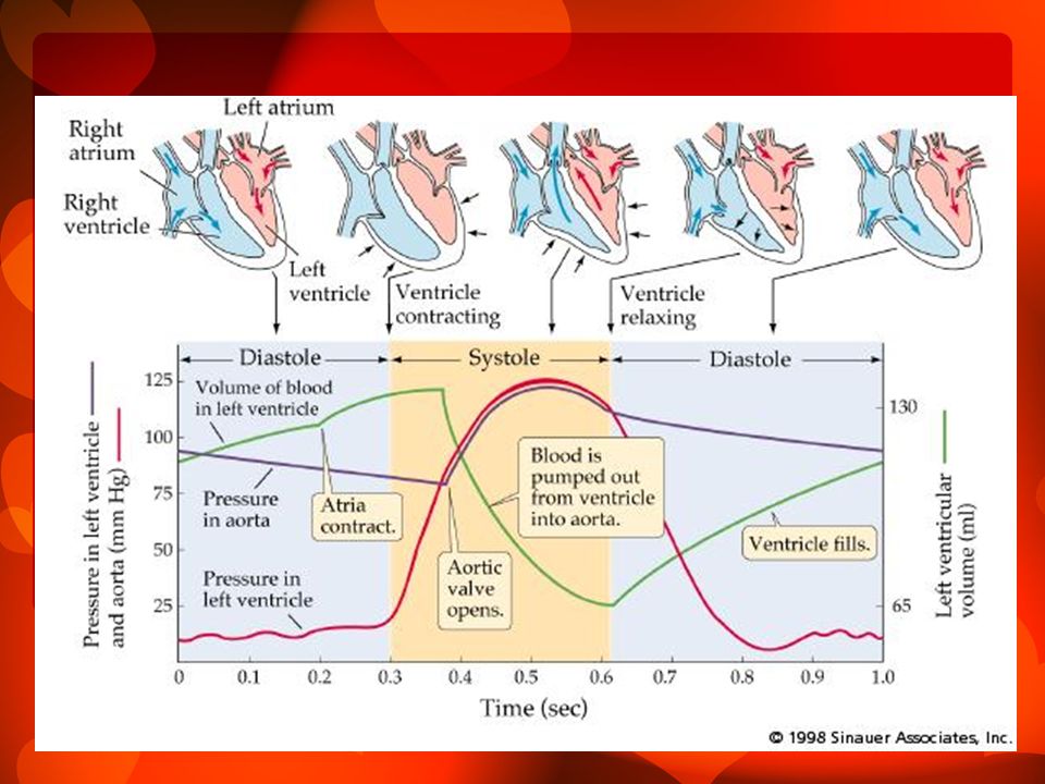
Cardiac Cycle and Heart Sounds

29
In a healthy heart the atria contract simultaneously
Systole – heart contraction Diastole – heart relaxation

30
The Cardiac Cycle refers to:
The events of one complete heart beat during which both atria and ventricles contract and then relax.

32
The average heart beats 75 beats/min.
The cardiac cycle lasts for .8 sec.

33
Heart Sounds When using a stethoscope, you can hear 2 distinct sounds during each cardiac cycle. These heart sounds are often described as lub and dub.

34
The first heart sound is made when: the AV valve closes - Lub
The second heart sound is made when: the semilunar valves close at the end of systole - Dub

35
The average heart rate is faster in females than males.
Females – beats per minute Males – bpm

36
Cardiovascular System: Blood Vessels

37
Blood circulates inside the blood vessels which is a closed transport system, the so called vascular system.

38
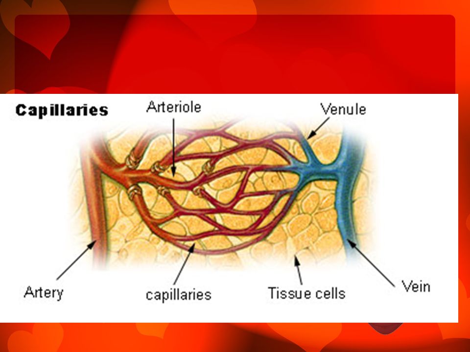
The Path of Blood: Arteries carry blood away from the heart (oxygen)
Veins carry blood to the heart (no oxygen) From the heart blood is propelled into: arteries smaller arteries arterioles capillary beds venules veins great veins
 From the heart blood is propelled into: arteries smaller arteries arterioles capillary beds venules veins great veins.")
40
Microscopic Anatomy of Blood Vessels

41
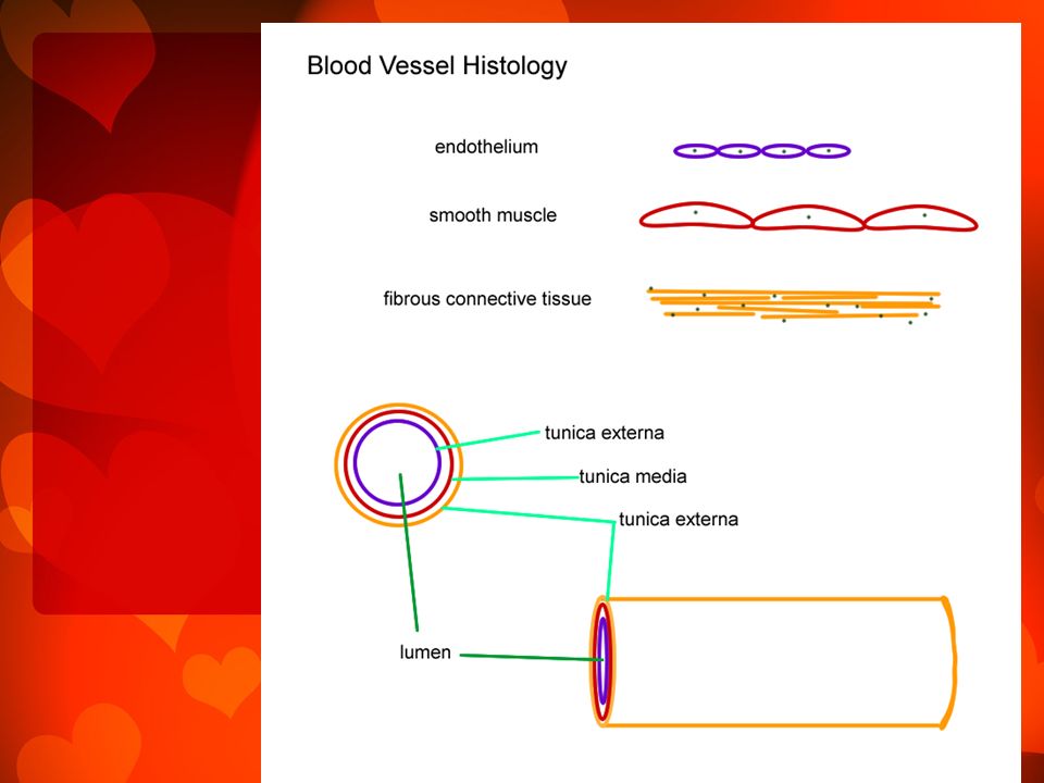
The walls of blood vessels have 3 coats or tunics
Tunica intima decreases friction Tunica media changes diameter of blood vessels Tunica externa function: support and protect blood vessels

43
The walls of arteries are usually thicker than the walls of veins
Artery walls must be strong and stretchy enough to take these continuous changes in pressure

44
Veins have thinner walls because they do not have to take the pressure
However, they are modified: to ensure the amount returning to the heart equals what is being pumped

45
Modifications Valves to prevent backflow
Muscles milk veins and move blood to the heart Breathing also helps

46
Location of Arteries and Veins in the body

47
Arteries are generally located in deep, well protected body areas.
Many veins are more superficial and some are easily seen and palpated on the body surface. Most deep veins follow the course of the major arteries.

48
Hepatic Portal Vein Delivers blood to the liver from the digestive organs

49
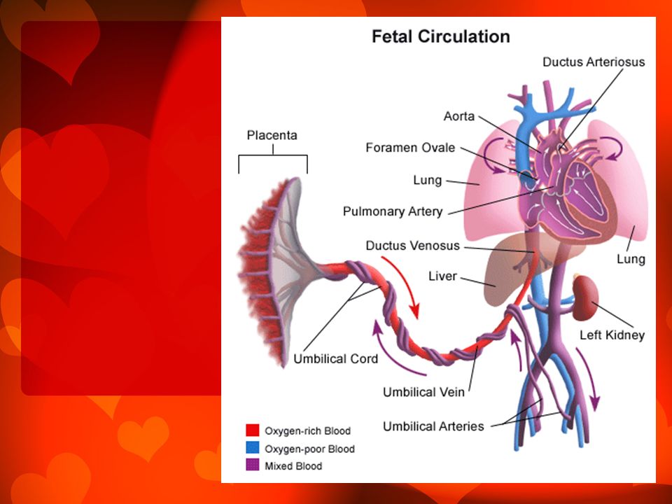
Fetal Circulation Since the lungs and digestive system are not yet functioning in a fetus, all nutrient, excretory and gas exchange occur through the placenta. Nutrients and oxygen move from the mother’s blood into the fetal blood and fetal wastes move in the opposite direction.

51
Physiology of Circulation
A good indication of the efficiency of a person’s circulatory system can be obtained by taking arterial pulse and blood pressure. Vital Signs: arterial pulse, blood pressure, respiratory rate, body temperature

52
Arterial Pulse Pulse: alternating expansion and recoiling of an artery that occurs with each ventricular beat Average pulse is a person at rest: BPM

53
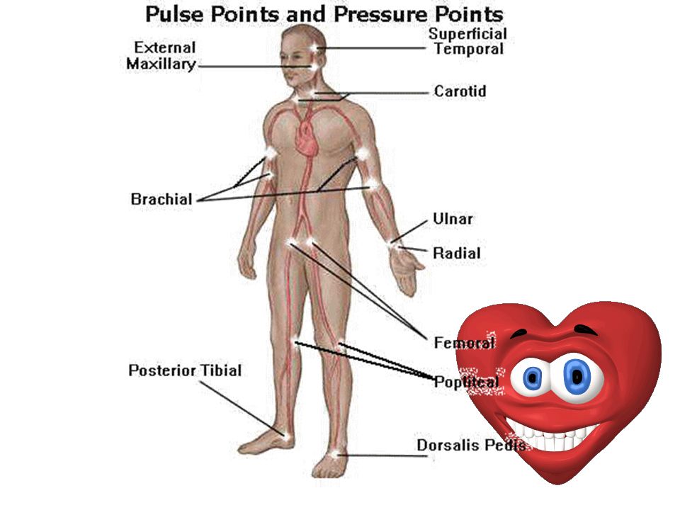
Pressure Points Areas where you can feel your pulse
These points can also be compressed to stop blood flow into distal tissue during hemorrhage

55
Blood Pressure Pressure the blood exerts against the inner wall of the blood vessels and it sis the force that keeps the blood circulation

56
Blood Pressure Gradient
Blood flows along a pressure gradient (from high to low) If a vein is cut the blood flows evenly from the wound If an artery is cut the blood comes out in rapid spurts
 If a vein is cut the blood flows evenly from the wound. If an artery is cut the blood comes out in rapid spurts.")
57
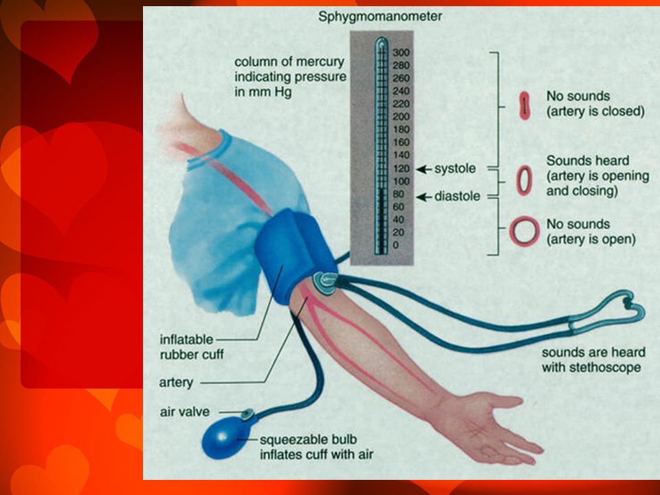
Measuring Blood Pressure
Because the heart alternately contracts and relaxes the off-and-on flow of blood into the arteries causes the blood pressure to rise and fall during each beat.

59
There are two arterial blood pressure measurements that are usually made:
Systolic Pressure – pressure in the arteries at the peak of ventricular contraction (first number) Diastolic Pressure – pressure when the ventricles are relaxing (second number)
 Diastolic Pressure – pressure when the ventricles are relaxing (second number)")
60
Blood pressure is reported in mmHg
Millimeters of mercury Average 120/80 mmHg

61
Effects of Various Factors on BP

62
Peripheral Resistance
Amount of friction encountered by the blood as it flows through blood vessels

63
1. Nervous System Can cause vasoconstriction (narrowing of blood vessels – pressure increases

64
2. Kidneys Role in blood pressure is alternating blood volume

65
3. Temperature Cold – vasoconstricting effect Heat vasodilating effect

66
4. Chemicals Epinephrine – increases heart rate and blood pressure
Nicotine – increase blood pressure by vasoconstriction Alcohol and Histamine – vasodilation – decreases blood pressure

67
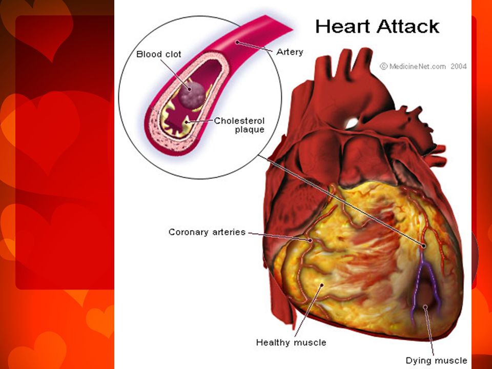
5. Diet Hypertension (high blood pressure)
Prevented by a diet low in salt, saturated fat and cholesterol

68
Variations in Blood Pressure
Normal adults systolic blood pressure varies between mmHg Normal adults diastolic blood pressure varies between mmHg Hypotension – low blood pressure Hypertension – high blood pressure

70
The End

Similar presentations


 Transport O 2, nutrients, hormones, cell wastes, etc…>")


, nutrient molecules and waste materials (from the digestive system) 2.Regulates.>")

 CVS consists of the heart and a series of blood vessels (arteries, veins and capillaries).>")