Download presentation
Presentation is loading. Please wait.

1
Chapter 18 Apoptosis Copyright © Garland Science 2008

2
Two distinct forms of cell death
Apoptosis M18.1 건강한 성인: Billions of cells die every hour Programmed cell death(PCD): idea in 1970s acceptance (20년) :apoptosis (PCD의 한종류) Two distinct forms of cell death apoptosis necrosis Swell Burst Spilling their contents Eliciting inflammatory response Cell shrinks, Cytoskeletin collapses, 핵막 분해, DNA 절단 Apoptic body-chemically altered:macrophage
: idea in 1970s acceptance (20년) :apoptosis (PCD의 한종류) Two distinct forms of cell death. apoptosis. necrosis. Swell. Burst. Spilling their contents. Eliciting inflammatory response. Cell shrinks, Cytoskeletin collapses, 핵막 분해, DNA 절단. Apoptic body-chemically altered:macrophage.")
3
Programmed cell death morphology in tobacco cells.

4
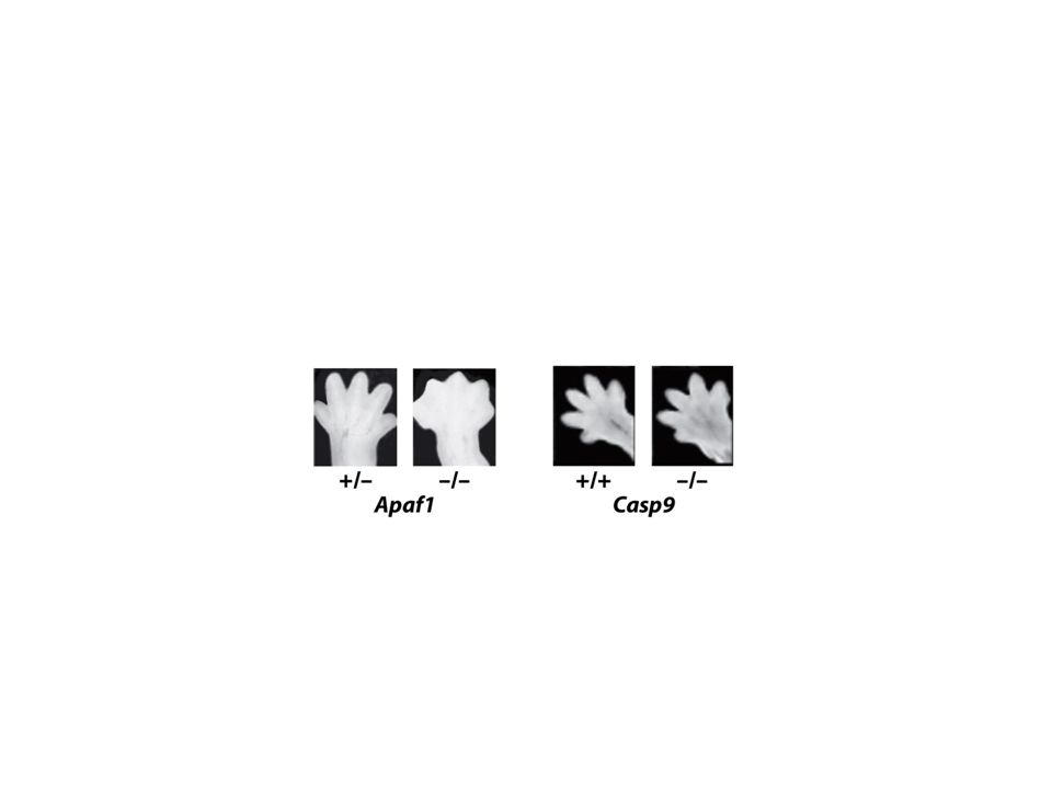
Programmed cell death eliminates unwanted cells
Sculpting the digits in the developing mouse paw by apotosis 불필요 세포/조직/기관제거 세포수 조절 (간세포 분열/phenobarbital) Quality control: B-/T-lymphocyte cell 제거 골수세포: 백혈구 제거 Figure Molecular Biology of the Cell (© Garland Science 2008)
 Quality control: B-/T-lymphocyte cell 제거. 골수세포: 백혈구 제거. Figure 18-2 Molecular Biology of the Cell (© Garland Science 2008)")
5
Apoptotic Cells Are Biochemically Recognizable
Markers of apoptosis TUNEL technique Phosphatidylserine: 세포막 안쪽 바깥: Annexin V로 검출 “eat me” signal “Don’t eat me” signal Figure 18-4a Molecular Biology of the Cell (© Garland Science 2008)
")
6
Apoptotic Cells Are Biochemically Recognizable
Markers of apoptosis Positively charged 형광색소 : 미토콘도리아 inner membrane 의 안쪽 Cytochrome C: released from the intermembrane space of mitochondria Figure 18-4b Molecular Biology of the Cell (© Garland Science 2008)
")
7
Procaspase activation during apoptosis
Apoptosis Depends on an Intracellular Proteolytic Cascade That Is Mediated by Caspases Caspase: Procaspase activation during apoptosis Figure 18-5a Molecular Biology of the Cell (© Garland Science 2008)
")
8
Procaspase activation during apoptosis
Apoptosis Depends on an Intracellular Proteolytic Cascade That Is Mediated by Caspases Procaspase activation during apoptosis Caspase recruitment domain: activation complex Cytoskeleton Cell-cell adhesion proteins endonuclease Figure 18-5b Molecular Biology of the Cell (© Garland Science 2008)
")
9
Cell-Surface Death Receptors Activate the Extrinsic Pathway of Apoptosis
The extrinsic pathway of apoptosis activated through Fas death receptors Tumor necrosis fctor receptor familiy DISC: Death-inducing signaling complex Decoy receptor FLIP blocking protein Figure Molecular Biology of the Cell (© Garland Science 2008)
")
10
The Intrinsic Pathway of Apoptosis Depends on Mitochondria
Release of cytochrome c from mitochondria during apoptosis bind procaspase-activating adapter protein(Apaf) apoptosome 형성 Recruit initiator procaspase proteins (procaspase-9) Figure Molecular Biology of the Cell (© Garland Science 2008)
 apoptosome 형성. Recruit initiator procaspase proteins (procaspase-9) Figure 18-7 Molecular Biology of the Cell (© Garland Science 2008)")
11
The Intrinsic Pathway of Apoptosis Depends on Mitochondria
Figure Molecular Biology of the Cell (© Garland Science 2008)
")
12
Apoptosis 효소?

13
Bcl2 Proteins Regulate the Intrinsic Pathway of Apoptosis
The three classes of Bcl2 Figure Molecular Biology of the Cell (© Garland Science 2008)
")
14
Bcl2 Proteins Regulate the Intrinsic Pathway of Apoptosis
The role of BH123 pro-apoptotic Bcl2 proteins(mainly Bax and Bak) in the release of mitochondrial intermembrane proteins in the intrinsic pathway of apoptosis Figure Molecular Biology of the Cell (© Garland Science 2008)
 in the release of mitochondrial intermembrane proteins in the intrinsic pathway of apoptosis. Figure Molecular Biology of the Cell (© Garland Science 2008)")
15
Bax, Bak Bax Bound to mitochondrial outer membrane Bak Located in cytosol, transport into mitochondira after apoptotic signaling Both work on ER and nuclear membrane, activated by ER stress release Ca++ and intrinsic apoptotic pathway

16
Bcl2 Proteins Regulate the Intrinsic Pathway of Apoptosis
How pro-apoptotic BH3-only and anti-apoptotic Bcl2 proteins regulate the intrinsic pathway of apoptosis Figure 18-11a Molecular Biology of the Cell (© Garland Science 2008)
")
17
Bcl2 Proteins Regulate the Intrinsic Pathway of Apoptosis
How pro-apoptotic BH3-only and anti-apoptotic Bcl2 proteins regulate the intrinsic pathway of apoptosis Figure 18-11b Molecular Biology of the Cell (© Garland Science 2008)
")
18
Bid is the link between the two pathways
Death receptor extrinsic pathway The initiator caspase, caspase-8 cleasve Bid producing tBid tBid translocates to mitochondria inhibit bcl2 Bid: a proapoptotic BHS-3-only protein

19
IAPs Inhibit Caspases A proposed model for the roles of IAPs and anti-IAPs in the control of apoptosis in mammalian cells IAP: Inhibitor of apoptosis first identified from vaculovirus. Figure 18-12a Molecular Biology of the Cell (© Garland Science 2008)
")
20
IAPs Inhibit Caspases A proposed model for the roles of IAPs and anti-IAPs in the control of apoptosis in mammalian cells Figure 18-12b Molecular Biology of the Cell (© Garland Science 2008)
")
21
Extracellular Survival Factors Inhibit Apoptosis in Various Ways
The role of survival factors and cell death in adjusting the number of developing nerve cells to the amount of target tissue Figure Molecular Biology of the Cell (© Garland Science 2008)
")
22
Extracellular Survival Factors Inhibit Apoptosis in Various Ways
Three ways that extracellular survival factors can inhibit apoptosis Figure Molecular Biology of the Cell (© Garland Science 2008)
")
23
Bak/bax double mutant in mouse
Cannot activate intrinsic apoptosis But without survival signal, nutrient import is not efficient Autophagedie from starvation

24
Figure Q18-1 Molecular Biology of the Cell (© Garland Science 2008)
")
26
UV illumination to human Hela Cells within 24h apoptosis in all cells from 6 h after illumination to 24 h Slow apoptosis? or Gradual apoptosis in cell population?

27
Figure Q18-2 Molecular Biology of the Cell (© Garland Science 2008)
")
28
Apoptotic-like regulation of programmed cell death in plants
Apoptosis (2010) 15:249–256 Apoptotic-like regulation of programmed cell death in plants Protoplast condensation, nuclear condensation, fragmentation A central regulatory role for the mitochondria The degradation of the cell and its contents by PCD associated proteases. Caspase-like Study using caspase inhibitor, substrate Lack in caspase ortholog from Arabidopsis genome No evidence that cyt c can directly activate AL-PCD proteases. No Bcl2 family homologs identified Involvement of chloroplast?
 15:249–256. Apoptotic-like regulation of programmed cell death in plants. Protoplast condensation, nuclear condensation, fragmentation. A central regulatory role for the mitochondria. The degradation of the cell and its contents by PCD associated proteases. Caspase-like. Study using caspase inhibitor, substrate. Lack in caspase ortholog from Arabidopsis genome. No evidence that cyt c can directly activate AL-PCD proteases. No Bcl2 family homologs identified. Involvement of chloroplast")
29
Schematic representation of AL-PCD
Fig. 1 Schematic representation of AL-PCD. Having perceived a death stimulus, plant cells can undergo AL-PCD resulting in a distinctive corpse morphology. It is thought that the mitochondrion plays a role in integrating signals generated through stress or developmental signals, thus determining whether the cell activates it’s PCD pathway or not and the chloroplast may also be an important integrator of stress responses in plant cells. A calcium influx is the earliest event detected and similar to animal cells, cyt c is released rapidly from the mitochondria in the early stages of AL-PCD but unlike the animal system, does not appear to be responsible for activating a caspase-driven cascade of events, which leads to PCD, but rather may serve to amplify the death process. Cyt c release will disrupt the electron transport chain resulting in generation of ROS. As well as a signalling molecule, which can lead to opening of the PTP and release of cyt c, more ROS can be generated in this way, causing a feedback loop that amplifies the original death signal. The chloroplast is also a source of ROS and most probably contributes to this feedback loop. We also know that a Mg2?-dependent nuclease which also normally resides in the mitochondrial intermembrane space can be released, causing HMW DNA cleavage (picture of cells courtesy of Siamsa Doyle) MMP: mitochondrial membrane permeablization PTP: the perability transition pore
 MMP: mitochondrial membrane permeablization. PTP: the perability transition pore.")
30
Caspase-independent DNA degradation
Endo G No homolog in plant Mito IMS contain strong Mg2+ dependent nuclease activity AIF/monodehydroascorbate reductase 5 homologs in Arabidospsis Antioxidant pathway DNA-binding defective AIF fails to induce cell death

31
Metacaspases 9 metacaspases in Arabidopsis
Arginine instead of Aspartic acid No caspase substrate clevage, no inhibition by caspase inhibitors Involvement in PCD of Arabidopsis Natural substrates? TSN(Tudor staphylococcal nuclease) in Norway spruce (유럽가문비) hTSN: caspase substrate, involved in PCD
 in Norway spruce (유럽가문비) hTSN: caspase substrate, involved in PCD.")
Similar presentations


, death domain, cytochrome.>")
 Apoptosis is a cell mechanism used to eliminate cells that are unnecessary to or that contain mutations that.>")
 2009- 2010 Dr Minnie O’Farrell Apoptosis in model systems. Molecular mechanisms of apoptosis in mammalian.>")


 In adult tissues cell death exactly balances cell division In apoptosis the cell destroys itself from within.>")


