Download presentation
Presentation is loading. Please wait.

1
EXCRETORY SYSTEM

2
Functions of the Urinary System Elimination of waste products –Nitrogenous wastes from protein and nucleic acid metabolism Urea is the main nitrogenous waste and is made in the liver –Toxins –Drugs

3
Regulate aspects of homeostasis –Water balance –Electrolytes –Acid-base balance in the blood –Blood pressure –Red blood cell production –Activation of vitamin D

4
Organs of the Urinary system Kidneys Ureters Urinary bladder Urethra

6
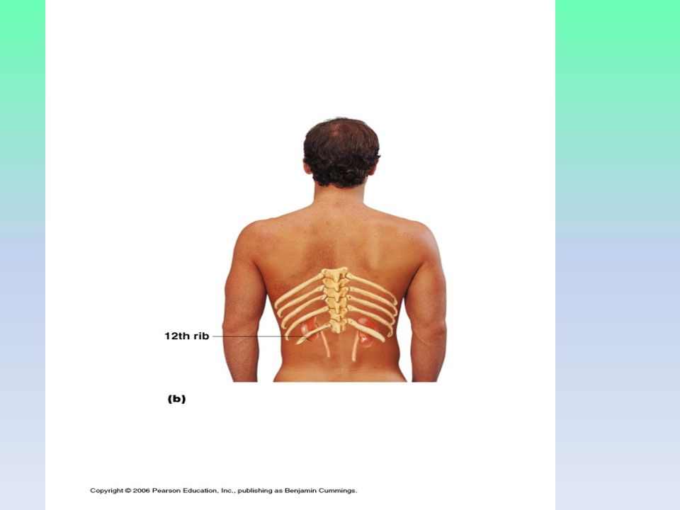
Location of the Kidneys Against the dorsal body wall Receive some protection from lower ribs The right kidney is slightly lower than the left Atop each kidney is an adrenal gland

8
Regions of the Kidney Renal cortex – outer region Renal medulla – inside the cortex Renal pelvis – inner collecting tube

9
Nephrons The structural and functional units of the kidneys Responsible for forming urine Main structures of the nephrons –Glomerulus –Renal tubule Dump urine into collecting ducts that lead to the renal pelvis

10
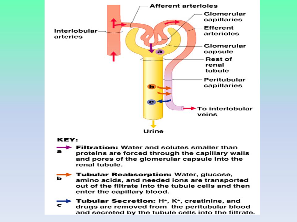
Glomerulus A specialized capillary bed Attached to arterioles on both sides (maintains high pressure) –Large afferent arteriole (takes blood into glomerulus) –Narrow efferent arteriole (takes blood out of glomerulus) The glomerulus sits within a capsule (the first part of the renal tubule)
 –Large afferent arteriole (takes blood into glomerulus) –Narrow efferent arteriole (takes blood out of glomerulus) The glomerulus sits within a capsule (the first part of the renal tubule)")
12
Renal Tubule Glomerular (Bowman’s) capsule Proximal convoluted tubule Loop of Henle Distal convoluted tubule
 capsule Proximal convoluted tubule Loop of Henle Distal convoluted tubule")
14
Peritubular Capillaries Arise from efferent arteriole of the glomerulus Normal, low pressure capillaries Attached to a venule Cling close to the renal tubule Reabsorb (reclaim) some substances from collecting tubes
 some substances from collecting tubes")
15
Urine Formation Processes Filtration Reabsorption Secretion

17
Filtration Nonselective passive process Pushed through via blood pressure Water and solutes smaller than proteins are forced through capillary walls Blood cells cannot pass out of the capillaries Filtrate is collected in the glomerular capsule and leaves via the renal tubule

18
Reabsorption Reabsorption moves materials back to the blood (body does not want to get rid of them) The peritubular capillaries reabsorb several materials –Some water –Glucose –Amino acids –Ions Some reabsorption is passive, most is active transport Most reabsorption occurs in the proximal tubule
 The peritubular capillaries reabsorb several materials –Some water –Glucose –Amino acids –Ions Some reabsorption is passive, most is active transport Most reabsorption occurs in the proximal tubule")
19
Materials Not Reabsorbed Nitrogenous waste products –Urea –Uric acid –Creatinine (from creatine metabolism in muscles) Excess water
 Excess water")
20
Secretion – Getting rid of extra stuff…last minute! Some materials move from the peritubular capillaries into the renal tubules –Hydrogen and potassium ions –Creatinine Materials left in the renal tubule move to a collecting duct

21
Formation of Urine

22
Characteristics of Urine Used for Medical Diagnosis Colored somewhat yellow due to the pigment urochrome (from the destruction of hemoglobin) and solutes Sterile Slightly aromatic Normal pH of around 6 Specific gravity of 1.001 to 1.035
 and solutes Sterile Slightly aromatic Normal pH of around 6 Specific gravity of to 1.035")
23
Abnormal Urine Constituents SubstanceName of conditionPossible causes GlucoseGlycosuria Nonpathological: excessive intake of sugary foods Pathological: diabetes mellitus ProteinsProteinuria Nonpathological: physical exertion, pregnancy Pathological: glomerulonephritis, hypertension PusPyuria Urinary tract infection RBCsHematuria Bleeding in urinary tract (due to trauma, kidney stones, infection) HemoglobinHemoglobinuria Various: transfusion reaction, hemolytic anemia Bile pigment Bilirubinuria Liver disease (hepatitis)
 HemoglobinHemoglobinuria Various: transfusion reaction, hemolytic anemia Bile pigment Bilirubinuria Liver disease (hepatitis)")
24
Ureters Slender tubes attaching the kidney to the bladder –Enter the posterior aspect of the bladder Peristalsis aids gravity in urine transport

25
Urinary Bladder Smooth, collapsible, muscular sac Temporarily stores urine

26
Urinary Bladder Wall Three layers of smooth muscle (detrusor muscle) Mucosa made of transitional epithelium Walls are thick and folded in an empty bladder Bladder can expand significantly without increasing internal pressure
 Mucosa made of transitional epithelium Walls are thick and folded in an empty bladder Bladder can expand significantly without increasing internal pressure")
27
Urethra Thin-walled tube that carries urine from the bladder to the outside of the body by peristalsis Release of urine is controlled by two sphincters –Internal urethral sphincter (involuntary) –External urethral sphincter (voluntary)
 –External urethral sphincter (voluntary)")
28
Urethra Gender Differences Length –Females – 3–4 cm (1 inch) –Males – 20 cm (8 inches) Location –Females – along wall of the vagina –Males – through the prostate and penis Function –Females – only carries urine –Males – carries urine and is a passageway for sperm cells
 –Males – 20 cm (8 inches) Location –Females – along wall of the vagina –Males – through the prostate and penis Function –Females – only carries urine –Males – carries urine and is a passageway for sperm cells")
29
Micturition (Voiding) Both sphincter muscles must open to allow voiding –The internal urethral sphincter is relaxed after stretching of the bladder –The external urethral sphincter must be voluntarily relaxed
 Both sphincter muscles must open to allow voiding –The internal urethral sphincter is relaxed after stretching of the bladder –The external urethral sphincter must be voluntarily relaxed")
30
Maintaining Water Balance Normal amount of water in the human body –Young adult females – 50% –Young adult males – 60% –Babies – 75% –Old age – 45% Water is necessary for many body functions and levels must be maintained

31
Distribution of Body Fluid Intracellular fluid (inside cells) Extracellular fluid (outside cells) –Interstitial fluid (fluid between cells) –Blood plasma
 Extracellular fluid (outside cells) –Interstitial fluid (fluid between cells) –Blood plasma")
32
The Link Between Water and Salt Changes in electrolyte balance causes water to move from one compartment to another –Alters blood volume and blood pressure –Can impair the activity of cells

33
Maintaining Water Balance Water intake must equal water output Sources for water intake –Ingested foods and fluids –Water produced from metabolic processes Sources for water output –Vaporization out of the lungs –Lost in perspiration –Leaves the body in the feces –Urine production

35
Dilute urine is produced if water intake is excessive Less urine (concentrated) is produced if large amounts of water are lost Proper concentrations of various electrolytes must be present
 is produced if large amounts of water are lost Proper concentrations of various electrolytes must be present")
36
Regulation of Water and Electrolyte Reabsorption Regulation is primarily by hormones –Antidiuretic hormone (ADH) prevents excessive water loss in urine by increasing water reabsorption in collecting ducts and distal tubules –Aldosterone increases blood volume and pressure by increasing reabsorption of sodium and water in distal tubules Cells in the kidneys and hypothalamus are active monitors
 prevents excessive water loss in urine by increasing water reabsorption in collecting ducts and distal tubules –Aldosterone increases blood volume and pressure by increasing reabsorption of sodium and water in distal tubules Cells in the kidneys and hypothalamus are active monitors")
39
Maintaining Acid-Base Balance in Blood Blood pH must remain between 7.35 and 7.45 to maintain homeostasis –Alkalosis – pH above 7.45 –Acidosis – pH below 7.35 Most ions originate as byproducts of cellular metabolism Most acid-base balance is maintained by the kidneys Other acid-base controlling systems –Blood buffers –Respiration

40
Blood Buffers Molecules react to prevent dramatic changes in hydrogen ion (H + ) concentrations –Bind to H + when pH drops –Release H + when pH rises Three major chemical buffer systems –Bicarbonate buffer system (only one we will look at) –Phosphate buffer system –Protein buffer system
 concentrations –Bind to H + when pH drops –Release H + when pH rises Three major chemical buffer systems –Bicarbonate buffer system (only one we will look at) –Phosphate buffer system –Protein buffer system")
41
The Bicarbonate Buffer System Mixture of carbonic acid (H 2 CO 3 ) and sodium bicarbonate (NaHCO 3 ) Bicarbonate ions (HCO 3 – ) react with strong acids to change them to weak acids Carbonic acid dissociates in the presence of a strong base to form a weak base and water
 and sodium bicarbonate (NaHCO 3 ) Bicarbonate ions (HCO 3 – ) react with strong acids to change them to weak acids Carbonic acid dissociates in the presence of a strong base to form a weak base and water")
42
Renal Mechanisms of Acid-Base Balance Excrete bicarbonate ions if needed Conserve or generate new bicarbonate ions if needed Urine pH varies from 4.5 to 8.0

43
Developmental Aspects of the Urinary System Functional kidneys are developed by the third month Urinary system of a newborn –Bladder is small –Urine cannot be concentrated Control of the voluntary urethral sphincter does not start until age 18 months Urinary infections are the only common problems before old age

44
Aging and the Urinary System There is a progressive decline in urinary function The bladder shrinks with aging Urinary retention is common in males

45
Medical Issues Ptosis – kidneys drop to a lower position causing ureters to be kinked; trouble draining urine Hydronephrosis – backed-up ureters (from ptosis) that can severly damage kidney Oliguria – abnormally low urinary output (100 – 400 ml/day) Anuria – extremely low urine output (less than 100 ml/day) – From crush injuries or low blood pressure
 that can severly damage kidney Oliguria – abnormally low urinary output (100 – 400 ml/day) Anuria – extremely low urine output (less than 100 ml/day) – From crush injuries or low blood pressure")
46
Renal calculi or kidney stones – crystals that from when urine is too concentrated Urethritis – inflammation of urethra; from bacteria in urethra Cystitis – inflammation of bladder Pyelonephritis – inflammation of kidney Incontinence – unable to voluntarily control external sphincter

47
Urinary retention – bladder unable to expel urine Hyperplasia – enlargement of prostate gland that can cause urinary retention Diabetes inspidus – lack of ADH causes excessive urination of very dilute urine Addison’s disease or hypoaldosteronism – low levels of aldosterone causing large amounts of urine and loss of salts and water

48
Polycystic kidneys – degenerative disease that runs in families; cysts interfere with normal kidney function Hypospadias – in male babies only; the urethral opening is on the under side of the penis instead of the end; corrected with surgery by 12 months old

Similar presentations