Download presentation
Presentation is loading. Please wait.

1
Sofiya Lypovetska MD PhD Ternopil state medical university
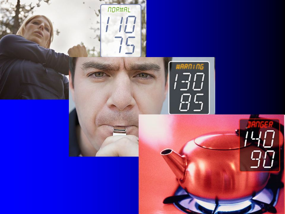
Hypertensive Crisis Sofiya Lypovetska MD PhD Ternopil state medical university Although hypertension may be epidemic, it rarely represents an emergency condition for the individual patient. In the absence of acute end-organ damage, it is rarely, if ever, necessary to lower a patient's BP acutely in the emergency department

2
SCOPE of the PROBLEM Hypertension is an increasingly important medical and public health issue. The prevalence of hypertension increases with advancing age to the point where more than half of people aged 60 to 69 years old and approximately three-fourths of those aged 70 years and older are affected Data from observational studies involving more than 1 million individuals have indicated that death from both ischemic heart disease and stroke increases progressively and linearly from BP levels

4
Definitions and classification of blood pressure levels (mmHg)
")
5
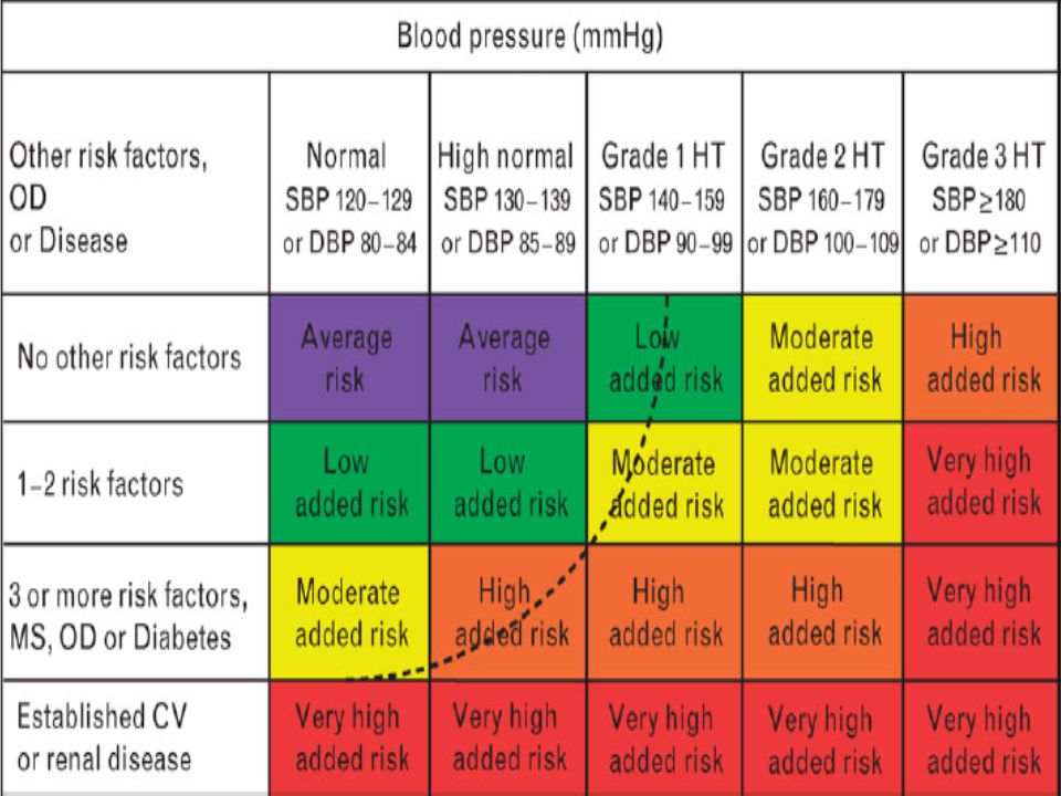
Factors influencing prognosis

6
Factors influencing prognosis

8
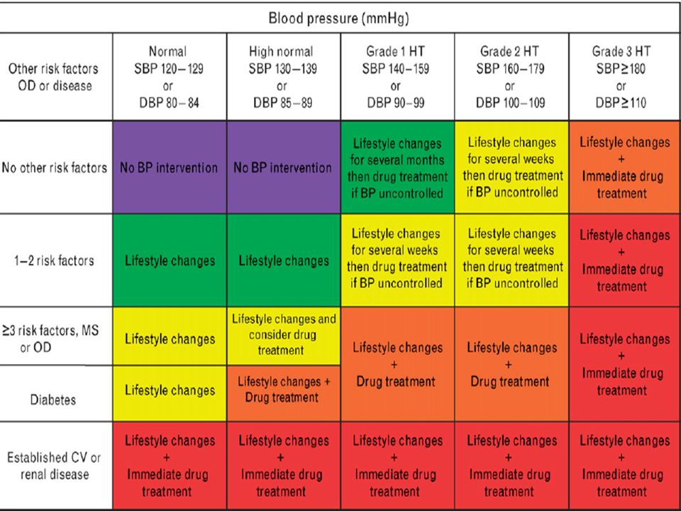
High/Very high risk subjects

9
Blood pressure measurement

10
Position statement: Ambulatory and home BP measurement

11
JNC –VII Guidelines The JNC-VIIis the seventh report of the National High Blood Pressure Education Program since it first published guidelines in 1972 and provides the most aggressive recommendations to date for reducing risk in hypertensive patients. The most severe stage of hypertension (stage 3) is classified as a systolic blood pressure (SBP) of greater than 180 mm Hg or a diastolic blood pressure (DBP) of greater than 110 mm Hg Unfortunately, the JNC-VII provides little guidance for the management of severely increased blood pressure in acute care settings. Of note is a statement placed in the JNC-VI in bold: “Elevated blood pressure alone, in the absence of symptoms or new or progressive target organ damage, rarely requires emergency therapy.”
 is classified as a systolic blood pressure (SBP) of greater than 180 mm Hg or a diastolic blood pressure (DBP) of greater than 110 mm Hg. Unfortunately, the JNC-VII provides little guidance for the management of severely increased blood pressure in acute care settings. Of note is a statement placed in the JNC-VI in bold: Elevated blood pressure alone, in the absence of symptoms or new or progressive target organ damage, rarely requires emergency therapy.")
13
Patient characteristics associated with resistant hypertension

14
Secondary causes of resistant hypertension

15
Medication that can interfere with blood pressure control

16
Conditions favouring use of some antihypertensive drugs versus others

17
Compelling and possible contraindications to use of antihypertensive drugs

18
Hypertensive Crisis Definitions- Is This : A Crisis? An Emergency?
An Urgency?… Clinical Presentations Treatments

19
Other Terminology Severely elevated BP (JNC VII) “accelerated HPT”
Defined as BP > 180/120 “accelerated HPT” term used to describe individuals with chronic hypertension with associated group 3 Keith-Wagener-Baker retinopathy “malignant HPT” describe those individuals with group 4 KWB retinopathy changes + papilledema

20
Definitions Hypertensive Crisis
Hypertensive Emergency………1-2 hours Rapid / progressive end organ damage Hypertensive Urgency………… hrs Inc. BP without evidence of end organ damage Uncontrolled Hypertension……..1 week Do not require acute intervention The presence or risk of progressive target-organ deterioration determines the urgency with which severely increased blood pressure needs to be controlled risk of a near-term complication of an increased blood pressure is multifactorial; age, chronicity of disease, rapidity of blood pressure increase, and type of previous end-organ disease are more important considerations than the actual severity of hypertension A hypertensive emergency involves rapid and progressive decompensation or damage of vital organ function caused by severely increased blood pressure. Require immediate tx with goal of BP dec in 1-2 hours. We propose that a better definition for hypertensive urgency is severely increased blood pressure in a patient at high risk for rapidly progressive end-organ damage but without evidence of new injury. In our opinion, high risk would include patients with a history of prior target-organ disease, such as congestive heart failure, unstable angina, coronary artery disease, renal insufficiency, transient ischemic attack, or stroke. Require tx with goal of BP dec in 24 – 48 hours. Uncontrolled hypertension refers to the vast majority of patients with hypertension who need timely and appropriate long-term management but do not require acute intervention. Therapy to dec BP w/in 1 week Shayne PH - Ann Emerg Med - 01-APR-2003; 41(4):
:")
21
Hypertensive Emergency
BP >180/120 with evidence of target organ dysfunction Hypertensive encephalopathy Intracerebral bleed Acute MI Acute CHF with pulm edema Unstable angina Aortic dissection Eclampsia Accelerated-malignant hypertension occurs most commonly in young black men with underlying renal parenchymal disease or renovascular disease.[20] When endothelial vasodilator responses are overwhelmed, endothelial decompensation causes further hypertension and endothelial damage, resulting in an inflammatory vasculopathy.[31] The diagnosis is based on a marked increase of blood pressure and characteristic eye-ground findings. Flame-shaped hemorrhages occur around the optic disk caused by high intravascular pressures, and soft exudates are caused by ischemic infarction of the nerve fibers after occlusion of supplying arterioles. Papilledema is considered by many to be the sine qua non of malignant hypertension. For this reason, accelerated hypertension has been used to describe the same condition (hemorrhages and exudates) without papilledema. Because absence of papilledema does not connote different clinical features or a better prognosis, the term “accelerated-malignant hypertension” is now recommended.[20] Common symptoms include headache (85%), visual blurring (55%), nocturia (38%), and weakness (30%). Laboratory evidence includes azotemia, proteinuria, hematuria, hypokalemia, and metabolic alkalosis. Accelerated-malignant hypertension is most commonly found in patients with long-standing hypertension and usually occurs without encephalopathy. Since the 1950s, the long-term prognosis of accelerated-malignant hypertension has improved from a 1-year survival rate of 10% to a 5-year survival rate of more than 75%.[47] Treatment for accelerated-malignant hypertension should begin immediately. Any of the potent parenteral antihypertensive agents are appropriate. In the absence of marked azotemia, nitroprusside is an excellent choice. Tx: parenteral agent
 without papilledema. Because absence of papilledema does not connote different clinical features or a better prognosis, the term accelerated-malignant hypertension is now recommended.[20] Common symptoms include headache (85%), visual blurring (55%), nocturia (38%), and weakness (30%). Laboratory evidence includes azotemia, proteinuria, hematuria, hypokalemia, and metabolic alkalosis. Accelerated-malignant hypertension is most commonly found in patients with long-standing hypertension and usually occurs without encephalopathy. Since the 1950s, the long-term prognosis of accelerated-malignant hypertension has improved from a 1-year survival rate of 10% to a 5-year survival rate of more than 75%.[47] Treatment for accelerated-malignant hypertension should begin immediately. Any of the potent parenteral antihypertensive agents are appropriate. In the absence of marked azotemia, nitroprusside is an excellent choice. Tx: parenteral agent.")
22
Cerebrovascular Hypertensive Emergencies
Cerebral Infarct Intracerebral Hemorrhage Cerebral Edema Cerebrovascular hypertensive emergencies include hypertensive encephalopathy, ischemia, and hemorrhage. An understanding of cerebrovascular physiology is helpful in determining the best treatment strategy.[48] Hypertensive Encephalopathy

23
Cerebral Perfusion Pressure
Cerebral blood flow a function of CPP Autoreg. Fails at 25% of MAP ICP CPP Vulnerable to MAP Cerebral blood flow is a function of the cerebral perfusion pressure, which is equal to the MAP minus the intracranial pressure (Cerebral perfusion pressure=MAP − Intracranial pressure). Cerebral blood flow is maintained by means of vasoconstriction and vasodilation of the cerebral vasculature. However, cerebral autoregulation fails at approximately 25% more than or less than the MAP. In addition, changes in intracranial pressure or brain injury can result in loss of the brain's ability to autoregulate blood flow. Increased intracranial pressure, commonly seen with hemorrhage or edema, decreases the cerebral perfusion pressure, making the brain more vulnerable to changes in MAP. CBF = blood flow; CPP = cerebral perfusion pressure; ICP = intracranial pressure; MAP = mean arterial pressure; TCA = total circulatory arrest. 23
. Cerebral blood flow is maintained by means of vasoconstriction and vasodilation of the cerebral vasculature. However, cerebral autoregulation fails at approximately 25% more than or less than the MAP. In addition, changes in intracranial pressure or brain injury can result in loss of the brain s ability to autoregulate blood flow. Increased intracranial pressure, commonly seen with hemorrhage or edema, decreases the cerebral perfusion pressure, making the brain more vulnerable to changes in MAP. CBF = blood flow; CPP = cerebral perfusion pressure; ICP = intracranial pressure; MAP = mean arterial pressure; TCA = total circulatory arrest. 23.")
24
Hypertensive Encephalopathy
Pathophysiology: - Loss of Cerebral Autoregulation of blood flow resulting in hyperperfusion of the brain, loss of integrity of the blood brain barrier, and vascular necrosis. Loss of Autoregulation occurs at a constant cerebral blood flow of above MAP 150 to 160 mmHg. Acute Onset Reversible

25
Hypertensive Encephalopathy
Symptoms: Headache, Nausea/Vomiting, Lethargy, Confusion, Lateralizing neurological symptoms that are not often in an anatomical distribution. Signs: Papilledema, Retinal Hemorrhages Decreased level of consciousness, Coma Focal neurological findings

26
Hypertensive encephalopathy
Clinical manifestation of cerebral edema and microhemorrhages seen with dysfunction of cerebral autoregulation Defined as an acute organic brain syndrome or delirium in the setting of severe hypertension

27
HPT Encephalopathy Not adequately treated – cerebral heamorrhage, coma and death. BUT with proper treatment – completely reversible Clinical diagnoses (exclusion)
")
28
Management of Hypertensive Encephalopathy
Reduce Mean Arterial Pressure (MAP) by 20 to 25% (T.397) and do not exceed this within first 30 to 60 min. Rosen recommends reduction of 30 to 40% (R.1759) MAP= 1/3(SBP-DBP) + DBP Treatment Reduces vasospasm that occurs at these high pressures Avoid excessive BP reduction to prevent hypoperfusion of the brain and further cerebral ischemia
 by 20 to 25% (T.397) and do not exceed this within first 30 to 60 min. Rosen recommends reduction of 30 to 40% (R.1759) MAP= 1/3(SBP-DBP) + DBP. Treatment Reduces vasospasm that occurs at these high pressures. Avoid excessive BP reduction to prevent hypoperfusion of the brain and further cerebral ischemia.")
29
Hypertensive Encephalopathy
Cerebral overperfusion MAP overwhelms autoregulation Vasodilation and Inc. Perm. Cerebral Edema Hemorrhage, Coma, Death Tx: Nipride, Fenoldopam, Labatalol, Nicardipine The mechanism is cerebral overperfusion; in effect, the MAP overwhelms the brain's ability to autoregulate cerebral blood flow. Overperfusion results in vasodilation and increased permeability of cerebral blood vessels, leading in turn to the development of cerebral edema. Hypertensive encephalopathy produces characteristic findings on computed tomography (CT), which should be performed to exclude other causes of altered mental status, such as intracranial bleeding. Schwartz et al[49] have described a posterior leukoencephalopathy in hypertensive encephalopathy that predominantly affects the white matter of the parieto-occipital regions bilaterally. If not adequately treated, hypertensive encephalopathy can progress to cerebral hemorrhage, coma, and death.[20]
, which should be performed to exclude other causes of altered mental status, such as intracranial bleeding. Schwartz et al[49] have described a posterior leukoencephalopathy in hypertensive encephalopathy that predominantly affects the white matter of the parieto-occipital regions bilaterally. If not adequately treated, hypertensive encephalopathy can progress to cerebral hemorrhage, coma, and death.[20]")
30
Hemorrhagic CVA causes
Hypertensive Vascular Disease Arteriovenous Anomalies (AVM) Arterial Aneurysms Tumors Trauma Theoretically, treatment for increased blood pressure in hemorrhagic cerebrovascular accidents and subarachnoid hemorrhage should be more aggressive than for patients with ischemic strokes. The rationale is to decrease the risk of ongoing bleeding from ruptured small arteries and arterioles[52] ; however, the relationship between rebleeding and systemic blood pressure is unproven. As with ischemic cerebral vascular accidents, overly aggressive treatment of hypertension might worsen brain injury by decreasing cerebral perfusion pressure, especially when intracranial pressure is increased. The American Heart Association guidelines for blood pressure control with hemorrhagic stroke are similar to those with ischemic stroke, decreasing the blood pressure only when the MAP is greater than 130 mm Hg or the SBP is greater than 220 mm Hg. Nimodipine might be given to decrease the incidence of vasospasm and rebleeding after subarachnoid hemorrhages, but the drug is not recommended for blood pressure control.[48]
 Arterial Aneurysms. Tumors. Trauma. Theoretically, treatment for increased blood pressure in hemorrhagic cerebrovascular accidents and subarachnoid hemorrhage should be more aggressive than for patients with ischemic strokes. The rationale is to decrease the risk of ongoing bleeding from ruptured small arteries and arterioles[52] ; however, the relationship between rebleeding and systemic blood pressure is unproven. As with ischemic cerebral vascular accidents, overly aggressive treatment of hypertension might worsen brain injury by decreasing cerebral perfusion pressure, especially when intracranial pressure is increased. The American Heart Association guidelines for blood pressure control with hemorrhagic stroke are similar to those with ischemic stroke, decreasing the blood pressure only when the MAP is greater than 130 mm Hg or the SBP is greater than 220 mm Hg. Nimodipine might be given to decrease the incidence of vasospasm and rebleeding after subarachnoid hemorrhages, but the drug is not recommended for blood pressure control.[48]")
31
Hemorrhagic CVA Management
Hemorrhagic CVA’s commonly results in a profound reactive rise in blood pressure Management is CONTROVERSIAL. Subarachnoid Hemorrhage: oral nimodipine (nimotop) 60mg po q 4 hours to reverse vasospasm. Nicardipine: 2mg IV boluses followed by an IV infusion of 4 to 15 mg/hr is used by some to treat Subarachnoid Hemorrhage.
 60mg po q 4 hours to reverse vasospasm. Nicardipine: 2mg IV boluses followed by an IV infusion of 4 to 15 mg/hr is used by some to treat Subarachnoid Hemorrhage.")
32
Ischemic CVA Pathophysiology:
Elevated Blood Pressure can be the cause of the central nervous system event, OR, it may be a normal physiologic response (Cushing’s Reflex) The treatment of increased blood pressure in the setting of ischemic cerebrovascular accidents is controversial. When systemic blood pressure is reduced, cerebral autoregulation might fail, producing an ischemic penumbra surrounding the infarct, leading to stroke extension. Alternatively, infarction can lead to edema, increasing intracranial pressure and reducing cerebral blood flow further.[31] [32] [48] Some believe that an increased MAP in the face of a stroke might be a protective measure. If true, decreasing the blood pressure might lead to further ischemic damage. However, acute antihypertensive therapy was not associated with a worse outcome at 3 months among control patients in the large National Institute of Neurological Disorders and Stroke Recombinant Tissue Plasminogen Activator (NINDS rt-PA) stroke trial.[50] The current American Heart Association guidelines recommend decreasing the blood pressure with stroke only when the MAP is greater than 130 mm Hg or the SBP is greater than 220 mm Hg.[51]
 The treatment of increased blood pressure in the setting of ischemic cerebrovascular accidents is controversial. When systemic blood pressure is reduced, cerebral autoregulation might fail, producing an ischemic penumbra surrounding the infarct, leading to stroke extension. Alternatively, infarction can lead to edema, increasing intracranial pressure and reducing cerebral blood flow further.[31] [32] [48] Some believe that an increased MAP in the face of a stroke might be a protective measure. If true, decreasing the blood pressure might lead to further ischemic damage. However, acute antihypertensive therapy was not associated with a worse outcome at 3 months among control patients in the large National Institute of Neurological Disorders and Stroke Recombinant Tissue Plasminogen Activator (NINDS rt-PA) stroke trial.[50] The current American Heart Association guidelines recommend decreasing the blood pressure with stroke only when the MAP is greater than 130 mm Hg or the SBP is greater than 220 mm Hg.[51]")
33
Ischemic CVA Management
Favors lowering MAP (mean arterial pressure) by 20%. Recommends IV Labetalol in small doses of 5mg increments IF Diastolic Blood Pressure is higher than 140 mmHg. (T. 398)
 by 20%. Recommends IV Labetalol in small doses of 5mg increments IF Diastolic Blood Pressure is higher than 140 mmHg. (T. 398)")
34
HPT Retinopathy

35
AV crossing changes

36
HPT retinopathy

37
HPT retinopathy

38
Cardiovascular Hypertensive Emergencies
Aortic Dissection Congestive Heart Failure Acute MI

39
Congestive Heart Failure
Pathophysiology: Increased Afterload with decreased Cardiac Output Blood pressure is frequently increased in patients with acute pulmonary edema, particularly when a high output state is the cause, as in volume-overloaded patients with renal failure, thyrotoxicosis, or severe anemia. Acute pulmonary edema with hypertension and congestive heart failure might be caused by transient diastolic dysfunction, which might or might not be a direct result of the increased blood pressure.[54] If the patient is critically ill, a nitroprusside infusion should be used. The goal is careful but rapid reduction of blood pressure to normal levels if necessary for symptom relief. In both critical and noncritical cases of congestive heart failure, an angiotensin-converting enzyme (ACE) inhibitor might be helpful; captopril might be given orally or sublingually, or enalaprilat can be given intravenously if the patient cannot take oral medications.[54] Large doses of furosemide are still popular. Although we agree that diuresis should eventually be instituted, it might initially exacerbate the underlying pressure natriuresis and further stimulate the renin-angiotensin axis. Scanty empiric evidence suggests that furosemide might even worsen clinical outcome, at least initially.[55] [56] There are clearly patients with volume overload in whom a diuretic is helpful. β-Blockers have recently been found to improve survival in patients with chronic congestive heart failure [57] ; however, this observation should not be extended to patients with acute pulmonary edema because the negative inotropic effects and bradycardia of β-blockade might precipitate immediate worsening. Intravenous nesiritide improves hemodynamic function and symptoms in decompensated heart failure and has a modest antihypertensive effect.[58]
 inhibitor might be helpful; captopril might be given orally or sublingually, or enalaprilat can be given intravenously if the patient cannot take oral medications.[54] Large doses of furosemide are still popular. Although we agree that diuresis should eventually be instituted, it might initially exacerbate the underlying pressure natriuresis and further stimulate the renin-angiotensin axis. Scanty empiric evidence suggests that furosemide might even worsen clinical outcome, at least initially.[55] [56] There are clearly patients with volume overload in whom a diuretic is helpful. β-Blockers have recently been found to improve survival in patients with chronic congestive heart failure [57] ; however, this observation should not be extended to patients with acute pulmonary edema because the negative inotropic effects and bradycardia of β-blockade might precipitate immediate worsening. Intravenous nesiritide improves hemodynamic function and symptoms in decompensated heart failure and has a modest antihypertensive effect.[58]")
40
CHF / Pulmonary Edema Shortness of Breath, Cough, Chest Pain
Symptoms: Shortness of Breath, Cough, Chest Pain Lower Extremity Swelling Signs: Jugular Venous Distension, Rales, S3 Gallop Hepatomegaly, Pedal Edema

41
CHF / Pulmonary Edema Treatment: Diuretics Nitroglycerin Vasodilators
Digitalis Beta-adrenoceptor agonists Other positive inotropic agents

42
Acute Coronary Syndrome
Pathophysiology: - Increased afterload, cardiac workload, and myocardial oxygen demand - Decreased coronary artery blood flow Acute coronary syndromes are frequently accompanied by hypertension. Reducing myocardial work by decreasing the blood pressure and heart rate has been demonstrated to reduce infarct size in patients not receiving thrombolytic therapy.[59] The ideal pharmacologic approach involves use of nitroglycerin rather than nitroprusside because the former is a potent coronary artery dilator in addition to reducing both preload and afterload. The goal of treatment is reduction of blood pressure to normal levels or even less if evidence of ischemia persists. However, this reduction should occur carefully, with the patient intensively monitored. Overly vigorous blood pressure decreasing can worsen ischemia because coronary perfusion depends on DBP.

43
Acute Coronary Syndrome / Acute MI
Symptoms: Chest Pain, Nausea / Vomiting, Diaphoresis, Shortness of Breath Signs: Congestive Heart Failure Signs, S4 Gallop (due to decreased ventricular compliance) Few physical findings in many patients Clinical History is very Important
 Few physical findings in many patients. Clinical History is very Important.")
44
Acute Coronary Syndrome/ Acute MI
Immediate Blood Pressure reduction is indicated to prevent Myocardial Damage No specific Defined BP target Management: Nitroglycerin IV or Sublingual Beta Blockers (Esmolol, Lopressor) Nitroglycerin is Drug of Choice
 Nitroglycerin is Drug of Choice.")
45
Aortic Dissection Pathophysiology:
- Atherosclerotic Vascular Disease, Chronic Hypertension, increased shearing force on the thoracic aorta, leading to intimal tear. - 50% begin in ascending aorta - 30% at aortic arch - 20% in descending aorta Acute aortic dissection is thought to occur through aortic dilation or high blood pressures superimposed on a structural weakness of the arterial wall. The result is a tear of the intimal layer of the aorta.[60] Pulsatile pressure extends the dissection by separating the layers of the arterial wall. Historical series report a mortality of 1% to 2% per hour. The stresses that extend the dissection are thought to be related as much to the aortic pulse wave or pulse pressure (dp/dt) as it is to MAP. Factors that contribute to increased pulse pressure include heart rate, myocardial contractility, and MAP. An arterial dilator, such as nitroprusside, or a calcium-channel blocker alone might decrease the MAP while simultaneously increasing the pulse pressure through reflex tachycardia. β-Blocking agents can control myocardial contractility and tachycardia and thus ought to be included to prevent progression of the dissection.
 as it is to MAP. Factors that contribute to increased pulse pressure include heart rate, myocardial contractility, and MAP. An arterial dilator, such as nitroprusside, or a calcium-channel blocker alone might decrease the MAP while simultaneously increasing the pulse pressure through reflex tachycardia. β-Blocking agents can control myocardial contractility and tachycardia and thus ought to be included to prevent progression of the dissection.")
46
Dissection of Thoracic Aorta
Symptoms: Chest pain radiating to the back (classic presentation) Neurological Symptoms (carotid artery dissection) Angina (coronary artery dissection) Shortness of breath (aortic insufficiency, cardiac tamponade) Signs: - Differential Blood Pressure (in UE) Bruit (interscapular) Neurological Deficits Acute Cardiac Tamponade (rare)
 Neurological Symptoms (carotid artery dissection) Angina (coronary artery dissection) Shortness of breath (aortic insufficiency, cardiac tamponade) Signs: - Differential Blood Pressure (in UE) Bruit (interscapular) Neurological Deficits. Acute Cardiac Tamponade (rare)")
47
Dissection of Thoracic Aorta
Optimal Blood Pressure in these patients is undefined and must be tailored for each patient, however, SBP of mmHg may be a intial starting point. (T.408)
")
48
Acute Renal Failure Pathophysiology:
Hypertensive Glomerulonephropathy, Acute Tubular Necrosis - Worsening renal function in the setting of severe hypertension with elevation of BUN/CR, proteinuria, or the presence of red cells and red cell casts in the urine.

49
Acute Renal Failure Symptoms: Signs:
- Many times there are few actual symptoms Facial or Peripheral Edema due to fluid overload or proteinuria may be present, shortness of breath Signs: Few findings unless edematous Pulmonary Edema

50
Acute Renal Failure Management: Nitroprusside is agent of choice
Dialysis (as needed) Lasix to enhance Sodium excretion; Also recommends Nitroprusside or Nifedipine Nitroglycerin is also a good agent in this setting since it is hepatically metabolized and gastrointestinally excreted.
 Lasix to enhance Sodium excretion; Also recommends Nitroprusside or Nifedipine. Nitroglycerin is also a good agent in this setting since it is hepatically metabolized and gastrointestinally excreted.")
51
Preeclampsia / Eclampsia
Pathophysiology: Systemic arterial vasoconstriction (including placental, leading to decreased uterine blood flow). Defined as SBP = 140/90 mmHg or greater, OR a 20 mmHg rise in SBP or mmHg rise in DBP from baseline and evidence of HELLP Syndrome Women who are pregnant for the first time, who are between 20 weeks' gestation and 2 weeks postpartum, and who have any degree of hypertension accompanied by peripheral edema and proteinuria should be considered to have preeclampsia. Hypertension is important mainly as a symptom of the underlying disorder rather than as a cause. Preeclampsia is important to recognize because it can progress suddenly to eclampsia defined by the occurrence of convulsions and can rapidly progress to coma or death. Magnesium infusion is more effective than other anticonvulsants in this setting. [66] Definitive treatment consists of delivery of the fetus, and therefore, the emergency physician usually collaborates with an obstetrician early in the patient's progress through the department. The mainstay of antihypertensive treatment in many institutions is hydralazine administered intravenously in boluses of 5 to 10 mg every 20 to 30 minutes. If treatment is refractory to hydralazine, second-line agents are diazoxide and β-blockers.[67] Calcium-channel blockers have been studied in chronic hypertension among pregnant patients, but they might not be effective with proteinuric hypertension.[68
. Defined as SBP = 140/90 mmHg or greater, OR a 20 mmHg rise in SBP or 10 mmHg rise in DBP from baseline and evidence of HELLP Syndrome. Women who are pregnant for the first time, who are between 20 weeks gestation and 2 weeks postpartum, and who have any degree of hypertension accompanied by peripheral edema and proteinuria should be considered to have preeclampsia. Hypertension is important mainly as a symptom of the underlying disorder rather than as a cause. Preeclampsia is important to recognize because it can progress suddenly to eclampsia defined by the occurrence of convulsions and can rapidly progress to coma or death. Magnesium infusion is more effective than other anticonvulsants in this setting. [66] Definitive treatment consists of delivery of the fetus, and therefore, the emergency physician usually collaborates with an obstetrician early in the patient s progress through the department. The mainstay of antihypertensive treatment in many institutions is hydralazine administered intravenously in boluses of 5 to 10 mg every 20 to 30 minutes. If treatment is refractory to hydralazine, second-line agents are diazoxide and β-blockers.[67] Calcium-channel blockers have been studied in chronic hypertension among pregnant patients, but they might not be effective with proteinuric hypertension.[68.")
52
Preeclampsia / Eclampsia
Symptoms: lower extremity swelling, headache, confusion, seizures, coma Signs: edema, hyperreflexia, elevation of blood pressure related to baseline BP prior to pregnancy (elevation may be mild 125/75) Management: IV Magnesium Sulfate, Hydralazine. May also use nifedipine or labetalol Delivery of Fetus is definitive treatment of pre-eclampsia
 Management: IV Magnesium Sulfate, Hydralazine. May also use nifedipine or labetalol Delivery of Fetus is definitive treatment of pre-eclampsia.")
53
Treatment of acute severe hypertension in preeclampsia

54
Pheochromocytoma Pathophysiology:
- Alpha and Beta stimulation of the cardiovascular system due to adrenergic excess states Symptoms: Episodic Headaches, flushing, tremor, diaphoresis, diarrhea, hyperactivity, and palpitations Signs: Tachycardia, tachypnea, tremor, hyperdynamic state (high output CHF)
")
55
Pheochromocytoma Management:
Alpha Blocker FIRST, followed by a Beta Blocker Phentolamine (alpha) + Esmolol (beta) Labetalol IV (combined alpha and beta blockade)
 + Esmolol (beta) Labetalol IV (combined alpha and beta blockade)")
56
Pharmacologic Agents Hypertensive Emergencies
Rapid Onset Rapid Maximal effect Rapid offset Ease of Titration Nitroprusside remains the mainstay of treatment for patients with hypertensive emergencies.[43] In aortic dissection, β-blockers should be added to nitroprusside to avoid the potential adverse effects of reflex tachycardia. Labetalol is a unique parenteral agent that achieves its maximal effect within minutes and then remains effective for several hours. This allows titration with small boluses, thus avoiding the constant monitoring and increased cost required with nitroprusside. Because labetalol does not dilate cerebral capacitance vessels, it is theoretically attractive in intracerebral disorders. Fenoldopam holds some promise as being equivalent to nitroprusside in efficacy without the rare side effects associated to nitroprusside's cyanide moiety and perhaps with less overshoot hypotension, but at present, its cost is often prohibitive. Compared with nitroprusside, fenoldopam might improve outcomes in patients with hypertension and acute renal failure. [43] Enalaprilat – IV ACE Hydralazine Adreniergic inhibitors labetalol esmolol phentolamine Sodium nitroprusside μg/kg per min as IV infusion‡ (maximal dose for 10 min only) Immediate Nausea, vomiting, muscle twitching, sweating, thiocyanate and cyanide intoxication Most hypertensive emergencies; caution with high intracranial pressure or azotemia Fenoldopam mcg/kg/min titrated in 0.1 mcg/kg/min Q 15 min: max dose 1.6mcg/kg/min periph dopamine receptor agonist located in sysemic and renal vac. Bed – improves renal function in pts with Malig HTN Nicardipine mg / hr inc infusion Q 15 min by 2.5mg up to 15 mg / hr. Onset 5-10 min durateion 4-6 hrs Nitroglycerin 5-100 μg/min IV infusion‡ 2-5 min 3-5 min Headache, vomiting, methemoglobinemia, tolerance with prolonged use Coronary ischemia Enalaprilat mg every 6 h IV 15-30 min 6 h Precipitous decrease in pressure in high-renin states; response variable Acute left ventricular failure; avoid in acute myocardial infarction Hydralazine hydrochloride 10-20 mg IV; mg IM 10-20 min; min 3-8 h Tachycardia, flushing, headache, vomiting, aggravation of angina Eclampsia Labetalol hydrochloride 20-80 mg IV bolus every 10 min; mg/min IV infusion 5-10 min 3-6 h Vomiting, scalp tingling, burning in throat, dizziness, nausea, heart block, orthostatic hypotension Most hypertensive emergencies except acute heart failure Esmolol hydrochloride μg/kg per min for 1 min, then μg/kg per min for 4 min; may repeat sequence 1-2 min 10-20 min Hypotension, nausea Aortic dissection, perioperative Phentolamine 5-15 mg IV 3-10 min Tachycardia, flushing, headache Catecholamine excess Parenteral Agents
 Immediate. Nausea, vomiting, muscle twitching, sweating, thiocyanate and cyanide intoxication. Most hypertensive emergencies; caution with high intracranial pressure or azotemia. Fenoldopam 0.1mcg/kg/min titrated in 0.1 mcg/kg/min Q 15 min: max dose 1.6mcg/kg/min. periph dopamine receptor agonist located in sysemic and renal vac. Bed – improves renal function in pts with Malig HTN. Nicardipine 5mg / hr inc infusion Q 15 min by 2.5mg up to 15 mg / hr. Onset 5-10 min durateion 4-6 hrs. Nitroglycerin μg/min IV infusion‡ 2-5 min. 3-5 min. Headache, vomiting, methemoglobinemia, tolerance with prolonged use. Coronary ischemia. Enalaprilat mg every 6 h IV min. 6 h. Precipitous decrease in pressure in high-renin states; response variable. Acute left ventricular failure; avoid in acute myocardial infarction. Hydralazine hydrochloride mg IV; mg IM min; min. 3-8 h. Tachycardia, flushing, headache, vomiting, aggravation of angina. Eclampsia. Labetalol hydrochloride mg IV bolus every 10 min; mg/min IV infusion min. 3-6 h. Vomiting, scalp tingling, burning in throat, dizziness, nausea, heart block, orthostatic hypotension. Most hypertensive emergencies except acute heart failure. Esmolol hydrochloride μg/kg per min for 1 min, then μg/kg per min for 4 min; may repeat sequence. 1-2 min min. Hypotension, nausea. Aortic dissection, perioperative. Phentolamine mg IV min. Tachycardia, flushing, headache. Catecholamine excess. Parenteral Agents.")
57
Parenteral drugs for treatment of hypertensive emergencies

58
Oral Regimens for Treatment of Hypertensive Urgency in the ED
Clonidine: 0.1 to 0.2mg PO, repeat 0.1mg q hour to desired BP reduction or max of 0.7mg. Labetalol: 200 to 400mg PO, repeat every 2 to 3 hours Captopril: 25mg PO Losartan: 50mg PO

59
Key Concepts Acute End-organ damage determines hypertensive emergency Be familiar with the agents of choice in specific emergencies Goal for most is careful reduction of MAP by 20-25% over minutes to hours DBP not less than 100 to 110 Except: Pregnancy, Dissection, MI, Patients without acute end-organ ischemia rarely require urgent intervention

60
Thank You!

Similar presentations


 Drug Therapy (Antihypertensives) ACEi B.B CCB D iuretics. Centrally acting agents: alphametyldopa, HTN + pregnancy.>")


