Download presentation
Presentation is loading. Please wait.

1
Rheological and Molecular Characterization of Equine Synovial Fluid
Nikki Buck Advisor: Dr. Skip Rochefort Oregon State University School of Chemical, Biological and Environmental Engineering Summer, 2008

2
Objectives Connect rheological properties to the molecular characterization of equine synovial fluid. Characterize the properties of hyaluronic acid within synovial fluid.

3
What are Polymers? Compound word derived from Greek Spaghetti Analogy
Poly: many Meros: part A polymer is a long chain of repeating units covalently bonded together. Spaghetti Analogy One polymer is one noodle entangled within a plate of spaghetti.

4
Polymer- Hyaluronic Acid
the main polymeric component of synovial fluid Repeating units of hyaluronan

5
Synovial Fluid Viscoelastic fluid that acts in both lubrication and shock absorption of articular joints. Equine synovial fluid is being studied from hock and stifle joints of racehorses.

6
Horse Anatomy Stifle (knee) Hock (ankle)

7
Rheology How do we study polymers?
Rheology: The study of the deformation and flow of matter Elasticity: The ability to return to its natural shape after deformation, restoring force Viscosity: Resistance to shear or extensional stress

8
Hypothesis: Part I The molecular weight of synovial fluid makes a difference in the viscosity and elasticity of samples. Prediction: samples with higher molecular weights will demonstrate more elasticity and viscosity at given shear rates and frequencies.

9
Rheometry: Dynamic Oscillation
The cone oscillates at a specific range of frequencies and the machine measures the viscosity and elasticity of the fluid. G’ = elastic modulus “stored energy” G’’ = viscous modulus “lost energy”

10
Dynamic Oscillation 40mm 2°cone Peltier plate geometry 25°Celcius
G’ Elasticity G’’ Viscosity

12
Rheometry: Steady Shear Flow
A cone or plate rotates at a constant shear rate (deformation rate), while the machine measures the shear stress exerted on the instrument by the fluid. w Fluid Image copywritten from Danielle Lieske, Oregon State University Viscosity = shear stress shear rate
, while the machine measures the shear stress exerted on the instrument by the fluid. w. Fluid. Image copywritten from Danielle Lieske, Oregon State University. Viscosity = shear stress. shear rate.")
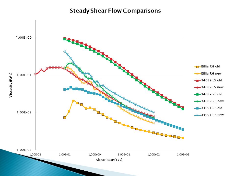
13
Steady Shear Flow Hyaluronic Acid
40mm 2°cone Peltier plate geometry 25°Celcius

15
Comparisons: Closer Look

16
GPC/MALLS Molecular Characterization
Two detector system: Sample first separated by size exclusion chromatography (porous columns) Refractive Index detector determines the concentration Light scattering determines the molecular weight Gel Permeation Chromatography Multi-angle laser light scatter Dn/dc differential refractive index needed to calculate the molecular weight. Change in refractivity as concentration changes.
 Refractive Index detector determines the concentration. Light scattering determines the molecular weight. Gel Permeation Chromatography. Multi-angle laser light scatter. Dn/dc differential refractive index needed to calculate the molecular weight. Change in refractivity as concentration changes.")
17
Light Scattering Detector measures the intensity of light as a function of deflection angle and concentration. Detector, I() Detector, Io Polymer Solution Light Source
 Detector, Io. Polymer Solution. Light Source. ")
18
GPC/MALLS hyaluronic acid
Light Scattering RI Injection volume: 0.2 mL Flow Rate: 0.2 mL/min

19
GPC/MALLS synovial fluid
Protein Peak Light Scattering RI HA Peak Injection volume: 0.2 mL Flow Rate: 0.2 mL/min

20
Light Scattering Read-Out
Sample ID: rstifle in 1:10 PBS August 1, 2008 Operator: Nikki Buck Collection Information Collection time : Fri Aug 01, :06 AM PST Solvent name : PBS pH 7 Solvent RI : 1.334 Calibration constants DAWN : e-06 » AUX2 : e-05 Flow rate : mL/min Calculation method : dn/dc + AUX Constant dn/dc (mL/g) : RESULTS: Molar Mass Moments (g/mol) Mw : 3.384e+05 (0.5%) 6.171e+04 (0.17%)
 : RESULTS: Molar Mass Moments (g/mol) Mw : 3.384e+05 (0.5%) 6.171e+04 (0.17%)")
21
Protease An enzyme that hydrolyzes the peptide bond between amino acids of a protein Enzyme used: Dipase from Bacillus polymyxa Protocol: Dilute synovial fluid 1:3 in PBS Add 0.78 units Protease per mL synovial fluid Incubate 15 min in 37°C water bath Filter Extract HA using phenol-chloroform

22
Hypothesis: Part 2 Proteins cause the second light scattering peak but do not interfere with the molecular weight reading of GPC/MALLS light scattering. Prediction: Synovial fluid samples allowed to incubate in protease will not demonstrate a protein peak during light scattering analysis, and will have molecular weights in the same range as that of the undigested samples.

23
Comparison: Pure Vs. Digested
Light Scattering 34089 Right Stifle MW: 3.384*105 g/mol 34089 Right Stifle digested in Protease MW: 3.819*105 g/mol RI Difference = 11.3% In a single sample we’ve seen differences of over 100% Light Scattering RI

24
Conclusions Viscosity and elasticity depend on more than the molecular weight of the hyaluronic acid within the synovial fluid. Samples with higher molecular weights did not necessarily exhibit more viscoelasticity. Concentration of hyaluronic acid must also be taken into account. Hyaluronic acid in the synovial fluid samples degrade at different rates over time when kept in a laboratory refrigerator. Molecular weights of the samples from horse are significantly lower now than they were two years ago, but this is not evident for or Billie. Proteins do not interfere with the hyaluronic acid molecular weight reading on a GPC/MALLS system. Protease may be used to digest proteins and purify synovial fluid to focus on the hyaluronic acid peak.

25
Acknowledgements Howard Hughes Medical Institute Dr. Kevin Ahern
Dr. Skip Rochefort, OSU School of Chemical Biological and Environmental Engineering Sara Tracy, M.S. Chemical Engineering Dr. Jill Parker, OSU College of Veterinary Medicine Haley Thompson, Coralie Backlund, and Jesse McKiernan

Similar presentations


>")


 Heidi Schmidt Advisors:>")


 Rheological Principles for Food Analysis Qingrong Huang Department of Food Science>")


. Sample components enter pores.>")


