Download presentation
Presentation is loading. Please wait.

1
KARYOTYPE Refers to the chromosome complement of a cell or a whole organism In particular, it shows the number, size, and shape of the chromosomes as seen during metaphase of mitosis Chromosomes numbers vary considerably among organisms and may differ markedly between closely related species Organism Chromosome number (2n) Drosophila8 Honey bee32 or 16 Goldfish94 Rat42 Rabbit44 Cat 38 Dog78 Gorilla48 Chimpanzee48 Human46
 Drosophila8 Honey bee32 or 16 Goldfish94 Rat42 Rabbit44 Cat 38 Dog78 Gorilla48 Chimpanzee48 Human46")
3
Chromosome structure This chromosome has two chromatids. Each chromatid contains an identical copy of the DNA molecule In non-dividing cells, chromosomes exist as single-armed structure with one chromatid The chromosome consists of a protein coated strand which coils in three ways during the time when the cell prepares to divide Figure 1. Chromatin and condensed chromosome structure during metaphase of cell division

4
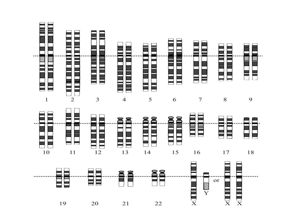
Human karyotype

5
Karyotyping Karyotyping is the process of finding the chromosomal characteristics of a cell Chromosomes can be stained to show banding. Chromosome structure and banding can be used to arrange the chromosome in their pairs Application of karyotyping can be found in an amniocentesis or chronic villus sampling How to read karyotype? Dark and light elements of the chromosome show which parts take part in cell division You may check the number of chromosome and the gender

7
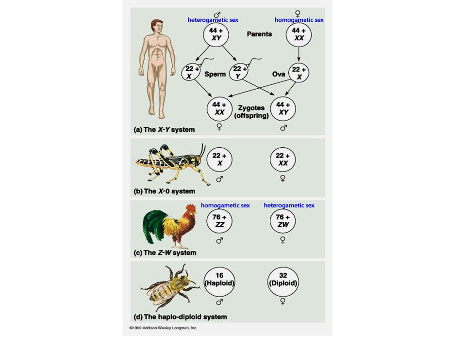
Sex inheritance In many animals sex is determined by a pair of sex chromosomes All the rest of the chromosomes are called autosomes In humans there are two types of sex chromosome, called X and Y chromosome The X chromosome is much larger than the Y, and carries many genes which are not present on the Y chromosome Figure 2. Sex chromosomes in human

9
Sex linkage A gene which is found on one of the sex chromosome and on the other is called a sex-linked gene Chromosome XChromosome Y Duchenne muscular dystrophysex determining factor Retinis pigmentosagenes involved in growth Kidney stonesand spermatogenesis genes Haemophilia Red-green colour blindness Spastic paraplegia Adrenoleukodystrophy

10
Sex linkage in Drosophila melanogaster

11
Red-green colour blindness Sex linked conditions such a red-green colour blindness are therefore much commoner in men than in women A – the normal dominant allele allows full colour vision a – the less common recessive allele produces red-green colour blindness

12
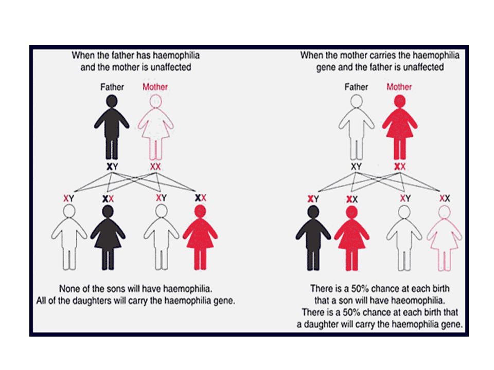
Haemophilia The most well-known sex-linked disease It is caused by a recessive allele of a gene which codes for the production of one of the proteins involved in blood clotting – Factor VIII Females with haemophilia are almost unknown. If a male has one defective allele he will have the haemophilia condition.

14
Sex-linkage – revision Sex-linkage occurs when the genes carried on the sex chromosomes Conditions like colour blindness and haemophilia are much more common in men than in woman and are said to be sex-linked Sex-linked genes are found on the X chromosome Since females have 2 X chromosomes, they can have 2 dominant alleles (homozygous dominant), 2 recessive alleles (homozygous recessive) or 1 dominant and 1 recessive allele (heterozygous). Males only have 1 X chromosome. This means that the terms homozygous or heterozygous do not apply Mendel's first law Law of segregation ‘Parental factors (genes) are in pairs and split so that one factor is present in each gamete’ Mendel's second law Principle of independent assortment ‘Any of one pair of characteristics may combine with either one of another pair’
 are in pairs and split so that one factor is present in each gamete’ Mendel s second law Principle of independent assortment ‘Any of one pair of characteristics may combine with either one of another pair’.")
15
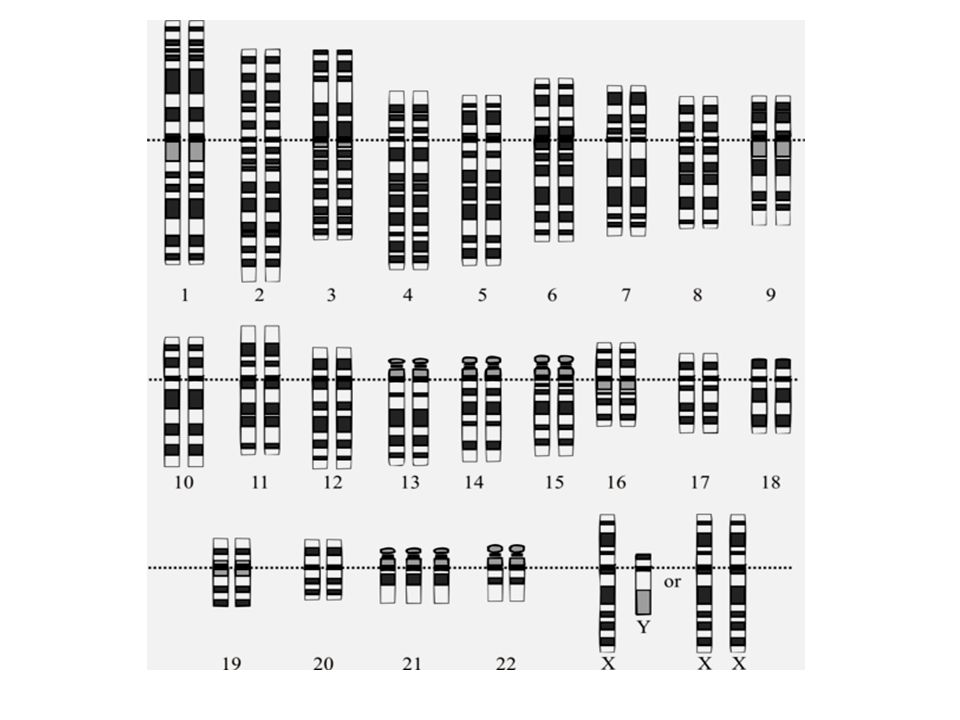
An example of a chromosome mutation – Down's syndrome Down's syndrome is caused by the possession of an extra chromosome 21 (an example of a trisomy) Chromosome 21 is one of the smaller chromosomes People with Down's syndrome therefore usually have 47 chromosomes in their cells The possession of extra chromosomes is known as polysomy The extra chromosome 21 usually comes from the mother's egg. This happens because of an error during meiosis in her ovary in which the 2 chromosome 21s fail to separate, both of them going into one daughter cell and none into the other. This error is called non-disjunction Children with Down's syndrome have characteristic facial features: slanting eyes, back of head flat, broad flat face, short nose, abnormal nose, small and arched palate, big wrinkled tongue, dental anomalies

18
Pre-natal testing There are two common pre-natal tests: Chorionic villus sampling Amniocentesis Chorionic villus sampling: It is a pre-natal test that can be done at 11-12 weeks of pregnancy It involves taking a sample of the chronic villi in order to obtain cells from tissue that originally came from the zygote The cells will therefore have the same genetic composition as the cells of the unborn baby so a karyotype can be made Amniocentesis Can be done around the 16th week of the pregnancy A sample of the amniotic fluid (containing fetal cells) is taken and a culture is made When sufficient cells have been obtained a karyotype can be done to detect chromosome abnormalities
 is taken and a culture is made When sufficient cells have been obtained a karyotype can be done to detect chromosome abnormalities")
19
Chorionic villus sampling

20
Amniocentesis

21
Multiple alleles

Similar presentations