Download presentation
Presentation is loading. Please wait.

1
SWEET HEART

2
THE HEART The heart is a cone shaped muscular organ It is normally placed on the left side of the chest and is of the size of a closed fist The heart has 4 cavities - 2 atria - 2 ventricles

4
HOW DOES THE HEART WORK? ‘Pacemaker of the heart’ (shown as 1 in the picture) Weak electrical activity starts from the Pacemaker Electrical activity travels along a certain path (shown in red) This stimulates all the parts of the heart, thereby causing the entire heart to contract and relax alternatively
 Weak electrical activity starts from the Pacemaker Electrical activity travels along a certain path (shown in red) This stimulates all the parts of the heart, thereby causing the entire heart to contract and relax alternatively.")
5
Contraction is called as ‘Systole’. Blood is pumped out of the heart in this phase. Relaxation is called as ‘Diastole’. Blood is collected in the heart in this phase. SYSTOLE DIASTOLE

16
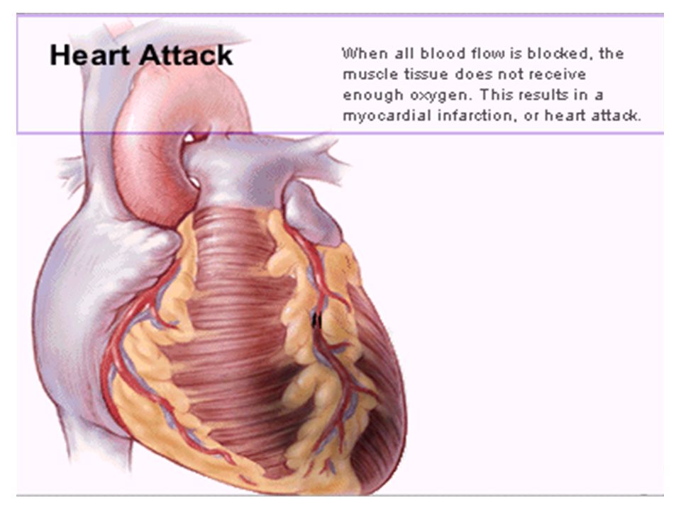
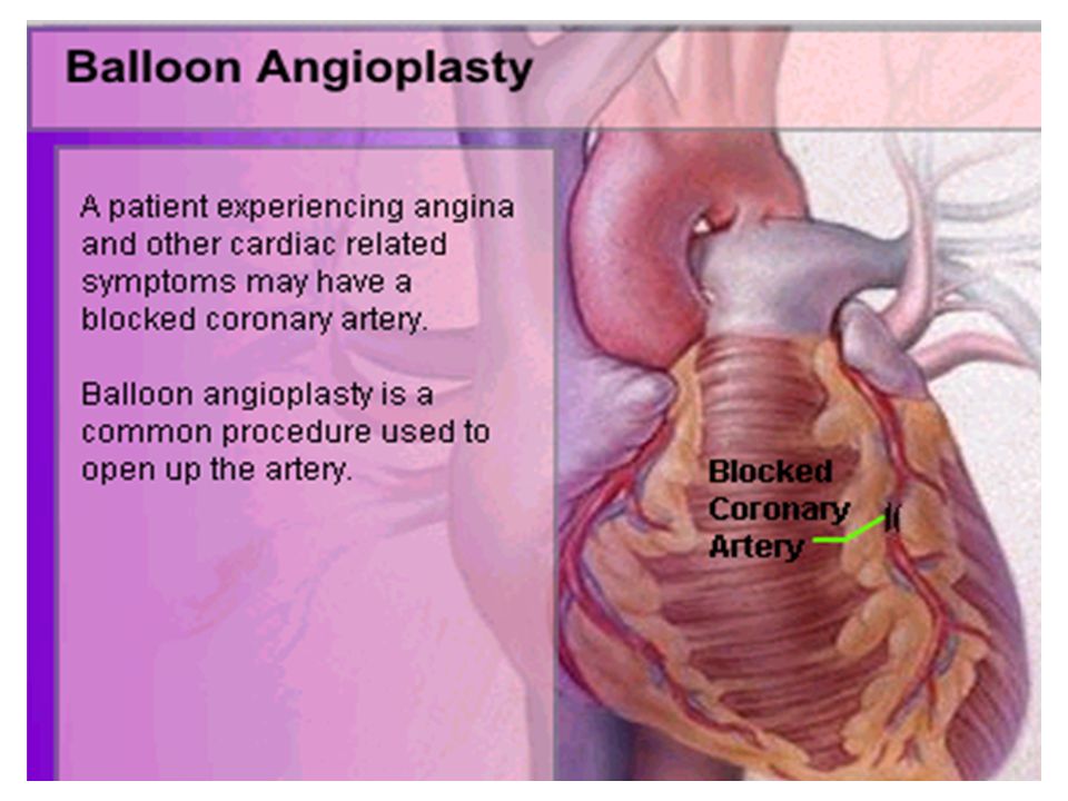
WARNING SYMPTOMS... Difficulty in breathing(Dysnpnoea) Chest Pain(Angina) Blue Discoloration (Cyanosis) Black out(Syncope) Swelling of the body, especially of the feet(Edema) Awareness of heart beats(Palpitation) Cough Coughing out blood(Hemoptysis) Fatigue
 Chest Pain(Angina) Blue Discoloration (Cyanosis) Black out(Syncope) Swelling of the body, especially of the feet(Edema) Awareness of heart beats(Palpitation) Cough Coughing out blood(Hemoptysis) Fatigue.")
17
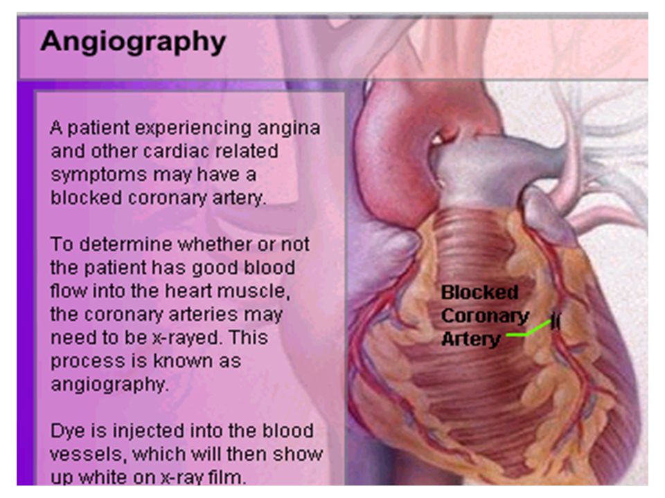
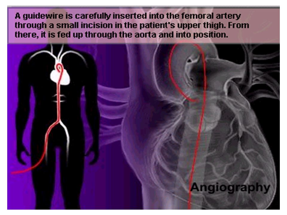
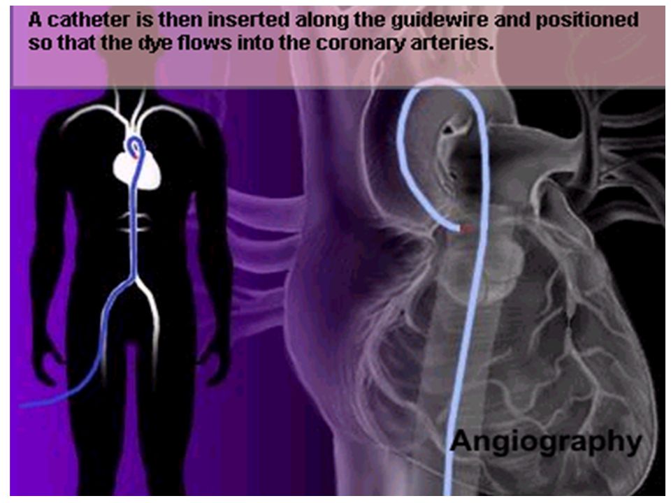
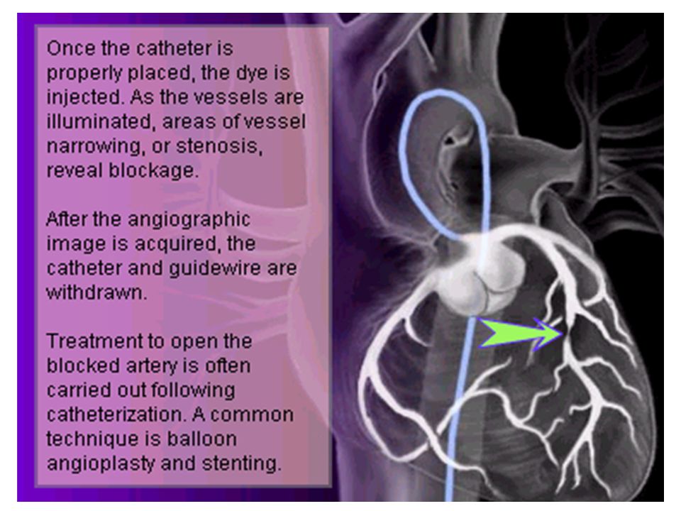
HOW TO KNOW OF HEART DISEASE? BP measurement Blood cholesterol measurements Blood sugar measurements E C G Echocardiogram / Doppler T M T / Stress Test X-ray Angiography

18
E C G (ELECTROCARDIOGRAM) An electrocardiogram (ECG) is a recording of the heart's electrical activity as a graph or series of wave lines on a moving strip of paper. This gives the doctor important information about the heart.

19
POSITION OF ECG LEADS 6 or 12 E C G leads are placed on the chest to obtain the ECG

20
AN E C G LOOKS LIKE THIS...

21
HOLTER MONITORING A Holter monitor is a portable ECG that monitors the electrical activity of a freely moving patient’s heart for one to five days, 24 hours around the clock

22
ECHOCARDIOGRAPHY An Echocardiogram uses sound waves to image the heart

23
Echo is used to examine the - size, shape, and motion of the heart, It is useful to diagnose abnormalities of the heart valves and to assess heart’s function

24
T M T (TREAD MILL TEST / STRESS TEST) An ECG performed while the patient exercises in a controlled manner on a treadmill or stationary bicycle at varied speeds and elevations. This test helps detect heart irregularities, disease and damage. The outcome of T M T is similar to the E C G

25
SCAN Depending on the precision required the following scans can be performed X Ray CT Scan MRI Scan Spect Thallium Scan Least Precise Most Precise All of these are painless procedures wherein X rays are taken

26
An X-ray shows The size of the heart and arteries The condition of the lungs The condition of surrounding structures

44
“Early in life people give up their health to gain wealth…. ….In later life, people give up their wealth to regain some of their health.” - KEN BLANCHART & MARJORIE

Similar presentations


 Courtesy of Graham and Emma.>")


>")


