Download presentation
Presentation is loading. Please wait.

1
Rad T 216 Venipuncture Anatomy and Equipment Overview

2
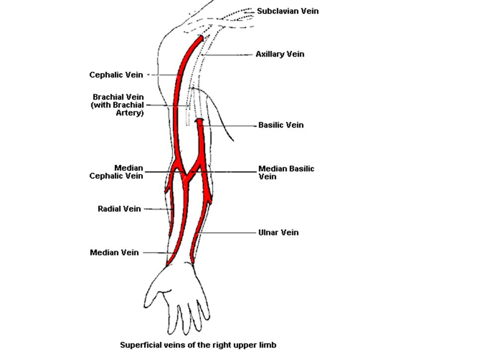
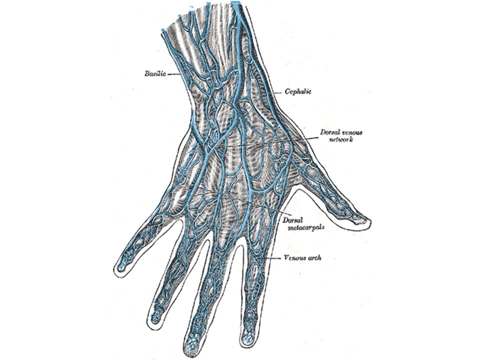
Limitations SB 571 limits Radiologic Technologists to performing venipuncture access on the upper extremity.

3
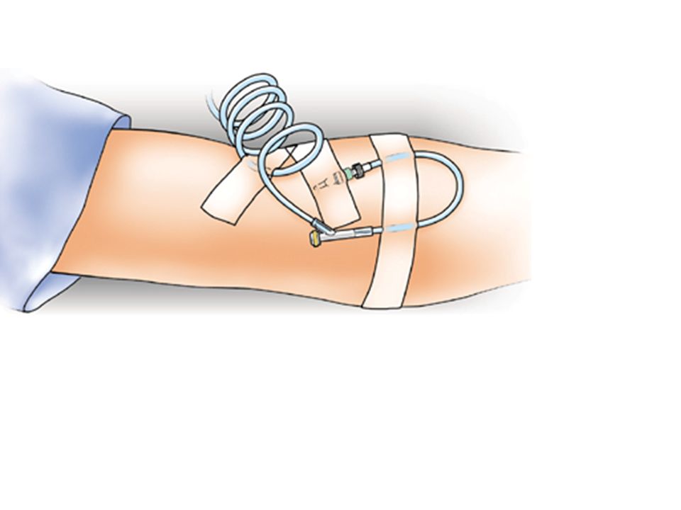
Over the needle catheter Butterfly needle

4
Types of Contrast Infusion Direct IV Push –This will be the primary method you use as a technologist. It can also be called a bolus injection. Drip Infusion –Used when you want to administer contrast over a period of time. Some MR contrast agents use this method.

5
Piggy back –For us, piggy backing would be to use an existing port for the injection. –To do this safely, you have to determine the patency of the existing line and what is already being administered through it. –Ringer’s, D5W, and normal saline are typically considered safe to inject with. NEVER inject with blood products or other medications.

12
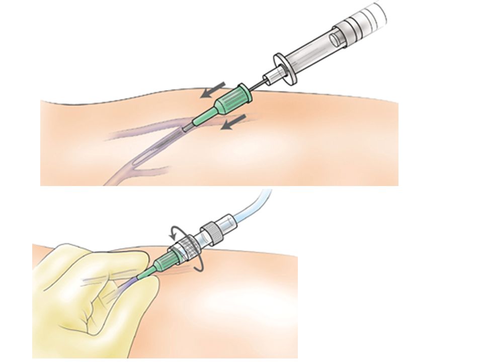
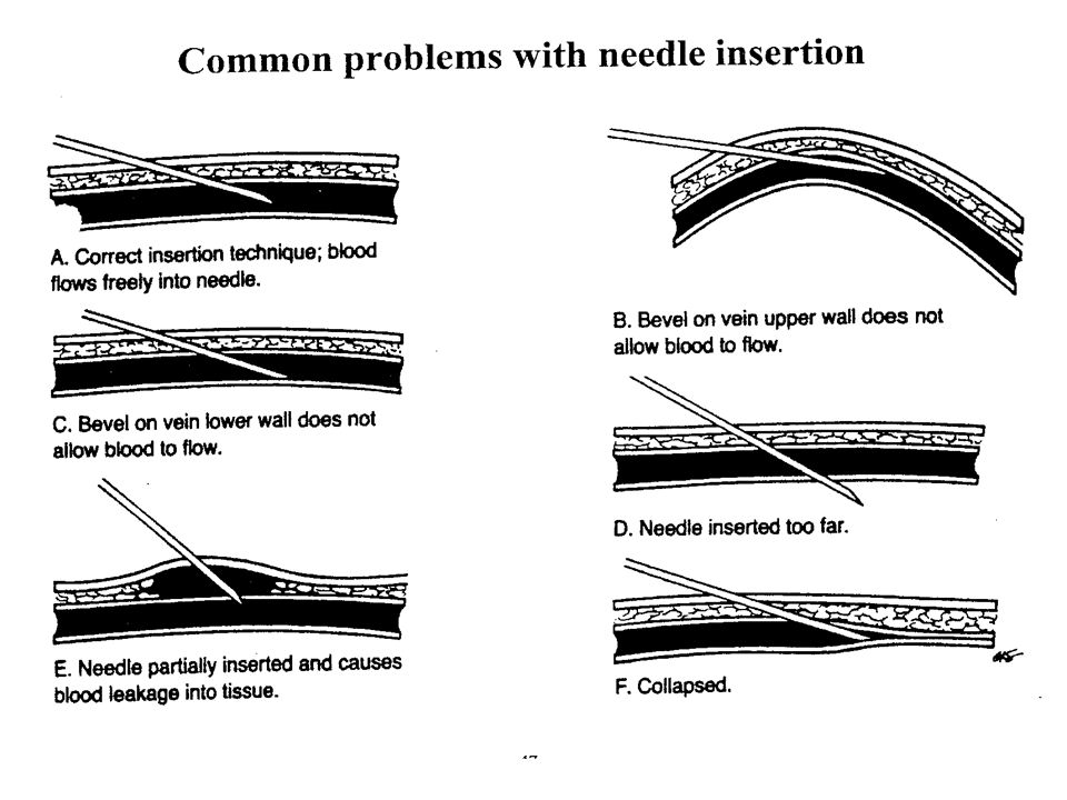
As you can see, the needle should enter at a 15 degree angle to the skin. The easiest way to get the angle is to ‘run’ the needle over your thumb. Larger patients may require a steeper angle to access the vein.

13
Problems with IV therapy Phlebitis

14
cellulitis bruising

15
Ulceration desquamination infiltration

Similar presentations


 involves injecting a medication directly into the blood via venous access devices IV products must be sterile.>")


 VTDRG: Chapter 8 (pg: 349-351)>")


