Download presentation
Presentation is loading. Please wait.

1
Management of Patients with Renal Disorders

2
Glomerular Diseases Acute Glomerulonephritis
Chronic Glomerulonephritis Nephrotic Syndrome

3
Glomerulonephritis

4
Acute Glomerulonephritis
Preceded (10 days) by an infection Assess for: Lesions Signs of circulatory overload Change in urine color and amount Mild to moderate hypertension Interventions: Treat cause: antibiotics, corticosteroids, immunosuppressants Restrict sodium, water, potassium, protein Dialysis, plasmapheresis Client education Three types: postinfectious, rapidly progressive glomerulonephritis, and membranous glomerulonephritis Manifestations: hematuria, edema, azotemia, proteinuria, and hypertension May be mild or progress to acute renal failure. Usually recover quickly and comletely. Medical management is supportive and focuses on treatment of underlying cause.
 by an infection. Assess for: Lesions. Signs of circulatory overload. Change in urine color and amount. Mild to moderate hypertension. Interventions: Treat cause: antibiotics, corticosteroids, immunosuppressants. Restrict sodium, water, potassium, protein. Dialysis, plasmapheresis. Client education. Three types: postinfectious, rapidly progressive glomerulonephritis, and membranous glomerulonephritis. Manifestations: hematuria, edema, azotemia, proteinuria, and hypertension. May be mild or progress to acute renal failure. Usually recover quickly and comletely. Medical management is supportive and focuses on treatment of underlying cause.")
5
Nursing Management- Acute Glomerulonephritis
Patient assessment Maintain fluid balance Fluid and dietary restrictions Patient education Follow-up care

6
Chronic Glomerulonephritis
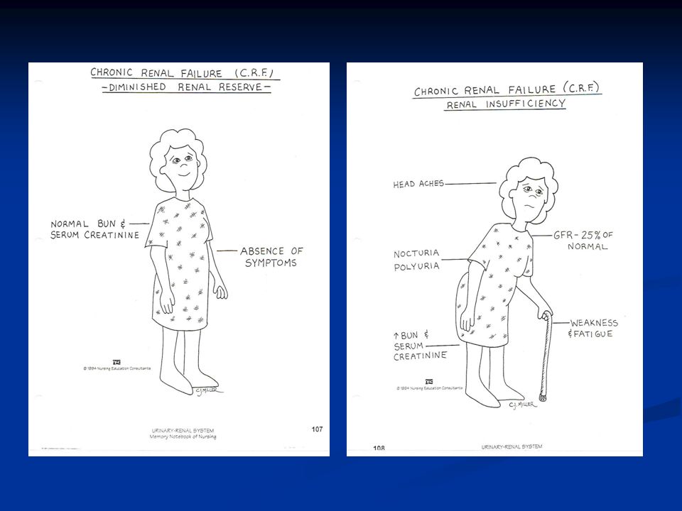
years to develop Diagnostics: Urine with fixed specific gravity, casts, and proteinuria Electrolyte imbalances hypoalbuminemia Causes: Repeated episodes of acute glomerulonephritis, hypertensive nephrosclerosis, hyperlipidemia, Manifestations: Mild proteinuria and hematuria, hypertension, and occasional edema

7
Nursing Management: Chronic Glomerulonephritis
Assessment Potential fluid and electrolyte imbalances Cardiac status Neurologic status Emotional support Teaching self-care

8
Nephrotic Syndrome Increased glomerular permeability
Severe loss of protein into urine Treatment: Immunosuppresive agents ACE Inhibitors Heparin Diet changes Mild diuretics

9
Nephrotic Syndrome

10
Nephrosclerosis Narrowing of vessel lumen from thickening in blood vessels of the nephron Occurs with hypertension, atherosclerosis and diabetes mellitus Collaborative management: Control hypertension Preserve renal function

11
Renal Failure Results when kidney’s cannot remove wastes or perform regulatory functions Systemic disorder resulting from many different causes Acute renal failure- reversible syndrome that results in decreased GFR and oliguria Chronic renal failure- progressive; irreversible deterioration of renal function resulting in azotemia

12
Acute Renal Failure Pathophysiology
Types of acute renal failure include: Prerenal Intrarenal Postrenal

13
Phases of Acute Renal Failure
Phases of rapid decrease in renal function lead to the collection of metabolic wastes in the body. Phases include: Onset Diuretic Oliguric Recovery Acute syndrome may be reversible with prompt intervention.

14
Assessment History Clinical manifestations Laboratory assessment
Radiographic assessment Other diagnostic assessments such as renal biopsy

15
Drug Therapy Cardioglycides Vitamins and minerals
Biologic response modifiers Phosphate binders Stool softeners and laxatives Monitor fluids Diuretics Calcium channel blockers Not parallel

16
Treatment Diet therapy Dialysis therapies Hemodialysis
Peritoneal dialysis

17
Renal Replacement Therapy
Standard treatment Dialysate solution Vascular access Continuous arteriovenous hemofiltration Continuous venovenous hemofiltration

18
Posthospital Care If renal failure is resolving, follow-up care may be required. There may be permanent renal damage and the need for chronic dialysis or even transplantation. Temporary dialysis is appropriate for some clients. S&P

19
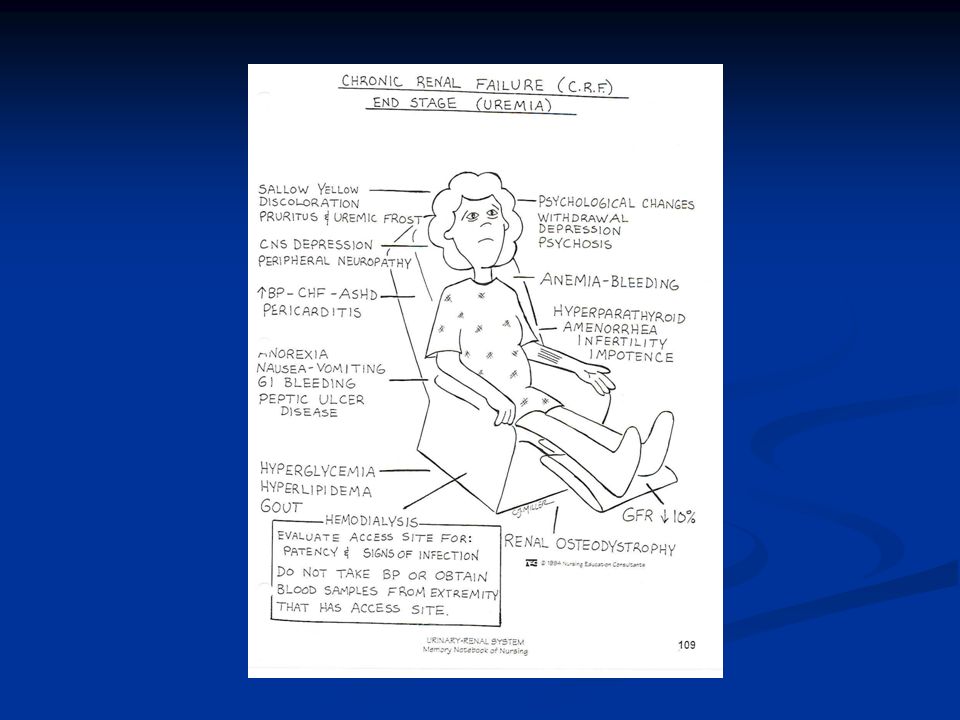
Chronic Renal Failure Progressive, irreversible kidney injury; kidney function does not recover Azotemia Uremia Uremic syndrome

20
Stages of Chronic Renal Failure
Diminished renal reserve Renal insufficiency End-stage renal disease

23
Changes R/T CRF Kidney Metabolic Electrolytes Acid-base balance
Urea and creatinine Electrolytes Sodium Potassium Acid-base balance Calcium and phosphorus (Continued)
")
24
Changes R/T CRF (Continued)
Cardiac Hypertension Hyperlipidemia Congestive heart failure Uremic pericarditis Hematologic Gastrointestinal

25
Clinical Manifestations
Neurologic Cardiovascular Respiratory Hematologic Gastrointestinal Urinary Skin

26
Imbalanced Nutrition: Less Than Body Requirements
Interventions include: Dietary evaluation for: Protein Fluid Potassium Sodium Phosphorus Vitamin supplementation

27
Excess Fluid Volume Interventions: Monitor client’s intake and output.
Promote fluid balance. Assess for manifestations of volume excess: Crackles in the bases of the lungs Edema Distended neck veins Drug therapy includes diuretics.

28
Decreased Cardiac Output
Interventions: Control hypertension with calcium channel blockers, ACE inhibitors, alpha- and beta-adrenergic blockers, and vasodilators. Instruct client and family to monitor blood pressure, client’s weight, diet, and drug therapy.

29
Risk for Infection Interventions include: Meticulous skin care
Preventive skin care Inspection of vascular access site for infection Monitoring of vital signs for manifestations of infection

30
Risk for Injury Interventions include: Drug therapy
Education to prevent fall or injury, pathologic fractures, bleeding, and toxic effects of prescribed drugs S&P

31
Fatigue Interventions:
Assess for vitamin deficiency, anemia, and buildup of urea. Administer vitamin and mineral supplements. Administer erythropoietin therapy for bone marrow production. Give iron supplements as needed. S&P

32
Anxiety Interventions include: Health care team involvement
Client and family education Continuity of care Encouragement of client to ask questions and discuss fears about the diagnosis of renal failure S&P

33
Potential for Pulmonary Edema
Interventions: Assess the client for early signs of pulmonary edema. Monitor serum electrolyte levels, vital signs, oxygen saturation levels, hypertension.

34
Hemodialysis Client selection Dialysis settings
Works using passive transfer of toxins by diffusion Anticoagulation needed, usually heparin treatment S&P

35
Vascular Access Arteriovenous fistula, or arteriovenous graft for long-term permanent access Hemodialysis catheter, dual or triple lumen, or arteriovenous shunt for temporary access Precautions Complications

36
Permanent Vascular Access

37
Hemodialysis Nursing Care
Postdialysis care: Monitor for complications such as hypotension, headache, nausea, malaise, vomiting, dizziness, and muscle cramps. Monitor vital signs and weight. Avoid invasive procedures 4 to 6 hours after dialysis. Continually monitor for hemorrhage.

38
Complications of Hemodialysis
Dialysis disequilibrium syndrome Infectious diseases Hepatitis B and C infections HIV exposure—poses some risk for clients undergoing dialysis S&P

39
Peritoneal Dialysis Procedure involves siliconized rubber catheter placed into the abdominal cavity for infusion of dialysate. Types of peritoneal dialysis: Continuous ambulatory peritoneal Automated peritoneal Intermittent peritoneal Continuous-cycle peritoneal

40
Complications Peritonitis Pain Exit site and tunnel infections
Poor dialysate flow Dialysate leakage Other complications

41
Nursing Care During Peritoneal Dialysis
Before treating, evaluate baseline vital signs, weight, and laboratory tests. Continually monitor the client for respiratory distress, pain, and discomfort. Monitor prescribed dwell time and initiate outflow. Observe the outflow amount and pattern of fluid.

42
Nursing Management of Hospitalized Client on Dialysis
Protect vascular access Monitor fluid balance indicators Monitor IV carefully Assess for s/s uremia Monitor cardiopulmonary status carefully Monitor BP Monitor medications Address pain and discomfort Infection control measures Monitor dietary e-lytes and fluids Skin care CAPD catheter care if appropriate

43
Renal Transplantation
Candidate selection criteria Donors Preoperative care Immunologic studies Surgical team Operative procedure

44
Postoperative Care Assessment Urologic management Complications
all body systems Pain Fluid and electrolyte status Urologic management Assessment of system patency Assessment of urine output hourly for 48 hours. Complications Rejection Acute tubular necrosis Thrombosis Renal artery stenosis Other complications Immunosuppressive drug therapy Psychosocial preparation

45
Post-transplantation Intervetions
Pain relief measures and analgesics Promote airway clearance and effective breathing pattern Strict asepsis Monitor for signs/symptoms of bleeding Encourage leg exercises, early ambulation, and monitor for signs of DVT

46
Renal Cell Carcinoma Healthy kidney tissue damaged and replaced by cancer cells Paraneoplastic syndrome: Anemia Erythrocytosis Hypercalcemia Liver dysfuntion Hormonal effects Increased sedimentation rate Hypertension

47
Renal Cell Carcinoma Management
Nonsurgical Radiofrequency ablation Chemotherapy Biological response modifiers and tumor necrosis factor lengthen survival time Renal artery embolization Surgical Pre-op care Nephrectomy Post-op care: Monitoring for hemorrhage and adrenal insufficiency Pain management Prevention of complications

48
Renal Trauma Minor injuries: Major injuries: Nonsurgical management:
Contusions, small lacerations Major injuries: Lacerations to cortex, medulla, or branches or renal artery Nonsurgical management: Drug and fluid therapy Surgical management: Nephrectomy or partial nephrectomy

Similar presentations
activate vitamin D (renal 1-alpha hydroxylase) produces erythropoietin.>")


 Dr. Belal Hijji, RN, PhD April 18 & 23, 2012.>")


>")


