Download presentation
Presentation is loading. Please wait.

1
LYMPHATIC SYSTEM.

2
I. What is the lymphoid system? A. System for returning fluid to cardiovascular system. C. Comprises an organ/vascular system that protects the body’s internal environment from the onslaught of foreign substances (i.e. bacteria, viruses, etc.), and malignant growths. D. 3 basic types of lymphoid tissue: a. unencapsulated b. incompletely encapsulated c. completely encapsulated. B. Pathway for cells of the immune system to move through.
, and malignant growths. D. 3 basic types of lymphoid tissue: a. unencapsulated b. incompletely encapsulated c. completely encapsulated. B. Pathway for cells of the immune system to move through..")
3
II. The lymphoid system consists of 2 major components, A. The lymph vascular network - two functions, 1. Acts to return extracellular fluids to blood circulatory system. 2. Pathway for certain cells of the immune system to move through, as well as re-enter the circulatory system. http://www.cs.stedwards.edu/~kswank/LymphSyst.html

4
http://rex.nci.nih.gov/PATIENTS/INFO_TEACHER/bookshelf/NIH_immune/html/imm04.html B. Lymph organs and regions of diffuse lymphoid tissue 1. These organs/tissues are 2. Serve at least two roles: a. Filter cellular and particulate debris b. Sites of residence, activation and proliferation of cells of the immune system. 3. Diffuse lymphoid tissue - aggregations of lymphocytes and other immune system cells - usually not permanent. These may or may not be nodular. a. lymph nodes b. spleen c. thymus d. tonsils and adenoids e. appendix f. aggregations of lymph nodules such as Peyer’s patches g. bone marrow

5
Encapsulated Thymus Spleen Lymph nodes Incompletely encapsulated Tonsils, adenoids Unencapsulated (sometimes called diffuse lymphoid tissue) MALT - e.g. Peyer’s patches Lymphoid tissue in appendix Temporary aggregations of lymphocytes and other immune system cells Classification based on encapsulation or lack thereof.

6
III. Tissues of lymphoid organs consist of two major cell types, A. Fixed cells 1. Reticular cells - connective tissue cells that secrete fine matrix of reticular fibers which these cells extend cytoplasmic processes through and around. (reticulocyte is a bad term for these cells since one stage of erythrocyte maturation is also called a reticulocyte). 2. Follicular dendritic cells (FDCs) - found in germinal centers of lymph nodules - bind antigens for later interaction with B-lymphocytes. http://www.finchcms.edu/anatomy/histology/organology/lymphoid/images/ff614.jpg
. 2. Follicular dendritic cells (FDCs) - found in germinal centers of lymph nodules - bind antigens for later interaction with B-lymphocytes.")
7
B. Motile cells 1. Macrophages - antigen presenting cells http://www.pbrc.hawaii.edu/~kunkel/catalog/by_category/medical/page001/08974a.html http://wsrv.clas.virginia.edu/~rjh9u/macro.htmlCopyright Dennis Kunkel Microscopy http://www.cellsalive.com/ a. differentiate from monocytes that leave blood. monocyte macrophage b. phagocytose particulates and bacteria c. involved in initiation of humoral and cell-mediated immune response

8
B. Motile cells 2. Various classes of T-lymphocytes http://www.cellsalive.com/ctl.htm b. involved in cell mediated immune response a. involved in initiation of humoral immune response 3. B-lymphocytes, including plasma cells that secrete specific antibodies.

9
IV. Immune system functions of the lymphoid system A. There are 3 major cell types involved in an immune response. 1. Antigen presenting cells (macrophages, dendritic cells) - process antigens and present them to T- lymphocytes, this will result in the activation of these cells which will then initiate humoral and cell-mediated immune responses. 2. T-lymphocytes - once activated as above, these cells may be responsible for cell-mediated immune responses or they may be involved in mediating the activation of B-lymphocytes to produce antibodies in a humoral immune response. 3. B- lymphocytes - responsible for antibody production - the humoral immune response to an antigen that results in the secretion of antibodies by B- lymphocytes that have become plasma cells.
 - process antigens and present them to T- lymphocytes, this will result in the activation of these cells which will then initiate humoral and cell-mediated immune responses. 2. T-lymphocytes - once activated as above, these cells may be responsible for cell-mediated immune responses or they may be involved in mediating the activation of B-lymphocytes to produce antibodies in a humoral immune response. 3. B- lymphocytes - responsible for antibody production - the humoral immune response to an antigen that results in the secretion of antibodies by B- lymphocytes that have become plasma cells..")
10
B. What is an immune response: 1. Involves the recognition of foreign antigens by certain lymphatic cells a. Antigen - a molecule that, in its make-up, has characteristics that will cause the activation of certain cells in the immune system. b. The portions of foreign molecules that immune system cells recognize are called epitopes. c. Epitopes are often parts of protein molecules that form part of the cell membrane or wall of organisms that invade the bodies tissues. 2. An immune response results in changes in the gene activity and metabolism of B- and/or T- lymphocytes that allows them to act in destroying the foreign substance or organism

11
C. An immune response may be either humoral or cell-mediated. 1. Humoral immune response - secretion of antibodies that bind to a foreign antigen by B-lymphocyte plasma cells. a. Mediated by interaction between macrophage, T-lymphocyte and B-lymphocyte b. Causes clonal proliferation of activated B-lymphocytes c. ”Mature” (activated) B-lymphocytes are plasma cells or memory B-lymphocytes http://www.cellsalive.com/antibody.htm Take a look at Figure 14-6 and corresponding text on pp. 260-261 in your text.
 B-lymphocytes are plasma cells or memory B-lymphocytes Take a look at Figure 14-6 and corresponding text on pp in your text..")
12
* Plasma cells are short lived and secrete copious amounts of antibody that are specific for a given antigen ** Antibodies act to identify foreign cells for attack by other components of the immune system such as T-lymphocytes and macrophages. ** Antibodies can also identify particulates and viruses for phagocytosis and destruction by other leucocytes such as neutrophils. * B-lymphocyte memory cells remain dormant and will rapidly respond to future encounters with the same antigen by rapid clonal proliferation and differentiation into plasma cells that produce the needed antibody.

13
2. Cell-mediated immune response - antigen activates T-lymphocytes to produce cytotoxic substances that cause the destruction of the antigen containing cell D. Without the activities of cells of the lymphoid system, your life on earth after birth would be brief indeed. a. Mediated by interaction between macrophage and T-cytotoxic (Tc) lymphocytes b. Causes clonal proliferation of Tc lymphocytes c. ”Mature" (activated) Tc lymphocytes differentiate into either Tc memory cells or Tc effector cells * Tc effector cells - actively kill invading foreign cells * Tc memory cells remain dormant and will rapidly respond to future invasions by foreign cells expressing the same antigen
 lymphocytes b. Causes clonal proliferation of Tc lymphocytes c. Mature (activated) Tc lymphocytes differentiate into either Tc memory cells or Tc effector cells * Tc effector cells - actively kill invading foreign cells * Tc memory cells remain dormant and will rapidly respond to future invasions by foreign cells expressing the same antigen.")
14
V. There are 2 basic types of lymphoid tissue, A. Non-nodular lymphoid tissue 1. Unencapsulated, sub-epithelial, aggregations of lymphocytes - diffuse lymphoid tissue - can occur anywhere in body 2. Non-nodular aggregations of lymphocytes in lymphoid organs such as the thymus, lymph nodes and spleen B. Nodular lymphoid tissue - “spherically” arranged aggregations of lymphocytes with a distinct cortex and medulla (germinal center) that are called lymphatic nodules. 1. Nonencapsulated lymph nodules (diffuse lymphatic tissue) 2. Nodules in lymph nodes * Some texts will identify the thymus as having nodular lymphatic tissue; however, this is not really correct if nodules are as described above. 3. Nodules in spleenic white pulp 4. MALT (mucosa-associated lymphoid tissue) – nodular aggregations of lymphoid tissue that are associated with the lining of the digestive tract, e.g., tonsils, appendix, Peyer’s patches,. These tissues comprise the largest (admittedly diffuse) lymphoid organ in the body and contain about 70% of the body’s immune cells.
 that are called lymphatic nodules. 1. Nonencapsulated lymph nodules (diffuse lymphatic tissue) 2. Nodules in lymph nodes * Some texts will identify the thymus as having nodular lymphatic tissue; however, this is not really correct if nodules are as described above. 3. Nodules in spleenic white pulp 4. MALT (mucosa-associated lymphoid tissue) – nodular aggregations of lymphoid tissue that are associated with the lining of the digestive tract, e.g., tonsils, appendix, Peyer’s patches,. These tissues comprise the largest (admittedly diffuse) lymphoid organ in the body and contain about 70% of the body’s immune cells..")
15
VI. Structure and function of lymphoid organs A. Lymphatic nodule 4. Mainly found in the lamina propria of the digestive tract, the respiratory tract, and urinary passages. Also a component of the lymph nodes, spleen, tonsils and appendix. 3. No connective tissue capsule directly surrounds individual lymphatic nodules 2. Nodule consists of a cortex and the medullary germinal center. 1. Consists of dense aggregation of lymphocytes that includes reticular cells, macrophages and follicular dendritic cells. ****

16
4. Nodules may be classified as “primary” or “secondary” http://www.finchcms.edu/anatomy/histology/organology/lymp hoid/o_l_7.html a. primary - germinal center not very evident b. secondary - germinal center very evident

17
3. Lymphatic nodules - general composition: a. Fixed cells - reticular cells in cortex and follicular dendritic cells in germinal center. b. Free cells - B-lymphocytes, including immunoblasts (clonally dividing B-lymphocytes) plasma cells, macrophages, a few eosinophils and mast cells. * Mostly small lymphocytes * Cortical cells are mostly B- lymphocytes. * Many of the medullary cells are activated B-lymphocytes undergoing clonal divisions - most of the resulting cells will mature into plasma cells. http://www.udel.edu/Biology/Wags/histopage/colorpage/cly/cly.htm
 plasma cells, macrophages, a few eosinophils and mast cells. * Mostly small lymphocytes * Cortical cells are mostly B- lymphocytes. * Many of the medullary cells are activated B-lymphocytes undergoing clonal divisions - most of the resulting cells will mature into plasma cells.")
18
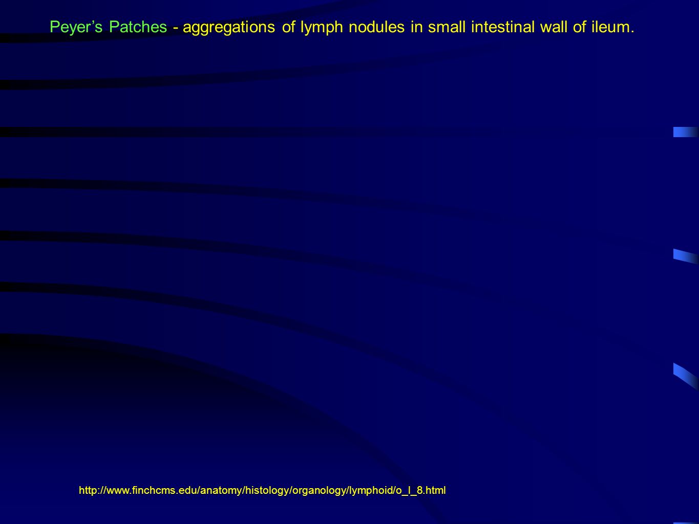
Peyer’s Patches - aggregations of lymph nodules in small intestinal wall of ileum. http://www.finchcms.edu/anatomy/histology/organology/lymphoid/o_l_8.html
19
B. Lymph nodes 1. Oval or bean shaped bodies present along the course of lymphatic vessels. Multiple lymph vessels may connect to a lymph node. 2. Contains a stroma consisting of reticular cells and fibers and large numbers of lymphocytes, all surrounded by a connective tissue capsule. 3. Reticular cells in the stroma provide a physical support network for the lymphocytes. Essentially an aggregation of lymphatic nodules with a connective tissue capsule around them.

20
6. Lymph node structure: c. The dense cortical stroma consists of numerous lymphatic nodules. d. A less dense medulla consisting of lymphocytes arranged in strands called medullary cords. * Lymph sinuses in medulla * Cords and sinuses extend toward a central hilus that is essentially a large trabecula connected to the connective tissue capsule. a. Surrounded by a connective tissue capsule - trabeculae that extend radially into the node b. A subcapsular sinus separates the cortical stroma from the connective tissue capsule.

21
** Arteries enter and veins and lymph vessels exit through the hilus ** Blood vessels branch into the cortical regions from arteries in the hilus - these give rise to "bulbs" of capillaries within the germinal centers of lymph nodules ** Lymph sinuses in the medulla empty into lymph vessels that exit via the hilus.

22
7. Between the cortex and medulla is the paracortical region or thymus dependent zone of the node that contains densely packed cells that are mainly T-lymphocytes. a. This region lacks lymphocytes in animals that have had the thymus removed at birth. 8. Cells outside the paracortical region are mostly B-lymphocytes.

23
Normal lymph node http://www.path.sunysb.edu/hemepath/tutorial/normal_node/nl_node_image.htm http://www.siumed.edu/~dking2/crr/CR034b.htm http://www.auburn.edu/academic/classes/zy/hist0509/index_histology.html

24
C. Tonsils - 3 types that are defined by their structure and their location in mouth and pharynx. 1. Palatine tonsils c. Overlying epithelium forms invaginations called crypts that penetrate into the band of nodules. a. On left and right in rear area of oral cavity. b. Dense lymphoid tissue that forms a band of lymphatic nodules that lie just below stratified squamous epithelium. http://commons.wikimedia.org/wiki/File:Throat_with_Tonsils_0011J.jpeg

25
d. These crypts act as collecting places for cellular debris and bacteria as well as a place where live lymphocytes and macrophages can wander about. e. The band of lymph nodules is separated from underlying tissues by a partial capsule of dense connective tissue. 1. Palatine tonsils http://www.entusa.com/oral_pictures_htm/acute_tonsillitis_2.htm

26
Portion of a palatine tonsil http://www.udel.edu/Biology/Wags/histopage/colorpage/cly/cly.htm

27
2. Pharyngeal tonsils (Adenoids) http://www.udel.edu/Biology/Wags/histopage/colorpage/cly/cly.htm a. Lie beneath a typical ciliated pseudostratified columnar respiratory epithelium in rear roof of pharynx. b. Diffuse lymphoid tissue containing nodules, but no crypts. c. A thin capsule of dense connective tissue underlies the lymphoid tissue. http://hcd2.bupa.co.uk/images/factsheets/Tons_aden_uvul_427x240.jpg
 a. Lie beneath a typical ciliated pseudostratified columnar respiratory epithelium in rear roof of pharynx. b. Diffuse lymphoid tissue containing nodules, but no crypts. c. A thin capsule of dense connective tissue underlies the lymphoid tissue.")
28
a. Situated at base of tongue. b. Each lingual tonsil consists of numerous. lymphoid nodules surrounding a single crypt c. The crypt is lined by a stratified squamous epithelium. non-keratinzed 3. Lingual tonsils http://www.georgetown.edu/dml/educ/micro/lymph/9.htm

29
D. Thymus 1. Location - situated over the heart where great vessels connect. 2. Important during early life when the cellular mediated component of the immune system develops. Undergoes atrophy in later life, at which time it loses its functional significance. 4. The thymus consists of multiple lobes each containing a cortical and medullary region. 5. A connective tissue capsule surrounds the thymus. http://www-micro.msb.le.ac.uk/MBChB/2b.html 3. The thymus can be considered a proliferation and maturation center for T- lymphocytes.

30
7. Thymus - general structure The cortical and medullary zones of lobules are continuous. thymic lobule

31
8. Cell types found in the active thymus are: a. T-lymphoblasts - stem cells for additional t- lymphocytes - mitotically dividing. b. T-lymphocytes c. Reticular cells d. Macrophages e. Plasma cells f. Mast cells g. Unidentified mesenchymal cells http://www.lab.anhb.uwa.edu.au/mb140/CorePages/Lymphoid1/Lymph1.htm

32
9. Cortical layer of thymus a. Site of T-lymphocyte production - divisions of t- lymphoblast cells. b. Thus, there is considerable mitotic activity of t- lymphoblasts c. Reticular cells are less numerous in this area and have thin and long processes that envelope groups of developing thymocytes. Reticular cells isolate groups of dividing and maturing t-lymphocytes in young animals.

33
10. Medullary zone a. Contains many reticular cells and fewer T- lymphoblasts and lymphocytes than the cortex. b. Also contains specialized structures known as Hassall's corpuscles - function not certain* Consist of a central, eosinophilic, hyaline core surrounded by concentric layers of reticular cells Potent source of cytokine -thymic stromal lymphopoietin (TSLP) that directs maturation of dendritic cells. http://www.lab.anhb.uwa.edu.au/mb140/CorePages/Lymphoid1/Lymph1.htm http://med-ed.med.virginia.edu/med-ed/ histology/imagedisplay.cfm?file=Cell0376
 that directs maturation of dendritic cells. histology/imagedisplay.cfm file=Cell0376.")
34
11. Blood supply a. Branches from the internal thoracic and inferior thyroid arteries penetrate the capsule surrounding the thymus b. Extend into thymus along interlobular septa. c. Capillaries branch into the cortico-medullary junction area and extend into the cortex. d. These eventually extend into the medulla where they drain into venules e. Venules connect to veins that exit thymus along connective tissue septa. f. There are no afferent lymphatic vessels connecting to the thymus. So it does not act as filter for lymphatic fluids. g. Only a few efferent lymphatic vessels are present, and these are associated with the blood vessels. Precursor t-lymphocyte stem cells migrate from bone marrow to thymus entering the organ via blood vessels in medullary zone. These cells undergo mitosis and maturation in cortical zone and then leave thymus through blood vessels of medullary zone to go about their various activities.

35
12. Blood-thymus barrier - only present in the cortex, acts to prevent most blood born foreign antigens from reaching developing thymocytes. - presumed important in allowing T- lymphocytes to develop properly. This barrier consists of : a. Non-fenestrated, continuous endothelium of blood capillaries b. Desmosome connections forming tight junctions between adjacent endothelial cells of capillaries, as well as similar connections between surrounding reticular cells c. Pericytes and reticular cells that form a sleeve around the capillaries in addition to surrounding connective tissue. d. Thick basal lamina from reticular cells that surround capillaries e. Macrophages that are present in the surrounding connective tissue. In the medulla, the sheath cells of the blood-thymus barrier are lost and vessels become permeable - cells can move in and out.

36
E. Spleen 1. General characteristics a. Largest piece of lymphatic tissue in body. b. Can be said to act as filter of blood both in an immunologic sense and also in the sense of removing worn out erythrocytes from circulation. http://www-micro.msb.le.ac.uk/MBChB/2b.html

37
2. Functions of the spleen a. Production of blood cells during embryogenesis * In embryo, erythrocytes, neutrophils, basophils, and eosinophils are produced in spleen. This stops about the time of birth. c. Recycling of ferritin from worn out erythrocytes for synthesis of hemoglobin * Bilirubin is returned to the blood and carried to the liver where it is excreted and passed out of body as part of the bile. b. Destruction of erythrocytes * Worn out erythrocytes are phagocytosed and digested by macrophages in the spleen. * Hemoglobin is broken down into bilirubin ( open chain of four pyrroles rather than ring of 4 pyrroles as in heme group ) and ferritin.pyrrole Bilirubin
 and ferritin.pyrrole Bilirubin.")
38
d. Immune response * Site of activation of both T- and B-lymphocytes * These cell types interact with dendritic cells (form from monocytes) that act as antigen presenting cells. e. Storage of erythrocytes that can be released into circulatory system when needed. 2. Functions of the spleen
 that act as antigen presenting cells. e. Storage of erythrocytes that can be released into circulatory system when needed. 2. Functions of the spleen.")
39
3. Structure of spleen a. Surrounded by a dense connective tissue capsule that extends processes (trabeculae) into lymphatic tissue of this organ. * Connective tissue contains nerves, blood vessels,lymph vessels, and smooth muscle. * A hilum (hilus) of connective tissue is present medially.
 into lymphatic tissue of this organ. * Connective tissue contains nerves, blood vessels,lymph vessels, and smooth muscle. * A hilum (hilus) of connective tissue is present medially..")
40
* Blood vessels and nerves run through the hilus and enter the spleenic pulp via the trabeculae. There are no lymph vessels in the pulp. * Pulp is divided into lymphatic nodules of white pulp, surrounded by a spongy lymphatic tissue called red pulp. * Color designations have to do with appearance in freshly cut open organ. http://www.lab.anhb.uwa.edu.au/mb140/

41
b. Red pulp * Forms spongy reticular tissue that is composed of cords of cellular (connective) tissue that surround blood sinusoids (cavities). http://www.vh.org/Providers/Textbooks/MicroscopicAnatomy/Section09/Plate09172.html http://medocs.ucdavis.edu/CHA/402/studyset/lab9/list.htm http://www.vh.org/Providers/Textbooks/MicroscopicAnatomy/Section09/Plate09175.html
 tissue that surround blood sinusoids (cavities)")
42
* The “strands” of reticular connective tissue between blood sinuses in red pulp are called the cords of Bilroth. The reticular C.T. supports the cells associated with it. Theodore Billroth, Austrian surgeon (1829 - 1894) - father of abdominal surgery. * Cell types present - macrophages, monocytes, lymphocytes, plasma cells, and various other blood cells (i.e. granulocytes and erythrocytes). * Blood sinusoids are present - sites of cellular exchange between spleen and circulatory system. Cells can enter or leave spleen through large spaces between endothelial cells lining sinusoids. * Supported by a network of reticular cells and their associated fibers. http://www.vh.org/Providers/Textbooks/MicroscopicAnatomy/ Section09/Plate09175.html http://www.lab.anhb.uwa.edu.au/mb140/ http://medocs.ucdavis.edu/CHA/402/studyset/lab9/list.htm
 - father of abdominal surgery. * Cell types present - macrophages, monocytes, lymphocytes, plasma cells, and various other blood cells (i.e. granulocytes and erythrocytes). * Blood sinusoids are present - sites of cellular exchange between spleen and circulatory system. Cells can enter or leave spleen through large spaces between endothelial cells lining sinusoids. * Supported by a network of reticular cells and their associated fibers. Section09/Plate09175.html")
43
http://www.lab.anhb.uwa.edu.au/mb140/ c. White pulp *Concentrations of lymphatic tissue within the red pulp that surround portions of central arteries and form lymphatic nodules. *Nodules consist of reticular mesh with spaces in mesh being filled with lymphocytes and some macrophages. “Central” artery is identifying characteristic. http://www.vh.org/Providers/Textbooks/MicroscopicAnatomy/Section09/Plate09172.html * Both B- and T-lymphocytes in white pulp. T-lymphocytes in sheath surrounding central artery (periarterial lymphatic sheath - PALS), B- lymphocytes in white pulp surrounding sheath (i.e. peripheral white pulp - PWP) and in the germinal center of the nodule. * Other cell types present - monocytes, plasma cells.
, B- lymphocytes in white pulp surrounding sheath (i.e. peripheral white pulp - PWP) and in the germinal center of the nodule. * Other cell types present - monocytes, plasma cells..")
44
*Marginal zone acts as a filter to pull foreign antigens out of blood so that lymphocytes can react to them and be induced to participate in an immune response. http://www.upei.ca/~morph/webct/Modules/Lymphoid/spleen.html *Marginal zone surrounds white pulp nodules - contains few lymphocytes, but many actively phagocytic cells with branching processes (dendritic cells - antigen presenting cells).
..")
45
The End

Similar presentations


 -mast cells -interdigitating.>")


 from the lamina propria. Absorb.>")