Download presentation
Presentation is loading. Please wait.

1
Announcements Tomorrow’s quiz on NEJM paper Bring two questions about NEJM Exam I provided today

2
Which of the following enzymes work most effectively at a very low pH?
Salivary amylase Trypsin Pepsin Pancreatic amylase Pancreatic lipase

3
In zero gravity environment of space, how does food swallowed by an astronaut reach her stomach?
Swallowing hard Running around wheel creates -> artificial gravity Involuntary muscle contractions IV administration of nutrients

4
Which of the following organs is incorrectly paired with its function?
Stomach –protein digestion Oral cavity -starch digestion Large intestine –bile production Small intestine –nutrient absorption Pancreas –enzyme production

6
Salivary glands Secretes enzymes that digest carbohydrates
1. Mouth Mechanical and chemical processing (chewing reduces size of food; saliva digests carbohydrates) Salivary glands Secretes enzymes that digest carbohydrates Liver Secretes molecules required for digestion of fats 2. Esophagus Transports food 3. Stomach Mechanical and chemical processing (digestion of proteins) Gall bladder Stores secretions from liver; empties into small intestine 4. Small intestine Chemical processing and absorption (digestion of proteins, fats, carbohydrates; absorption of nutrients and water) Figure 40.7 The Alimentary Canal Caption: The alimentary canal is a tube that runs from the mouth to the anus. The salivary glands, liver, gallbladder, and pancreas are not part of the canal itself. Instead, they secrete material into the tube at specific points. Pancreas Secretes enzymes and other materials into small intestine 5. Large intestine Water absorption and feces formation
 Salivary glands Secretes enzymes that digest carbohydrates. Liver Secretes molecules required for digestion of fats. 2. Esophagus Transports food. 3. Stomach Mechanical and chemical processing (digestion of proteins) Gall bladder Stores secretions from liver; empties into small intestine. 4. Small intestine Chemical processing and absorption (digestion of proteins, fats, carbohydrates; absorption of nutrients and water) Figure 40.7 The Alimentary Canal. Caption: The alimentary canal is a tube that runs from the mouth to the anus. The salivary glands, liver, gallbladder, and pancreas are not part of the canal itself. Instead, they secrete material into the tube at specific points. Pancreas Secretes enzymes and other materials into small intestine. 5. Large intestine Water absorption and feces formation.")
7
Stomach End of esophagus Sphincter seals off stomach from small
stomach from esophagus Sphincter seals off stomach from small intestine Figure: 40.8a Caption: The stomach is a muscular outpocketing of the alimentary canal. Muscle contractions mix food and break it into smaller pieces. Layers of muscle Beginning of small intestine Lumen (interior)
")
8
Stomach lining Canal empties to lumen Parietal cells (secrete HCl)
Chief cells (secrete pepsinogen) Figure: 40.8b Caption: This transmission electron micrograph shows a section through the stomach wall. Exercise: In part (b), draw the route of molecules and ions secreted by parietal and chief cells.
 Figure: 40.8b. Caption: This transmission electron micrograph shows a section through the stomach wall. Exercise: In part (b), draw the route of molecules and ions secreted by parietal and chief cells.")
9
Secretion of HCI by parietal cells
HCl to lumen CO H2O H2CO3 To blood H+ H+ HCO3– HCO3– K+ H+/K+ pump Cl– Figure: 40.8c Caption: According to available data, this is how parietal cells secrete hydrochloric acid. Exercise: In part (c), label the reaction catalyzed by carbonic anhydrase. Cl– Cl– Cl– Chloride channel From blood Canal empties to lumen Parietal cell
, label the reaction catalyzed by carbonic anhydrase. Cl– Cl– Cl– Chloride channel. From blood. Canal empties. to lumen. Parietal cell.")
10
Enzymes, Hormones, other?
Enzymes: pepsinogen (inactive)-> pepsin (protease) from chief cells Other: HCl -> denature ECM bacteria, from parietal cells, activator of pepsin Other: Mucins/mucus protective from goblet or mucus cells Other: mechanical churning -> acid chyme Hormones: Gastrin (+), CCK/secretin (-), enterogastrone (-- pyloric sphinct)
-> pepsin (protease) from chief cells. Other: HCl -> denature ECM bacteria, from parietal cells, activator of pepsin. Other: Mucins/mucus protective from goblet or mucus cells. Other: mechanical churning -> acid chyme. Hormones: Gastrin (+), CCK/secretin (-), enterogastrone (-- pyloric sphinct)")
11
Enzymes, Hormones, other?
Your turn

13
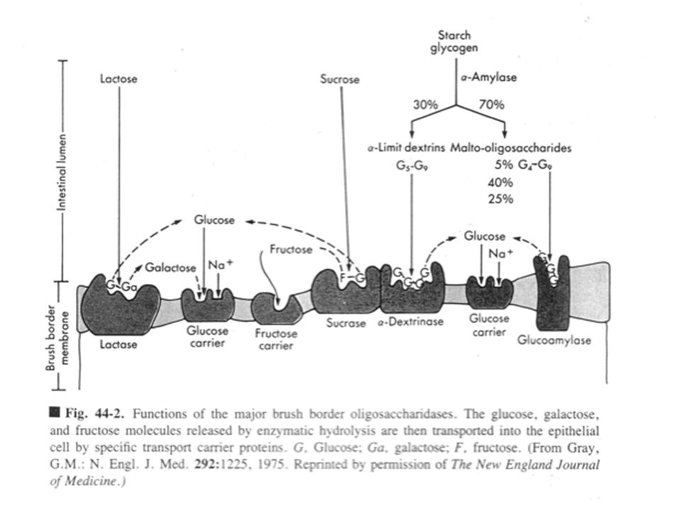
DIGESTION OF LIPIDS IN SMALL INTESTINE
Glycerol Lipase Fatty acids 1. Large fat globules are not digested efficiently by lipase. 2. Bile salts (produced in liver) act as emulsifying agents. 3. Small fat droplets result from emulsification. 4. Lipase digests the small fat droplets into glycerol and free fatty acids. Figure 40.10 Caption: Bile salts are small lipids that act as emulsifying agents. Once they break up large fat globules, lipase can digest fats efficiently.
 act as. emulsifying agents. 3. Small fat droplets result from emulsification. 4. Lipase digests the small fat droplets into glycerol and free fatty acids. Figure Caption: Bile salts are small lipids that act as emulsifying agents. Once they break up large fat globules, lipase can digest fats efficiently.")
14
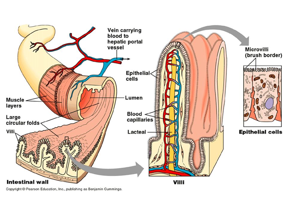
Cross-section of small intestine
Fold Villi Figure: left Caption: The transmission electron micrograph shows a cross section through the small intestine. 115 µm Cross-section of small intestine

15
Lumen of small intestine
Apical side Na+/glucose cotransporter H2O Osmosis Na+ Glucose K+ GLUT-2 transport protein Na+/K+-ATPase K+ ATP Basolateral side ADP Blood H2O Na+ Glucose Osmosis

18
Capillaries are small and extremely thin walled.

19
Insulin causes cells in the liver and skeletal
Glycogen Glucose Insulin causes cells in the liver and skeletal muscle to synthesize glycogen; fat storage cells synthesize triglycerides. Pancreas secretes INSULIN If glucose levels too high Glucose levels fall HOMEOTASIS (normal glucose levels in blood) If glucose levels too low Glucose levels rise Figure 40.13 Caption: Insulin and glucagon are both secreted by cells in the pancreas, but have opposite effects on blood glucose concentrations. EXERCISE Draw a large X through the arrow that is disrupted in individuals with diabetes mellitus. Next to the X, jot notes indicating the nature of the defect in individuals with type I diabetes mellitus versus type II diabetes mellitus. Pancreas secretes GLUCAGON Glucagon causes cells in liver and skeletal muscle to catabolize glycogen; fat storage cells catabolize fatty acids. Glucose Glycogen
 If glucose. levels too low. Glucose. levels rise. Figure Caption: Insulin and glucagon are both secreted by cells in the pancreas, but have opposite effects on blood glucose concentrations. EXERCISE Draw a large X through the arrow that is disrupted in individuals with diabetes mellitus. Next to the X, jot notes indicating the nature of the defect in individuals with type I diabetes mellitus versus type II diabetes mellitus. Pancreas secretes. GLUCAGON. Glucagon causes cells in liver and skeletal. muscle to catabolize glycogen; fat storage. cells catabolize fatty acids. Glucose. Glycogen.")
Similar presentations


>")
 100 120140160180 200 220 240 260 410 50 52 54 56 58 510.>")


, proteins, carbohydrates,>")


 into small molecules. Allows absorption of nutrients (ions and.>")


