Download presentation
Presentation is loading. Please wait.

1
Number of Entry Tears Is Associated With Aortic Growth in Type B Dissections Ann Thorac Surg March 28, 2013 Thoracic Aortic Research Center, University of Milano

2
Background Aortic growth rate in acute type B aortic dissection (ABAD) is a significant predictor for aortic complications and death. ABAD can be clinically classified as complicated or uncomplicated, and therapy given is based on this classification. Prompt intervention in patients with an ABAD is currently required in only complicated cases. For those uncomplicated ABAD patients managed conservatively by antihypertensive treatment. However, their medium-term and long-term outcome is less favorable due to aortic-related complications.

3
Previous clinical and bioengineering studies have demonstrated that increased inflow and impaired outflow of the false lumen can lead to a significant increase in the mean and diastolic pressure in the false lumen. In the number of entry tears might be important in predicting which ABAD patients will develop aortic dilatation. This study investigated whether the number of identifiable entry tears in ABAD patients is associated with aortic growth.

4
Patients and Methods Patients We retrospectively analyzed all computed tomographic angiography (CTA) data of ABAD patients who were medically treated in 4 referral centers between 2005 and 2010. CTA was obtained within 7 days of the initial diagnosis. A second CTA (follow-up CTA) was obtained at least 3 months after the baseline CTA and before any possible intervention. Exclude the patients who with a complete false lumen thrombosis at follow-up CTA.
 was obtained at least 3 months after the baseline CTA and before any possible intervention. Exclude the patients who with a complete false lumen thrombosis at follow-up CTA..")
5
Imaging All CTA images were acquired on multislice CT-scanners with 16-slice or 64-slice detector configuration. Noncontrast images were obtained first, from the top of the aortic arch to the bifurcation of the abdominal aorta. Biphasic protocols were used, with the acquisition of an arterial and venous phase. The arterial phase was started by a bolus-tracking technique in the descending aorta. The acquisition was started 7 seconds after a region of interest had reached 100 HU. 50 mL intravenous injection of nonionic contrast. The venous phase was scanned for 60 seconds after the start of the contrast medium injection.

6
Analysis The acquired CTA data sets were transferred to a 3Mensio Vascular 4.2 workstation for analysis. All CTA images were investigated by 2 investigators for the presence and number of entry tears over the entire length of the aorta. An entry tear was defined at least 2 different views. (transversal, coronal, sagittal, or center vessel reconstruction)
.")
7
Defined as a discontinuity of the aortic flap with a clear flow of contrast between the true and false lumen

8
Minimum and maximum diameters were calculated. (Diameter measurements were performed from the outer to the outer vessel wall). Aortic diameters were measured in the follow levels. 2 cm below the origin of the left subclavian artery (LSCA), 10 cm below the LSCA, 20 cm below the LSCA, 8 cm below the most distal renal artery, at the level of the largest point of the descending aorta.
. Aortic diameters were measured in the follow levels. 2 cm below the origin of the left subclavian artery (LSCA), 10 cm below the LSCA, 20 cm below the LSCA, 8 cm below the most distal renal artery, at the level of the largest point of the descending aorta..")
9
The annual growth rates at each affected level was calculated by dividing the difference between the diameter on the baseline scan and the last available CTA scan by the time interval between the 2 CTAs.

10
Statistics Statistical analysis of the number of entry tears in the different patient groups was performed with the use of the Kruskal-Wallis test and post hoc with the Mann- Whitney U test. Linear regression was used to analyze the influence of the distance of the proximal entry tear on aortic growth rate. Data are presented as mean standard deviation and range. Interobserver and intraobserver reliability was calculated using the multiobserver k statistic. Statistical analysis was performed using SPSS 20.0 software.

11
Results 60 ABAD patients (39 male) ; a median age of 59.7 years. The last available CTA was obtained after a median of 22 months (range 3 to 130 months). Of the 300 analyzed aortic segments, 243 (81%) were dissected.
. Of the 300 analyzed aortic segments, 243 (81%) were dissected..")
13
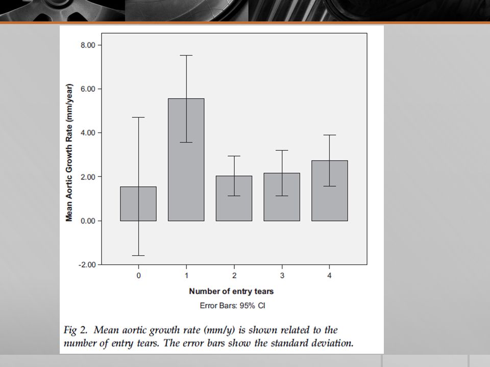
The median distance between the left subclavian artery (LSCA) and the primary entry tear was 38 mm (interquartile range, 24 to 137) and did not have a significant effect on aortic growth rate (p =0.434). Patients with 1 entry tear located within 5 cm of the LSCA showed significant more growth than their counterparts (5.8 + 7.7 vs 2.5 + 2.7 mm/y; p =0.003). Intraobserver agreement was substantial.
. Intraobserver agreement was substantial..")
14
Comment Our study demonstrated that the number of patent entry tears in patients with ABAD, on the baseline CTA scan, is associated with aortic growth. Previous clinical and bioengineering studies showed that in patients with ABAD, an impaired outflow of the false lumen might lead to a significant increase in mean arterial and diastolic pressure. Increased blood pressure in the false lumen is likely to result in a higher growth rate of the false lumen, potentially associated with aortic dilatation and rupture.

15
Although the mortality rate was higher in patients with a partially thrombosed false lumen than in patients with a completely patent false lumen, the peak aortic growth rate of these subsets were comparable. Sueyoshi and colleagues introduced the concept of the “sac formation,” defined as a partially closed false lumen at the reentry site resulting in a blind pouch with a persistent proximal entry tear. This imaging sign was associated with the largest increase in aortic diameter and these findings correlate with our study.

16
Currently, the role of partial false lumen thrombosis as predictor for aortic growth is not completely understood nor is its importance as cause of aortic- related death. In this context, the number of patent entry tears may further clarify this issue, because a limited number of entry tears between the true and false lumen might induce false lumen thrombosis.

17
Endovascular treatment for type B aortic dissection is focused on coverage of the proximal entry tear to reduce the false lumen pressurization, to promote false lumen thrombosis, and to favor aortic remodeling. Studies have shown that distal entry tears might serve as a renewed entry tear, which is associated with persistent patency of the false lumen and may result eventually in failure of therapy.

18
Our study showed that the location of an entry tear did not influence aortic growth directly, and aortic growth is still prone to develop in patients with an entry tear located more downstream. However, patients with 1 entry tear located within 5 cm of the LSCA tended to have higher aortic growth rates.

19
Theoretically, the same mechanism might result in the development of aortic dilatation in patients with 1 residual entry tear after thoracic endovascular aortic repair.

20
To minimize potential bias related to the reduced detection of entry tears, intraobserver and interobserver variability were conducted. This radiologic sign might be used at the primary risk assessment in uncomplicated ABAD patients, especially with improving imaging modalities.

21
The analysis has been based on the evaluation of the dissected aortic segments. The same methods adopted in previous studies on predictors of aortic growth in ABAD. This method allows the evaluations all the segments that are involved in the dissection, taking into consideration their differences in aortic growth rate.

22
Study limitations Clinical information of the study population was not available due to the multicenter character of the study. A certain number of entry tears could have been missed on the baseline CTAs ( because entry tears could conceivably be smaller than the slice thickness of our CTA scans). It is also possible that other imaging predictors might influence the aortic growth.
. It is also possible that other imaging predictors might influence the aortic growth..")
23
conclusion This study shows that the number of entry tears on a baseline CTA scan influence aortic growth in patients with conservatively treated ABAD. In those with 1 entry tear, an impaired outflow and changed flow pattern might be the cause of higher growth rates.

Similar presentations


 LECT7 ALI B ALHAILIY.>")


