Download presentation
Presentation is loading. Please wait.

1
Anatomy and Physiology Blood vessels

2
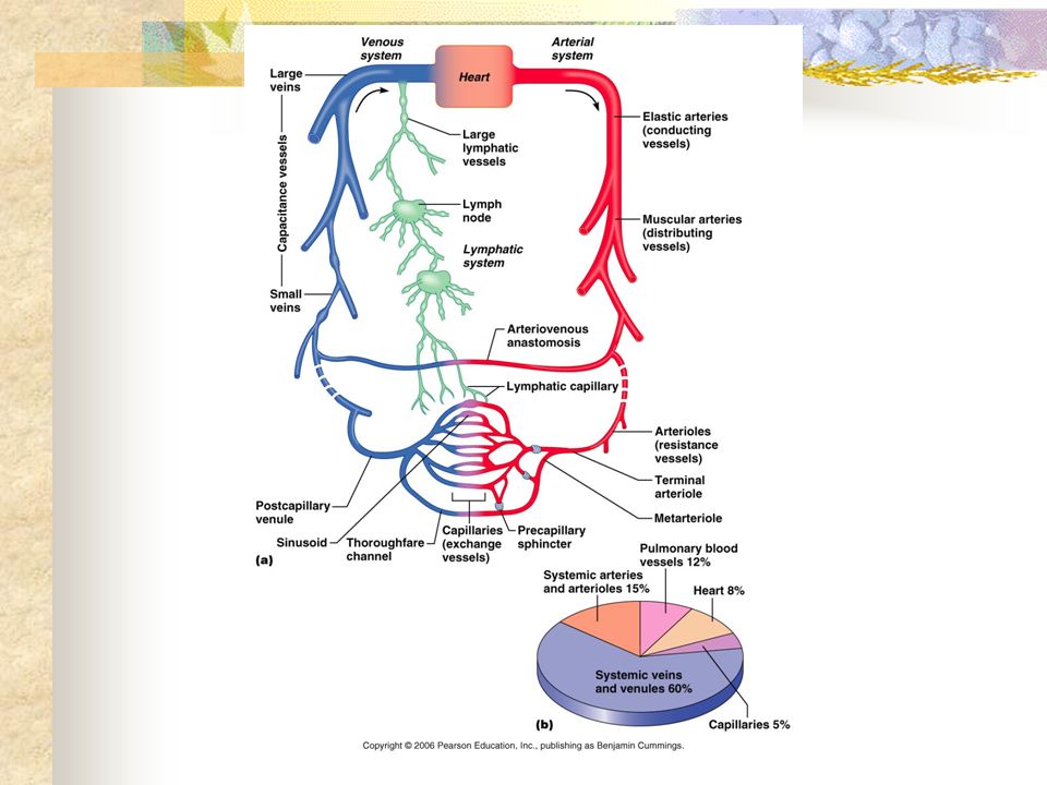
Blood vessel overview Blood travels from the heart through arteries. Initially these are large and very elastic. They soon become muscular and these arteries deliver blood to organs. Arteries become smaller until they become arterioles and finally they enter a network of capillaries. The blood moves through this capillary bed and then flows into small venules, then larger venules before becoming veins and delivering the blood back to the heart.

5
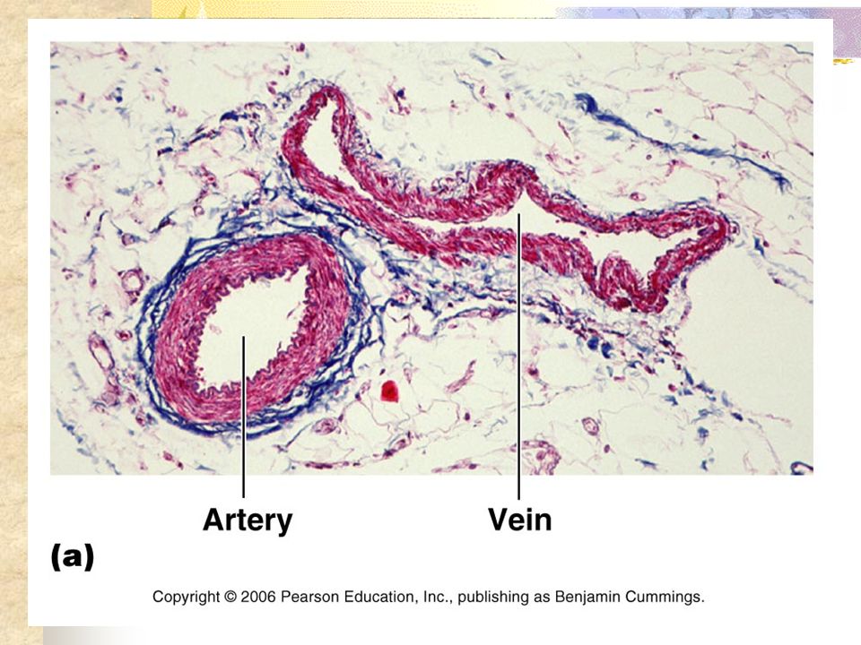
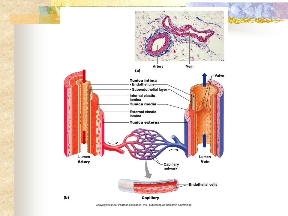
Blood vessel structure Arteries and veins have the same basic structure. The inside layer of the artery (left hand side) is composed of simple squamous epithelial cells called the endothelial layer or tunica intima. Next to that layer is the tunica media. The outside layer is called the tunica adventitia. Look carefully at this slide to visualise the three layers.
 is composed of simple squamous epithelial cells called the endothelial layer or tunica intima. Next to that layer is the tunica media. The outside layer is called the tunica adventitia. Look carefully at this slide to visualise the three layers..")
7
Arteries There are two types of arteries and these are elastic and muscular. Elastic arteries have thick walls and contain lots of elastin. You will remember that this is the main protein of elastic connective tissue. This gives the elastic arteries greater flexibility and blood pressure in these arteries remains relatively constant. On the other hand muscular arteries (which distribute blood to the organs) contain smooth muscle and it is in these arteries that blood pressure can change.
 contain smooth muscle and it is in these arteries that blood pressure can change..")
8
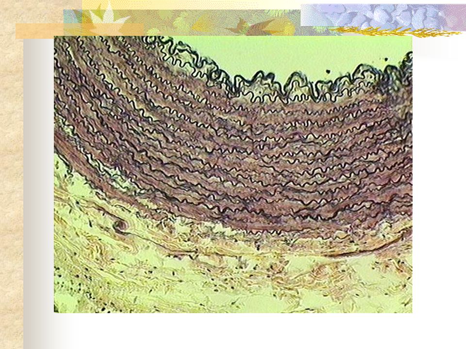
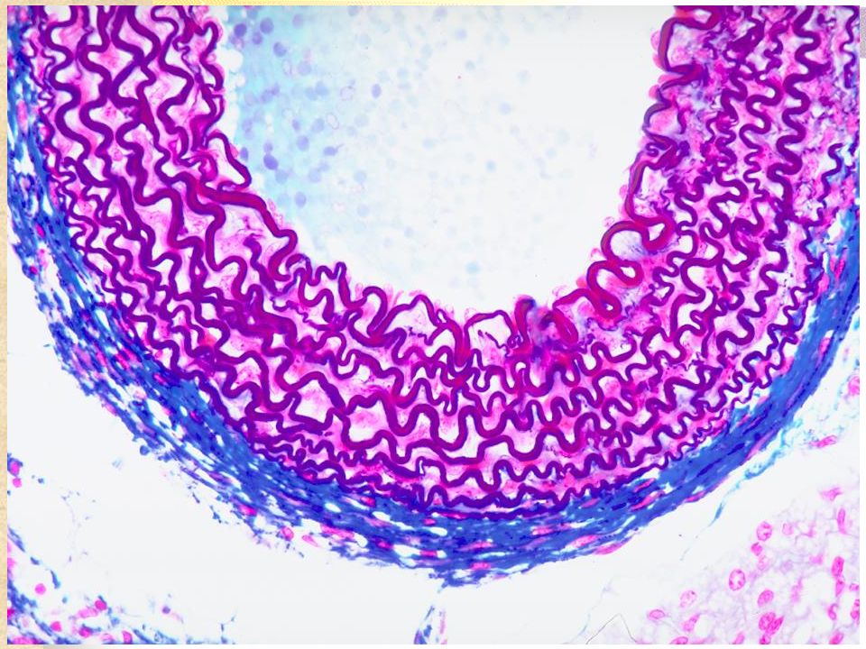
Large elastic artery In this slide you can see the tunica media as the layer with lots of coiled elastic tissue. The outside layer is called the tunica adventitia. This is the green layer and it is composed of connective tissue. This section of elastic artery has been stained with a special stain to show the layers and magnified about 500 times.

10
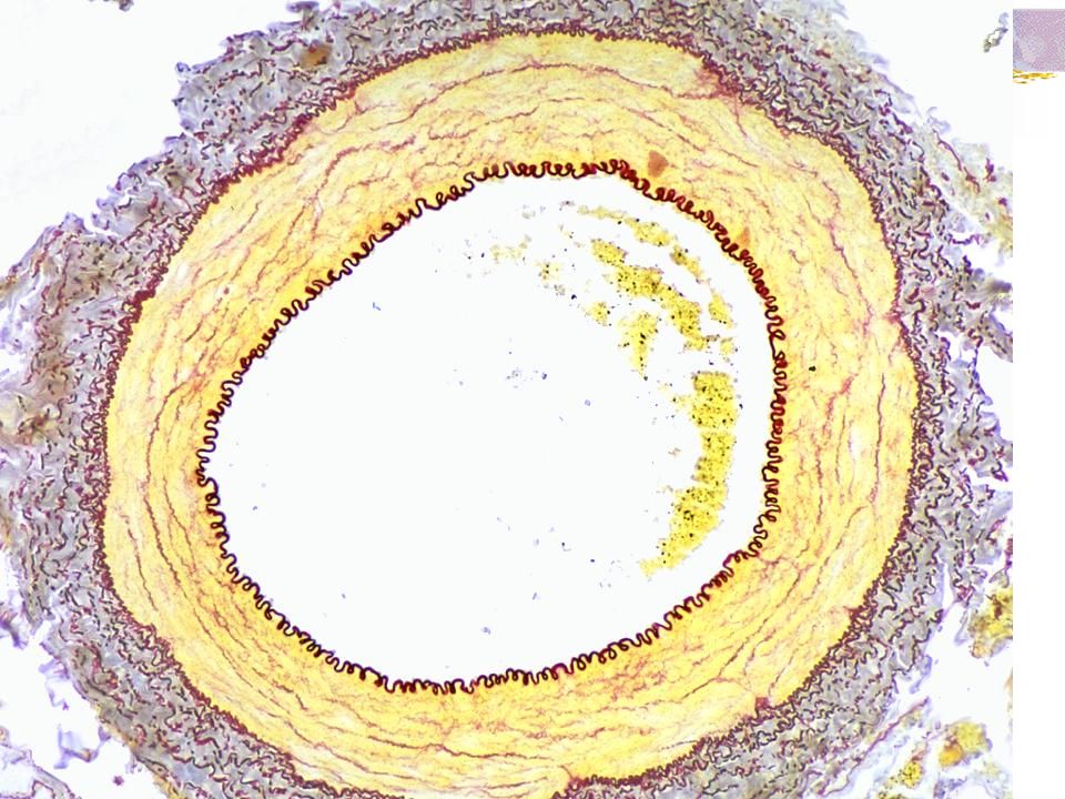
Small Elastic Artery (such as renal artery) As arteries become smaller the adventitia becomes smaller as does the tunica media. You cannot see the endothelial layer in this highly magnified small elastic artery but you can see the purple tunica media and the blue tunica adventitia.

12
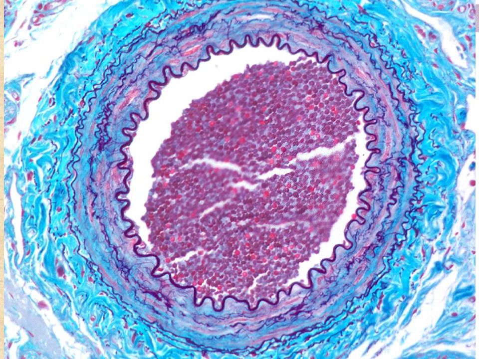
Muscular Artery Muscular arteries deliver blood to organs. The elastin of the elastic arteries is mostly replaced by smooth muscle cells and this limits the capacity for the artery to change shape. Unfortunately, damage to the endothelial layer through stress, cigarette smoking and a number of other causes can lead to cholesterol plaques building up and restricting blood flow. This can lead to what is called ischaemia or reduced blood flow.

14
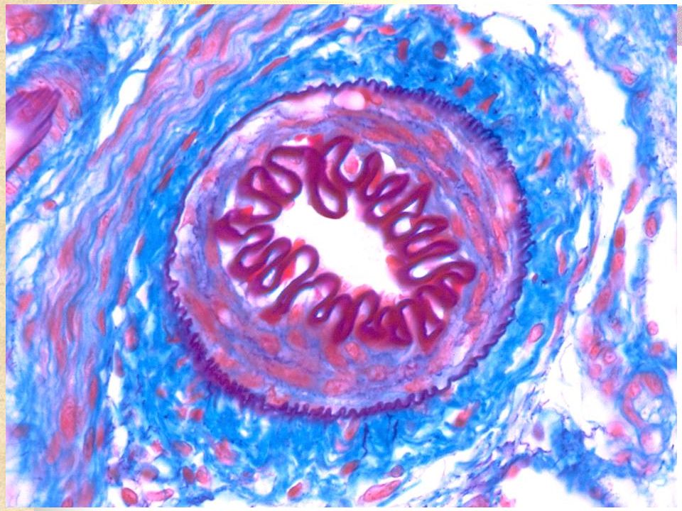
Arterioles Muscular arteries become arterioles when the tunica media is reduced in size to one or a few layers of smooth muscle cells. At the same time the tunica adventitia also reduces in size.

16
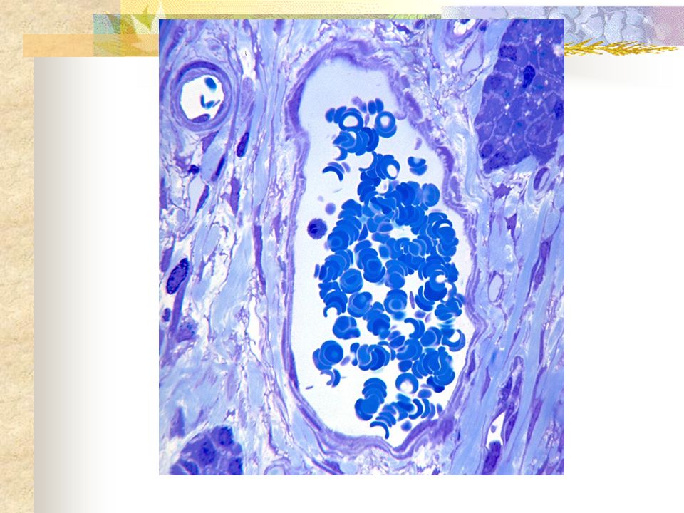
Arterioles Arterioles control blood flow by dilation or constriction. They contribute to peripheral resistance and a significant fall in blood pressure. You can see in this slide that the blood vessel has become quite small.

18
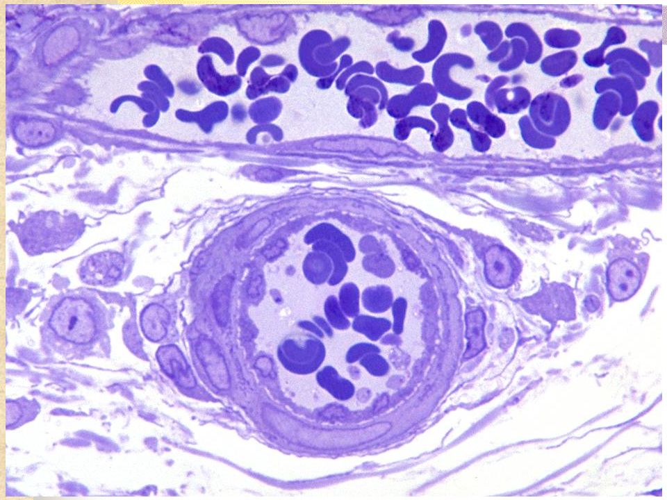
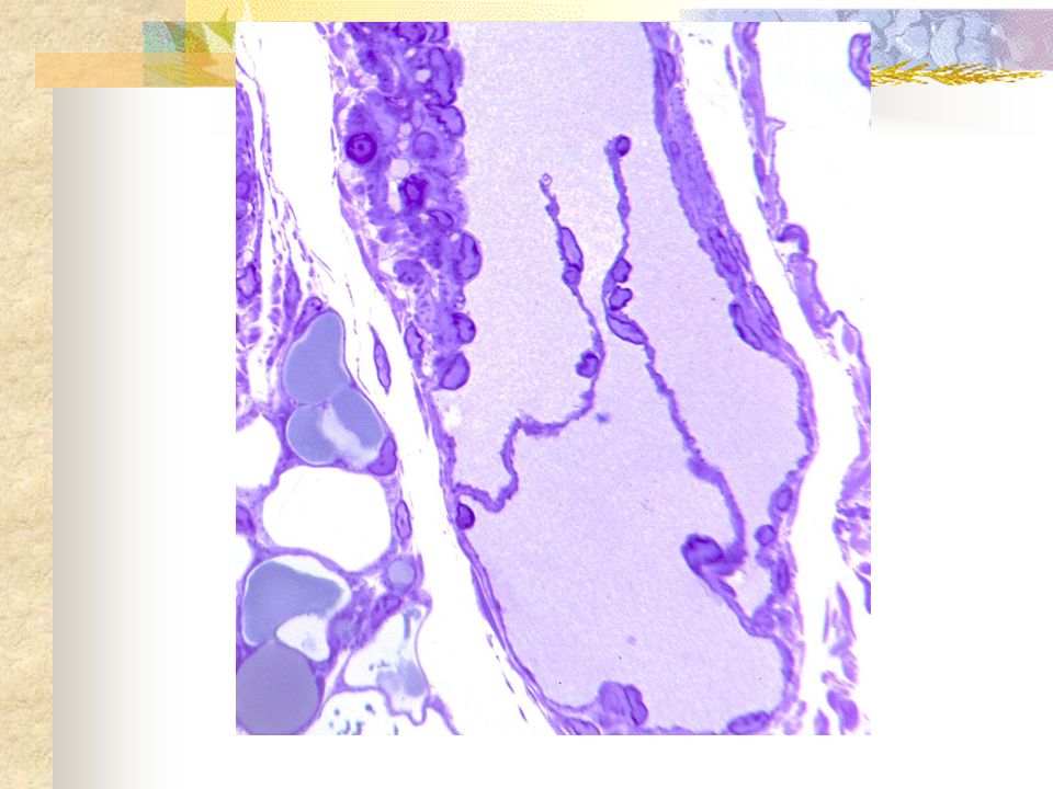
Small arterioles A small arteriole has one layer of tunica media smooth muscle and the endothelial layer. You can see the single layer of squamous endothelial cells in both blood vessels in this picture. Can you see the nuclei bulging out into the lumen of the blood vessel? This is because the nuclei of squamous cells is fatter than the cell itself. The cells in the middle of the pipes are red blood cells.

20
Capillaries Arteries carry blood away from the heart and veins carry blood back to the heart. These are connected by the capillary system. Capillaries consist of one layer of squamous epithelial cells. Blood flow is small and substance exchange occurs by osmosis and diffusion. Blood pressure drops significantly in capillaries because the blood that was running through one pipe is now running through many.

22
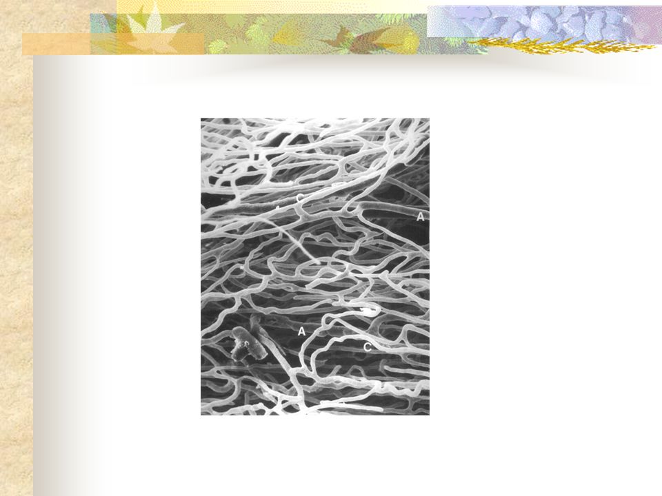
Capillaries of the heart Capillaries are all through our body. This is a section of heart to show you the dense capillary structure in the heart.

24
Venules The blood then leaves the capillaries and enters the venules on its way back to the heart. These have very thin walls and the endothelium is associated with a thin smooth muscle layer.

26
Venules As the venule becomes larger it adds smooth muscle. Here you can see a valve. When these are pushed together by blood flow they stop back flow. Have a look at your arm when flexed. You might see the valves running down the arm. These stop the blood running back down the arm between heartbeats

28
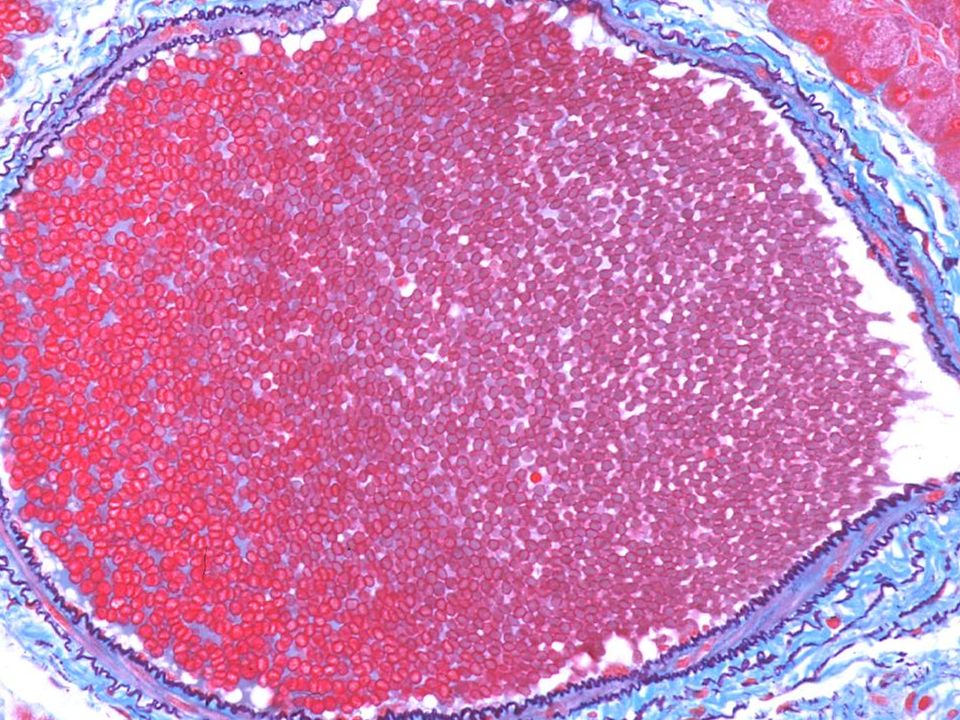
Medium Sized Vein A sympathetic nerve supply to the tunica media causes smooth muscle contraction and this assists blood flow back to the heart. Blood pressure in the veins is much lower than the arteries. On this slide you can see the smooth muscle layer.

30
Activity Make a schematic diagram of the blood vessel network in our body.

Similar presentations

. The Circulatory System is known as a CLOSED SYSTEM because the blood is contained within either the heart.>")

 Inner layer of arteries and veins Endothelium made of simple squamous epithelial cells.>")


