Download presentation
Presentation is loading. Please wait.

1
Chapt. 9 Regulation of Enzymes Regulation of Enzymes Student Learning Outcomes : Explain that enzyme activities must be regulated for proper body function Explain three general mechanisms: Reversible binding in active site: substrate, inhibitors Changing conformation of active site of enzyme: Allosteric effectors, covalent modification, Protein-protein interactions, zymogen cleavage (Changing concentration of enzyme) Synthesis, degradation
 Synthesis, degradation.")
2
Regulation of metabolic pathways Fig. 9.1 Metabolic pathway analogous to cars on highway: Flux of substrates affected by rate-limiting enzyme (barrier) Removal of barrier increases flow Activating rate-limiting enzyme
 Removal of barrier increases flow Activating rate-limiting enzyme.")
3
Regulation of glucose metabolism pathway Ex. Regulation of glucose metabolism pathway: Hexokinase & glucokinases convert glucose -> G-6-P in cells Glycolysis for energy Feedback regulation by ATP Store G-6-P as glycogen Feedforward by insulin

4
II. Regulation by substrate, product concentration Michaelis-Menten equation describes kinetics: More substrate gives more reaction, to maximal V i (initial velocity) relates to concentration of substrate [S] to V max (maximal velocity ) and K m ([S] for 1/2 V max Applies to simple reactions: E + S ES E + P ; k 1 = forward, k 2 back; k 3 for E+P V i = V max [S]/ K m + [S] K m = k 2 + k 3 /k 1 ; V max = k3[E t ]
 relates to concentration of substrate [S] to V max (maximal velocity ) and K m ([S] for 1/2 V max Applies to simple reactions: E + S ES E + P ; k 1 = forward, k 2 back; k 3 for E+P V i = V max [S]/ K m + [S] K m = k 2 + k 3 /k 1 ; V max = k3[E t ].")
5
II. Regulation by substrate, product concentration: Fig. 9.2 Ex. Graph of Michaelis-Menten equation has limit of V max at infinite substrate. K m = [S] where V max /2 Ex. Glucokinase Km 5 mM: If blood glucose 4 mM -> Vi = 0.44 Vmax (V m x 4mM/ (5mM + 4 mM) Blood glucose 20 mM -> Vi = 0.8 Vmax (V m x 20mM/ 5 + 20 mM
 Blood glucose 20 mM -> Vi = 0.8 Vmax (V m x 20mM/ mM.")
6
Different isozymes have different Km for glucose Fig. 9.3 Different hexokinases differ in K m for glucose: glucose + ATP -> G-6-P + ADP Hexokinase I (rbc) only glycolysis Glucokinase (liver, pancreas) storage Fasting blood sugar about 5 mM (90 mg/dL) so rbc can function even if low blood sugar of glucose S 0.5 = half-max for S-shape curve
 only glycolysis Glucokinase (liver, pancreas) storage Fasting blood sugar about 5 mM (90 mg/dL) so rbc can function even if low blood sugar of glucose S 0.5 = half-max for S-shape curve.")
7
Reversible inhibitors decrease reaction velocity Regulation through active site: reversible inhibitors A.Competitive inhibitors compete with substrate Overcome by excess substrate (increase apparent K m ) B.Noncompetitive do not compete with substrate Not overcome by substrate (lowers [E] and V max ) Fig. 9.4 Products can also inhibit enzyme activity
![Reversible inhibitors decrease reaction velocity Regulation through active site: reversible inhibitors A.Competitive inhibitors compete with substrate Overcome by excess substrate (increase apparent K m ) B.Noncompetitive do not compete with substrate Not overcome by substrate (lowers [E] and V max ) Fig.](http://images.slideplayer.com/24/6967747/slides/slide_7.jpg "9.4 Products can also inhibit enzyme activity.")
8
III. Regulation through conformational changes Regulation through conformational changes of enzyme can affect catalytic site: Allostery – ex. Glycogen phosphorylase Phosphorylation – ex. Glycogen phosphorylase kinase Protein-protein interactions - ex. Protein kinase A Proteolytic cleavage - ex. chymotrypsinogen

9
A. Allosteric Activators and inhibitors Allosteric enzymes : Often multimeric, Exhibit positive cooperativity in substrate binding (ex. Hemoglobin and O 2 ) T (taut state) inactive without substrate R (relaxed) state active with substrate Fig. 9.5
 T (taut state) inactive without substrate R (relaxed) state active with substrate Fig")
10
Allosteric activators and inhibitors Fig. 9.6 Allosteric enzymes often cooperative S binding Allosteric activators and inhibitors: Bind at allosteric site, not catalytic site Conformational change Activators often bind R (relaxed) state decrease S 0.5 Inhibitors often bind T (taut state ) increase S 0.5
 state decrease S 0.5 Inhibitors often bind T (taut state ) increase S 0.5.")
11
B. Conformational change by covalent modification Fig. 9.7 Phosphorylation can activate or inhibit enzymes: Protein kinases add phosphate Protein phosphatases remove PO 4 2- adds bulky group, negative charge, interacts with other amino acids

12
Muscle glycogen phosphorylase regulation Fig. 9.8 Muscle glycogen phosphorylase is regulated by both phosphorylation and/or allostery: Rate-limiting step glycogen -> glucose-1-PO 4 ATP use increases AMP - allostery phosphorylation increases activity Signal from PKA

13
Ex. Protein kinase A Protein kinase A: Regulatory, catalytic subunits: Ser/thr protein kinase, phosphorylates many enzymes Including glycogen phosphorylase kinase Adrenline increase cAMP, dissociates R subunits, Starts PO 4 cascade Fig. 9.9 cAMP activates PKA

14
Other covalent modifications affect proteins Covalent modifications affect protein activity, location in cell: acetyl- (on histones) ADP-ribosylation (as by cholera toxin on G subunit) Lipid addition (as on Ras protein) Fig. 6.13 modified amino acids

15
Conformational changes from Protein-Protein interactions Fig. 9.10 CaM kinase family activated by Ca 2+ /calmodulin; phosphorylate metabolic enzymes, ion channels, transcription factors, regulate synthesis, release of neurotransmitters. Ca-Calmodulin family of modulator proteins activated when [Ca 2+ ] increases. Ca 2+ /calmodulin binds to targets e.g. protein kinases, allosteric result

16
Small monomeric G proteins Small (monomeric) G proteins affect conformation of other proteins: GTP bound form binds and activates or inhibits GDP bound form inactive Other intermediates regulate the G proteins (GEF, GAP, etc) Ras family ( Ras, Rho, Rab, Ran, Arf ) diverse roles in cells Fig. 9.11

17
Proteolytic cleavage is irreversible Proteolytic cleavage is irreversible conformational change: Some during synthesis and processing Others after secretion: Proenzymes inactive: Ex. Precursor protease is zymogen: (chymotrypsinogen is cleaved by trypsin in intestine) Ex. Blood clotting factors fibrinogen, prothrombin
 Ex. Blood clotting factors fibrinogen, prothrombin.")
18
Regulation of pathways Regulation of metabolic pathways is complex: Sequential steps, different enzymes, rate-limiting one Match regulation to function of path Fig. 9.12

19
Lineweaver-Burk plot Fig. 9.13 Lineweaver-Burk transformation converts Michaelis-Menten to straight line (y = mx + b) double reciprocal plot Ease of determining K m and V max
 double reciprocal plot Ease of determining K m and V max.")
20
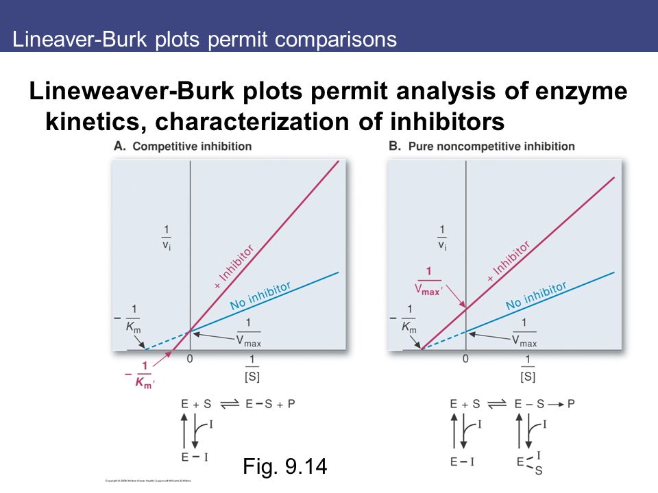
Lineaver-Burk plots permit comparisons Lineweaver-Burk plots permit analysis of enzyme kinetics, characterization of inhibitors Fig. 9.14
21
Key concepts Key concepts: Enzyme activity is regulated to reflect physiological state Rate of enzyme reaction depends on concentration of substrate, enzyme Allosteric activators or inhibitors bind sites other than the active site: conformational Mechanisms of regulation of enzyme activity include: feedback inhibition, covalent modifications, interactions of modulator proteins (rate synthesis, degradation)
")
22
Review questions 3. Methanol (CH 3 OH) is converted by alcohol dehydrogenases (ADH) to formaldehyde (CH 2 O), a highly toxic compound. Patients ingested toxic levels of methanol can be treated with ethanol (CH 3 CH 2 OH) to inhibit methanol oxidation by ADH. Which is the best rationale for this treatment? a.Ethanol is structural analog of methanol – noncompetitive inhibitor b.Ethanol is structural analog of methanol – will compete with methanol for binding enzyme c.Ethanol will alter the V max of ADH for oxidation of methanol. d.Ethanol is effective inhibitor of methanol oxidation regardless of the concentration of methanol e.Ethanol will inhibit enzyme by binding the formadehyde- binding site on the enzyme, even though it cannot bind the substrate binding site for methanol.
 is converted by alcohol dehydrogenases (ADH) to formaldehyde (CH 2 O), a highly toxic compound. Patients ingested toxic levels of methanol can be treated with ethanol (CH 3 CH 2 OH) to inhibit methanol oxidation by ADH. Which is the best rationale for this treatment. a.Ethanol is structural analog of methanol – noncompetitive inhibitor b.Ethanol is structural analog of methanol – will compete with methanol for binding enzyme c.Ethanol will alter the V max of ADH for oxidation of methanol. d.Ethanol is effective inhibitor of methanol oxidation regardless of the concentration of methanol e.Ethanol will inhibit enzyme by binding the formadehyde- binding site on the enzyme, even though it cannot bind the substrate binding site for methanol..")
Similar presentations


 –Stimulation and inhibition by control proteins –Reversible covalent modification.>")


 Enzyme Regulation.>")


